1. Disease Overview
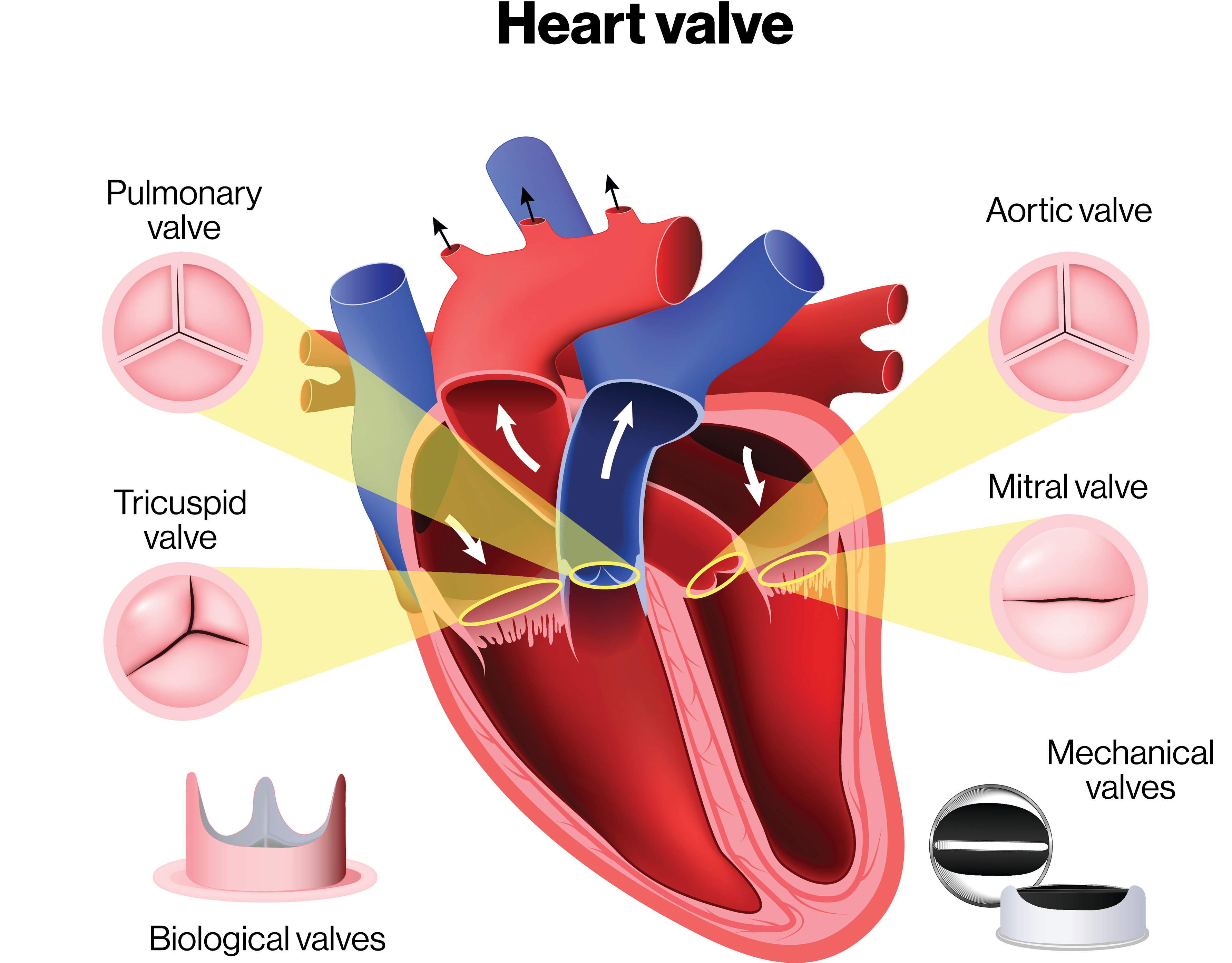
Combined multi-valvular disease (also called multiple valve disease or mixed valvular disease) occurs when two or more of the heart’s four valves are significantly diseased at the same time. The heart has four valves — the aortic, mitral, tricuspid and pulmonary — that keep blood moving in one direction. When more than one becomes narrowed (stenosis), leaky (regurgitation) or both, the heart faces several simultaneous mechanical problems, and the resulting strain is greater than the sum of any single lesion.
The most common combinations involve the aortic and mitral valves (the two left-sided, high-pressure valves), frequently as a legacy of rheumatic heart disease. Functional tricuspid regurgitation commonly accompanies long-standing left-sided disease as the right heart dilates. Because the lesions interact — one valve can mask or worsen another — diagnosis and treatment planning are more complex than for isolated valve disease.
Combined valve disease matters because it tends to be more advanced, more symptomatic, and higher-risk to operate on than single-valve disease, and surgery often means treating more than one valve in a single operation. With modern imaging, experienced heart-valve teams and improving transcatheter options, most patients can be helped substantially. Explore treatment options and top hospitals as you research.
2. Key Facts at a Glance
| Fact | Detail |
|---|---|
| Also known as | Multiple valve disease, mixed/combined valvular heart disease, polyvalvular disease |
| Body system affected | Cardiovascular system — heart valves, cardiac chambers, lungs, right heart |
| Common in | Adults with rheumatic heart disease; older adults with degenerative valve disease; people with prior endocarditis |
| Severity range | Mild and asymptomatic to severe, disabling heart failure |
| Key treatments | Medication, valve repair, single- or double-valve replacement, transcatheter therapies |
| Main tests | Echocardiography (transthoracic and transoesophageal), ECG, chest X-ray, cardiac CT/MRI |
| Outlook | Good with timely, well-planned intervention; poor if severe multi-valve disease is left untreated |
3. Alternative Names and Medical Terminology
- Multiple valve disease / multivalvular disease / polyvalvular disease
- Combined valvular heart disease or mixed valve disease
- Aortic-mitral valve disease (the most common left-sided combination)
- Mixed valve lesion — a single valve that is both stenotic and regurgitant
- Common abbreviations: AS (aortic stenosis), AR (aortic regurgitation), MS (mitral stenosis), MR (mitral regurgitation), TR (tricuspid regurgitation), RHD (rheumatic heart disease), VHD (valvular heart disease)
4. Relevant Heart, Lung or Vascular Anatomy
The heart has four one-way valves. On the left side, oxygen-rich blood flows from the lungs into the left atrium, through the mitral valve into the left ventricle, and out through the aortic valve into the aorta and body. On the right side, returning blood passes through the tricuspid valve into the right ventricle and out through the pulmonary valve to the lungs.
Because these valves work in series, disease in one affects the pressures and volumes seen by the others. Mitral valve disease, for example, raises pressure in the left atrium and lungs, straining the right heart and stretching the tricuspid valve. The aortic and mitral valves share the same fibrous skeleton (the aortic-mitral curtain), so rheumatic or infective processes often damage both together.
5. How the Disease Affects the Body
Each affected valve imposes its own burden, and in combination these burdens interact in ways that can be additive or offsetting. A stenotic valve makes the upstream chamber work harder to push blood through a narrow opening, causing it to thicken (hypertrophy). A regurgitant valve lets blood leak backward, so the chamber handles extra volume and eventually dilates.
When two valves are diseased, the heart may face pressure and volume overload at once. In combined aortic stenosis and mitral regurgitation, the left ventricle generates high pressure to overcome the tight aortic valve, but that same pressure drives more blood backward through the leaky mitral valve, worsening lung congestion. Conversely, some combinations mask each other — mitral stenosis limits filling of the left ventricle, so a co-existing aortic stenosis can look less severe because flow across it is reduced.
Over time these strains raise pressure in the lungs (pulmonary hypertension), dilate the left atrium (promoting atrial fibrillation), and stress the right heart, producing fluid retention, breathlessness and fatigue. Because the burden is shared and interlinked, combined valve disease often produces symptoms earlier and progresses faster than a single lesion of equal severity — so accurate assessment means weighing all lesions together.
6. Types and Classification
Combined valve disease is classified by which valves are involved and the type of dysfunction in each:
- By valves affected: aortic + mitral (most common), mitral + tricuspid, aortic + mitral + tricuspid (triple-valve), or combinations involving the pulmonary valve.
- By lesion type: stenosis, regurgitation, or mixed (both in the same valve).
- Organic vs functional: organic disease means the valve is structurally damaged; functional disease means a normal valve leaks because the chamber or annulus is dilated — typical of functional tricuspid regurgitation.
- By cause: rheumatic, degenerative, infective, congenital or radiation-related.
Each lesion is graded mild, moderate or severe, and the picture is summarised by the dominant and secondary lesions.
7. Causes of the Disease
- Rheumatic heart disease — the leading global cause, especially in low- and middle-income regions; a delayed consequence of streptococcal throat infection that scars several valves, classically the mitral and aortic.
- Degenerative (age-related) disease — calcification of the aortic valve and mitral annulus, and myxomatous (floppy) degeneration, increasingly common in older adults.
- Infective endocarditis — infection can destroy more than one valve, particularly with intravenous drug use or prosthetic valves.
- Congenital abnormalities — such as a bicuspid aortic valve combined with other defects.
- Connective-tissue disorders — Marfan and related syndromes weaken valve tissue.
- Radiation and drugs — prior chest radiotherapy and certain medications can affect multiple valves.
8. How the Disease Develops
How combined valve disease develops depends on its cause. In rheumatic disease, an abnormal immune reaction to streptococcal infection in childhood inflames the valve tissue; over years, repeated or smouldering inflammation causes the leaflets to thicken, fuse and scar. The mitral valve is usually affected first and most severely, followed by the aortic; the tricuspid may be damaged directly or become leaky as the right heart dilates. This slow scarring explains why rheumatic patients often present in their 30s to 50s with several valves already diseased.
In degenerative disease, gradual calcium deposition and tissue wear stiffen the aortic valve and mitral annulus over decades, so an older adult may accumulate aortic stenosis and mitral regurgitation together. Functional lesions develop downstream: severe left-sided disease raises lung and right-heart pressures, the tricuspid annulus stretches, and previously mild tricuspid regurgitation becomes severe. The result is a self-reinforcing cycle in which one diseased valve strains the heart and worsens another, so combined disease tends to accelerate once established.
9. Risk Factors
Modifiable / environmental:
- History of untreated or recurrent streptococcal throat infections and rheumatic fever
- Living in or originating from regions with high rheumatic-fever burden and limited access to care
- Intravenous drug use (endocarditis risk)
- Cardiovascular risk factors that accelerate calcification — high blood pressure, kidney disease, diabetes
Non-modifiable:
- Older age (degenerative disease)
- Congenital valve abnormalities (e.g., bicuspid aortic valve)
- Connective-tissue disorders and family history
- Prior chest radiotherapy or previous valve infection/surgery
10. Genetic and Family-History Factors
Most combined valve disease is acquired rather than directly inherited, but genetics play a role. Bicuspid aortic valve clusters in families and predisposes to early aortic disease that may combine with other valve problems. Connective-tissue disorders such as Marfan, Loeys-Dietz and Ehlers-Danlos syndromes are inherited and can cause multi-valve regurgitation and aortic disease. A family history of mitral valve prolapse also raises risk. If you have a first-degree relative with early valve disease or a connective-tissue syndrome, tell your cardiologist, as screening echocardiography may be advised.
11. Who Is Most at Risk?
- People with a history of rheumatic fever or who grew up in high-prevalence regions (parts of South Asia, sub-Saharan Africa, the Pacific, and Indigenous communities)
- Older adults, in whom degenerative aortic and mitral disease coexist
- Those with a bicuspid aortic valve or connective-tissue disorder
- People who have had infective endocarditis or previous valve surgery
- Patients with chronic kidney disease or prior chest radiation
- Individuals with long-standing left-sided valve disease who are at risk of developing functional tricuspid regurgitation
12. Prevalence and Epidemiology
Valvular heart disease affects a substantial minority of the population, and its frequency rises steeply with age. Combined lesions are common within valve disease — a significant proportion of patients referred for valve surgery have more than one valve affected. In high-income countries, degenerative disease dominates and combined aortic-mitral disease is seen mainly in older adults. In low- and middle-income countries, rheumatic heart disease remains widespread and typically produces multi-valve involvement in younger adults, with women often affected more than men. Exact figures vary by region, so these patterns are approximate and population-dependent.
13. Signs and Symptoms
Symptoms depend on which valves are affected, the type and severity of each lesion, and how well the heart is compensating. Because the burdens combine, patients often feel worse than the individual lesions might suggest. Common symptoms include:
- Breathlessness — on exertion first, later at rest or lying flat (orthopnoea), sometimes waking at night gasping
- Fatigue and reduced exercise tolerance from low cardiac output
- Palpitations — often from atrial fibrillation, common when the left atrium is enlarged
- Chest discomfort — particularly with aortic stenosis
- Dizziness or fainting (syncope), especially with severe aortic stenosis
- Swelling of the ankles, legs and abdomen from fluid — signs of right-heart involvement
- Cough, sometimes with pink frothy sputum in acute congestion
Symptoms usually build slowly over years, but combined disease can also decompensate suddenly, for instance when atrial fibrillation develops. Any new or worsening breathlessness, chest pain or fainting should prompt medical review.
14. Early-Stage Symptoms
Early combined valve disease may cause no symptoms at all, and the first clue is often a heart murmur found on routine examination. When symptoms do appear, they are usually subtle:
- Getting more breathless than before on stairs or hills
- Tiredness and reduced stamina
- Occasional palpitations or an awareness of an irregular heartbeat
- Mild ankle swelling by the end of the day
Because these are easy to attribute to ageing or being unfit, early disease is frequently missed until an echocardiogram is performed.
15. Advanced-Stage Symptoms
- Severe breathlessness at rest or with minimal activity; inability to lie flat
- Marked fatigue and frequent hospital admissions for heart failure
- Persistent or permanent atrial fibrillation with a fast, irregular pulse
- Significant fluid retention — swollen legs, abdominal bloating (ascites), enlarged liver
- Fainting or near-fainting, chest pain
- Cyanosis (bluish lips/fingers) and signs of end-organ under-perfusion in advanced cases
- Features of pulmonary hypertension and right-heart failure when the disease is long-standing
16. Symptoms in Women, Men and Older Adults
Women are disproportionately affected by rheumatic mitral disease and may first present during pregnancy, when extra circulatory demand unmasks a silent lesion. Men more often have degenerative or bicuspid aortic disease. Older adults frequently have coexisting problems — coronary disease, high blood pressure, kidney disease — and may report vague symptoms such as fatigue or reduced appetite rather than classic breathlessness. Because these are easily blamed on ageing, a low threshold for echocardiography is important.
17. Emergency Warning Signs
Seek emergency care (call local emergency services) for:
- Fainting or collapse
- Severe or crushing chest pain
- Sudden, severe breathlessness or inability to breathe when lying down, especially with pink frothy sputum (acute pulmonary oedema)
- A very fast, irregular heartbeat with dizziness or chest pain
- Signs of stroke — facial droop, arm weakness, slurred speech (valve disease and atrial fibrillation raise clot risk)
18. When to Seek Medical Help
Arrange a prompt, non-emergency appointment if you notice new or worsening breathlessness, reduced exercise tolerance, palpitations, ankle swelling or unexplained fatigue — particularly if you have a known heart murmur, a history of rheumatic fever, or previous valve disease. Anyone told they have a murmur should have an echocardiogram. If you already have diagnosed valve disease, report any change in symptoms early, as timing of treatment matters. You can contact us for guidance on finding a cardiologist.
19. Disease Stages, Grades and Severity
Each valve lesion is graded individually — usually mild, moderate or severe — using echocardiographic measurements such as valve area, pressure gradients and regurgitant volume. Guideline frameworks (ACC/AHA) also describe stages A to D: at-risk (A), progressive (B), asymptomatic severe (C) and symptomatic severe (D). In combined disease, clinicians grade every affected valve and then judge the overall burden, because two moderate lesions together can be as disabling as one severe lesion. A key challenge is that one lesion can distort the measurement of another — for example, low flow from mitral stenosis can under-estimate aortic stenosis — so severity is judged using multiple methods and the patient’s symptoms.
20. Disease Progression
Combined valve disease is usually slowly progressive, worsening over years, but the pace varies with cause. Rheumatic disease advances steadily; degenerative calcific disease speeds up once calcification is established. Progression accelerates when atrial fibrillation develops, pulmonary hypertension sets in, or a functional tricuspid leak becomes severe — each adding strain that hastens decline. Untreated severe disease eventually leads to irreversible heart-muscle damage and right-heart failure, which is why intervening before the ventricle or right heart is permanently harmed is so important.
21. Possible Complications
- Heart failure — left-sided, right-sided or both
- Atrial fibrillation and other arrhythmias
- Stroke and systemic embolism from clots in a dilated, fibrillating left atrium
- Pulmonary hypertension and progressive right-heart failure
- Infective endocarditis — abnormal valves are prone to infection
- Liver congestion, kidney dysfunction and ascites from chronic right-heart failure
- Sudden cardiac death, particularly with severe aortic stenosis
22. Related and Associated Medical Conditions
- Atrial fibrillation — extremely common and both a cause and consequence of symptom worsening
- Coronary artery disease — frequently coexists in older patients and is assessed before surgery; see combined valvular and coronary artery disease
- Pulmonary hypertension and right-heart failure
- Infective endocarditis
- Rheumatic heart disease as the underlying process
- Chronic kidney disease, stroke and anaemia as contributors and complications
23. Screening and Early Detection
There is no mass screening programme for valve disease in the general population, but targeted detection is valuable. Auscultation (listening for murmurs) during routine check-ups is the simplest tool, and any murmur warrants an echocardiogram. In regions with high rheumatic-fever burden, echocardiographic screening programmes for schoolchildren can catch early rheumatic valve disease before symptoms appear. People with a bicuspid aortic valve, connective-tissue disorders, prior endocarditis or a strong family history should have periodic echocardiographic surveillance.
24. How the Disease Is Diagnosed
Diagnosis combines clinical assessment with imaging. It usually begins when a doctor hears abnormal heart sounds or murmurs, or a patient reports breathlessness, palpitations or fatigue. The cornerstone is echocardiography — an ultrasound of the heart — which shows each valve’s structure and movement, grades the severity of stenosis and regurgitation, and assesses chamber sizes, ventricular function and pulmonary pressures.
Because lesions interact, transthoracic echocardiography (TTE) is often supplemented by transoesophageal echocardiography (TOE/TEE) for detailed mitral and clot views, and by 3D echocardiography. An ECG detects atrial fibrillation and chamber enlargement; a chest X-ray shows heart size and lung congestion; cardiac CT quantifies calcification and defines the aorta; and cardiac MRI measures regurgitant volumes when echo is unclear.
Before surgery, at-risk patients undergo coronary angiography to check for coexisting coronary disease. The whole picture — all valves graded, chambers measured, coronary and pulmonary status defined, and symptoms documented — is then reviewed by a multidisciplinary heart-valve team. Accurate, integrated assessment is the single most important step in managing combined valve disease.
25. Physical Examination and Medical History
The doctor asks about symptoms, rheumatic fever, previous endocarditis, drug use, family and pregnancy history. On examination they may find:
- Multiple or complex murmurs — combined lesions produce overlapping sounds that are hard to separate
- An irregular pulse (atrial fibrillation)
- Raised jugular venous pressure, leg and abdominal swelling, and an enlarged liver in right-heart involvement
- A displaced apex beat and signs of pulmonary congestion (lung crackles)
Because auscultation alone cannot untangle multi-valve disease, findings are always confirmed with echocardiography.
26. Diagnostic Tests and Imaging
- Transthoracic echocardiography (TTE) — first-line; grades each valve and assesses ventricular and pulmonary status
- Transoesophageal (TOE/TEE) and 3D echocardiography — detailed valve anatomy, clot detection and repair planning
- ECG — arrhythmia and chamber enlargement
- Chest X-ray — heart size, lung congestion, valve calcification
- Cardiac CT — calcium scoring and access assessment for transcatheter therapy
- Cardiac MRI — accurate regurgitant and ventricular volumes when echo is inconclusive
- Exercise testing — to unmask symptoms in “asymptomatic” severe disease
- Coronary angiography — pre-operative assessment of coronary arteries
27. Blood Tests, Biomarkers and Genetic Testing
Blood tests support planning rather than making the diagnosis. Useful tests include natriuretic peptides (BNP/NT-proBNP), which rise with heart strain and help gauge severity; full blood count (anaemia worsens symptoms); and kidney and liver function, important before surgery. If endocarditis is suspected, blood cultures and inflammatory markers are essential. Genetic testing is reserved for suspected connective-tissue disorders. Patients on warfarin need regular INR monitoring.
28. Understanding Test Results
Your echocardiogram report lists each valve with a severity grade and the type of problem (stenosis, regurgitation or mixed), plus ejection fraction (pumping strength), chamber sizes and pulmonary artery pressure. In combined disease, one lesion can mask another, so your cardiologist interprets the numbers together and may repeat tests if flow conditions distort the readings. Rising natriuretic peptides, falling ejection fraction, an enlarging heart or new pulmonary hypertension all signal that intervention may be needed. Ask your doctor to explain which valves are involved and how severe each is.
29. Differential Diagnosis
Symptoms overlap with other conditions that must be excluded or identified alongside it:
- Coronary artery disease and angina
- Cardiomyopathy and other causes of heart failure
- Lung disease — COPD, asthma or pulmonary embolism causing breathlessness
- Primary pulmonary hypertension
- Anaemia and thyroid disorders causing fatigue and palpitations
- Isolated single-valve disease — echocardiography distinguishes how many valves are truly involved
30. Specialist and Multidisciplinary Evaluation
Combined valve disease is best managed by a heart-valve team (Heart Team) — typically a cardiologist, imaging specialist, cardiac surgeon, interventional cardiologist and anaesthetist, supported by heart-failure and rhythm specialists. Because multi-valve intervention is complex and higher-risk, guideline bodies recommend these decisions be made collectively rather than by one clinician. The team weighs the severity of each lesion, symptoms, other illnesses, and the risks and durability of different strategies. Browse our network of cardiac specialists and hospitals with dedicated valve programmes.
31. Treatment Goals
- Relieve symptoms such as breathlessness, fatigue and fluid overload
- Prevent complications — heart failure, stroke, endocarditis and sudden death
- Correct the mechanical problems in each significantly diseased valve, ideally in one operation
- Preserve heart-muscle and right-heart function by intervening before irreversible damage
- Control associated conditions — atrial fibrillation, coronary disease, pulmonary hypertension
- Restore quality of life and life expectancy as close to normal as possible
32. When Is Treatment Required?
Intervention is considered when at least one lesion is severe and causing symptoms, or when severe disease is starting to damage the heart even without symptoms (a falling ejection fraction or enlarging ventricle). A distinctive feature of combined disease is that moderate lesions are treated more readily: guidelines support fixing a moderately diseased second valve during surgery for a severe primary lesion, because a second operation later carries much higher risk. The decision balances benefit against the higher risk of multi-valve surgery, individualised by the Heart Team.
33. Active Monitoring and Watchful Waiting
Patients with mild or asymptomatic moderate combined valve disease are often managed with regular monitoring rather than immediate intervention. This means periodic clinical review and echocardiography — often every 6 to 12 months for significant lesions, longer for mild disease. Monitoring aims to catch the point at which disease becomes severe or begins to harm the heart, so treatment can be timed optimally. Patients are taught which symptoms to report, and risk factors and blood pressure are managed meanwhile.
34. Medications
Medicines do not cure valve disease or reverse structural damage, but they control symptoms and complications and are essential while awaiting surgery or when surgery is not appropriate:
- Diuretics — reduce fluid overload and breathlessness
- ACE inhibitors/ARBs and beta-blockers — manage blood pressure, heart failure and heart rate
- Anticoagulants (warfarin or DOACs) — prevent stroke in atrial fibrillation; warfarin is required for mechanical valves and rheumatic mitral disease with AF
- Antibiotic prophylaxis — in selected high-risk patients to prevent endocarditis
- Penicillin prophylaxis — long-term, to prevent recurrent rheumatic fever
35. Minimally Invasive Treatments
For patients needing surgery, minimally invasive cardiac surgery can sometimes repair or replace valves through small incisions rather than a full breastbone division. Benefits include less pain, smaller scars, lower transfusion needs and faster recovery. However, these approaches are more challenging when several valves must be addressed, and suitability depends on the valves involved and the patient’s anatomy. Some centres also offer endoscopic and robotically assisted valve surgery. See minimally invasive cardiac surgery and endoscopic heart surgery.
36. Catheter-Based and Endovascular Treatments
Transcatheter (percutaneous) therapies treat valves through a catheter, usually via the groin, without open-heart surgery:
- TAVI/TAVR — transcatheter aortic valve implantation for aortic stenosis
- Transcatheter edge-to-edge repair (TEER) — clip-based repair of the mitral or tricuspid valve for regurgitation
- Balloon mitral valvuloplasty — widens a rheumatic stenotic mitral valve in suitable patients
- Transcatheter tricuspid therapies — an evolving option for severe tricuspid regurgitation
For combined disease, transcatheter options are especially valuable in high-risk or elderly patients, and can be staged — treating one valve percutaneously and another surgically, or in sequence. See hybrid cardiac procedures.
37. Surgical Treatment Options
Open-heart surgery remains the mainstay for most patients with severe combined valve disease, typically correcting all significantly diseased valves in a single operation using cardiopulmonary bypass. Options for each valve include:
- Valve repair — reconstructing the patient’s own valve, preferred when feasible (especially for the mitral and tricuspid valves) because it preserves function and avoids long-term anticoagulation
- Valve replacement — with a mechanical prosthesis (very durable but needs lifelong warfarin) or a tissue prosthesis (no long-term anticoagulation but limited lifespan)
- Double- or triple-valve procedures — for example, aortic replacement with mitral repair or replacement, often adding tricuspid repair (an annuloplasty ring) when the tricuspid is leaking
Concomitant procedures are often done at the same time — coronary bypass if there is coronary disease, a maze/ablation for atrial fibrillation, and left atrial appendage closure to reduce stroke risk. The choice of repair versus replacement, and mechanical versus tissue valves, is individualised by age, lifestyle, bleeding risk and preference. Multi-valve surgery carries higher risk than single-valve surgery, so it is best done at high-volume centres. Explore mitral valve procedures and aortic valve procedures.
38. Advanced and Emerging Treatments
- Expanding transcatheter therapies — newer transcatheter mitral and tricuspid replacement devices are in development and trials, potentially offering non-surgical options for patients previously limited to open surgery.
- Hybrid and staged strategies — combining surgical and catheter techniques to reduce the risk of correcting several valves.
- Improved tissue valves and durable repair techniques, including tissue-engineered and anti-calcification designs aimed at longer prosthesis life.
- Advanced 3D and fusion imaging and computer modelling to plan complex multi-valve interventions with greater precision.
These options are best accessed at specialist and academic centres; ask whether a clinical trial is appropriate for you.
39. Treatment Options Compared
- Medication — controls symptoms and complications but does not fix the valves; used for mild disease or when intervention is not suitable.
- Transcatheter therapy — less invasive, faster recovery, well suited to high-risk/elderly patients, but not yet possible for every valve or combination; durability data still maturing for some devices.
- Valve repair — preserves the native valve, avoids long-term anticoagulation, excellent durability for suitable mitral/tricuspid valves.
- Mechanical replacement — very durable, but requires lifelong warfarin and INR monitoring; favoured in younger patients.
- Tissue replacement — no long-term anticoagulation, but wears out over 10–20 years and may need re-intervention; favoured in older patients.
The right combination is chosen per valve and per patient by the Heart Team.
40. How Doctors Choose the Right Treatment
Decisions weigh the severity and type of each lesion, symptoms, ejection fraction and chamber sizes, other illnesses, age and frailty, bleeding risk, and the patient’s values. A central principle is minimising the number of operations — correcting a second, moderately diseased valve during surgery for the primary lesion, because reoperation is high-risk. Surgical risk scores (STS, EuroSCORE) and the Heart Team’s judgement guide whether surgery, transcatheter therapy, a hybrid approach or monitoring is best. Shared decision-making with a well-informed patient is essential.
41. Benefits and Risks of Treatment
Benefits: relief of breathlessness and fatigue, prevention of heart failure, stroke and sudden death, and improved survival and quality of life when severe disease is corrected in time.
Risks: multi-valve surgery carries higher operative risk than single-valve surgery — bleeding, infection, stroke, kidney injury, arrhythmias needing a pacemaker, and, uncommonly, death. Prosthetic valves carry small ongoing risks of clot, bleeding, infection and eventual wear. Overall, for severe symptomatic disease, the benefits of well-timed intervention usually substantially outweigh the risks, especially at experienced centres.
42. What Happens If the Disease Is Left Untreated?
Untreated severe combined valve disease progresses to worsening heart failure, with escalating breathlessness, fluid retention and repeated hospital admissions. The combined mechanical load causes progressive, eventually irreversible damage to the heart muscle and right heart, and drives pulmonary hypertension. Risks of atrial fibrillation, stroke, endocarditis and sudden death rise. Once the ventricle or right heart is permanently damaged, even successful surgery gives a poorer result — which is why timely intervention matters.
43. Treatment Success and Expected Outcomes
Most patients who undergo well-planned surgery or transcatheter treatment experience marked improvement in symptoms and quality of life, and better life expectancy than if severe disease were left untreated. Outcomes are best when treatment is performed before advanced heart or right-heart damage, at high-volume centres, and when all significant lesions are addressed. Because multi-valve procedures are more complex, early risks are somewhat higher than for single-valve operations, but long-term results are good for the majority. Your Heart Team can give you a personalised estimate.
44. Prognosis and Long-Term Outlook
Prognosis depends on which and how many valves are involved, severity, timing of treatment, heart-muscle and right-heart function, and coexisting conditions. With timely, well-executed intervention, many patients return to near-normal activity and enjoy an outlook far better than the natural course of untreated severe disease. Long-term results are influenced by the durability of any prosthetic valve, control of atrial fibrillation, adherence to anticoagulation, and prevention of endocarditis and recurrent rheumatic fever. Patients treated late, with advanced pulmonary hypertension or right-heart failure, have a more guarded outlook, underscoring the value of acting at the right time. With modern care, most patients can expect meaningful improvement in both how long and how well they live.
45. Recovery and Rehabilitation
After open multi-valve surgery, most patients spend a day or two in intensive care and around a week in hospital, with full recovery over 6 to 12 weeks. Recovery from transcatheter procedures is usually much quicker. Cardiac rehabilitation — a supervised programme of graded exercise, education and risk-factor management — improves stamina, confidence and outcomes and is strongly recommended. Wound care, gradual return to activity, breathing exercises and careful anticoagulation management are key early priorities. Most patients return to normal life and work within a few weeks to a couple of months.
46. Follow-Up Tests and Long-Term Monitoring
Lifelong follow-up is important. It typically includes regular clinical review and echocardiography to check valve repairs and prosthetic function, ventricular function and any remaining or new lesions (such as progressive tricuspid regurgitation). Patients on warfarin need ongoing INR checks. Follow-up also monitors for atrial fibrillation, endocarditis risk and heart-failure control. The frequency is individualised — often an early post-operative visit, then annual reviews, with more frequent checks as a tissue valve ages.
47. Managing Recurrence or Disease Progression
Valve disease can recur or progress after treatment — a repaired valve may leak again, a tissue prosthesis wears out over 10–20 years, and an untreated moderate valve may worsen. Management includes regular surveillance to detect these early, optimising medication, controlling atrial fibrillation, and re-intervention when needed. Increasingly, a degenerated tissue valve can be treated with a valve-in-valve transcatheter procedure, avoiding repeat open surgery. Preventing recurrent rheumatic fever with long-term penicillin and preventing endocarditis are also essential.
48. Living with the Disease
Living well means partnering with your care team and staying alert to changes. Key habits include taking medicines exactly as prescribed (especially anticoagulants), attending all follow-up appointments and echocardiograms, monitoring weight and symptoms for fluid overload, and good dental hygiene to reduce endocarditis risk. Staying active within advised limits, eating well, not smoking, and managing blood pressure all help. Carry a record of your valve type and medications, and tell any dentist or doctor before procedures. Many people with treated valve disease lead full, active lives.
49. Diet and Nutrition Guidelines
- Follow a heart-healthy diet — plenty of vegetables, fruit, whole grains, and lean protein; limit saturated fat and processed foods
- Limit salt to reduce fluid retention and breathlessness, especially if you have heart failure
- Monitor fluid intake if advised, and watch for sudden weight gain
- If taking warfarin, keep your intake of vitamin-K-rich foods (green leafy vegetables) consistent rather than avoiding them, and limit alcohol
- Maintain a healthy weight and manage diabetes, cholesterol and blood pressure through diet
50. Exercise and Physical-Activity Guidelines
Regular, moderate activity such as walking is encouraged for most patients and forms the core of cardiac rehabilitation. However, recommendations depend on the severity of your valves: those with severe aortic stenosis or severe symptomatic disease should avoid strenuous or competitive exertion until treated. After surgery, activity is increased gradually, avoiding heavy lifting while the breastbone heals. Always agree an exercise plan with your cardiologist, tailored to your specific lesions and heart function.
51. Medications, Activities and Habits to Avoid
- Do not stop anticoagulants without medical advice — this risks life-threatening valve thrombosis or stroke
- Avoid NSAIDs (ibuprofen and similar) where possible, as they cause fluid retention and interact with heart medicines
- Avoid strenuous or competitive sport with severe untreated disease
- Stop smoking and limit alcohol, which can trigger atrial fibrillation and worsen heart failure
- Avoid excess salt and fluid if prone to congestion
- Do not neglect dental or skin infections; treat them promptly to reduce endocarditis risk
- Check with your doctor or pharmacist before starting any new medicine or supplement
52. Preventing the Disease or Reducing Its Risks
The most important preventable cause worldwide is rheumatic heart disease, reduced by prompt antibiotic treatment of streptococcal throat infections and long-term penicillin prophylaxis after rheumatic fever. Good dental and skin hygiene and prompt treatment of infections lower endocarditis risk. Controlling blood pressure, diabetes and kidney disease may slow degenerative calcification. For those with known valve disease, regular monitoring allows timely treatment. Congenital or inherited abnormalities cannot be prevented, but early detection improves outcomes.
53. Pregnancy and the Disease
Pregnancy places major extra demands on the heart and can unmask or worsen combined valve disease, particularly rheumatic mitral stenosis, which may cause severe breathlessness or heart failure. Women with known valve disease should have pre-pregnancy counselling, ideally before conceiving. Severe symptomatic disease may need treatment (sometimes balloon valvuloplasty) before or during pregnancy. Anticoagulation needs careful management, as warfarin can harm the fetus. Pregnancy with significant valve disease should be managed by a joint cardiac-obstetric team.
54. Disease in Children and Young Adults
In children and young adults, combined valve disease is most often rheumatic or congenital. Young rheumatic patients may need balloon valvuloplasty or valve repair to preserve their own valves and delay prosthetic replacement, plus long-term penicillin to prevent recurrence. Congenital multi-valve problems are managed by paediatric and congenital cardiac specialists. Preserving native valves and timing surgery to allow for growth are important. See congenital heart procedures.
55. Disease in Older Adults
Older adults more often have degenerative combined disease — for example, calcific aortic stenosis with mitral regurgitation — frequently alongside coronary disease, kidney disease and frailty. These raise surgical risk, so transcatheter therapies (TAVI, mitral/tricuspid clips) and hybrid or staged strategies are especially valuable. Tissue valves are usually preferred, avoiding lifelong anticoagulation. Decisions weigh benefit against frailty and quality of life, with the Heart Team playing a central role.
56. Emotional Health and Patient Support
A diagnosis of multi-valve disease and the prospect of major surgery can cause anxiety, low mood and fear — common and understandable feelings. Talking with your care team, joining patient support groups and involving family all help, and cardiac rehabilitation provides emotional as well as physical support. If anxiety or depression persists, ask about counselling — mental wellbeing is an important part of recovery. Reading other patient stories can be reassuring.
57. Preparing for Your Specialist Appointment
- Write down your symptoms, when they started and what makes them better or worse
- List all medications, doses and allergies, and bring previous echocardiogram or test reports
- Note your medical history — rheumatic fever, endocarditis, previous heart surgery, other illnesses
- Record your family history of heart or valve disease
- Prepare your questions in advance and bring a family member or friend to help remember information
- Be ready to discuss your priorities and preferences for treatment
58. Questions to Ask Your Doctor
- Which of my heart valves are affected, and how severe is each?
- Are my lesions stenosis, regurgitation, or both?
- Do I need treatment now, or can we safely monitor?
- Would valve repair be possible, or will I need replacement?
- If I need surgery, will more than one valve be treated in the same operation?
- Am I a candidate for a transcatheter or minimally invasive approach?
- What are the risks of treatment versus leaving it untreated in my case?
- Will I need lifelong anticoagulation, and what does that involve?
- What is my expected recovery, and when can I return to normal activity?
- How experienced is this centre with multi-valve procedures?
59. Cost of Diagnosis and Treatment
Costs vary widely by country, hospital, valve type and whether one or several valves are treated. The figures below are approximate ranges for planning only.
| Country / Region | Approx. cost of valve surgery (USD) | Notes |
|---|---|---|
| United States | $60,000–$200,000+ | Higher for double/triple-valve or TAVI |
| United Kingdom (private) | £25,000–£60,000+ | NHS free at point of care for residents |
| India | $6,000–$15,000 | Popular medical-tourism destination |
| Turkey | $10,000–$25,000 | JCI-accredited centres |
| Thailand | $12,000–$28,000 | Established medical tourism |
| Singapore | $25,000–$55,000 | High-quality regional hub |
Medical-tourism destinations such as India, Turkey and Thailand are often 50–90% less expensive than the US or UK for comparable, high-quality care. Explore destinations and hospitals for detailed quotes.
60. Factors Affecting Treatment Cost
- Number of valves treated and whether repair or replacement is used
- Type of prosthesis — mechanical vs tissue, and device costs for transcatheter valves
- Approach — open surgery, minimally invasive, or transcatheter (TAVI/clips are device-intensive)
- Additional procedures — coronary bypass, maze/ablation, appendage closure
- Hospital and surgeon reputation, accreditation and country
- Length of ICU and hospital stay and any complications
- For medical travellers: flights, accommodation, and follow-up costs
61. Choosing the Right Specialist
Look for a cardiologist and cardiac surgeon experienced specifically in multi-valve disease, ideally working within a formal Heart Team. Helpful indicators include board certification, a high personal and institutional volume of valve operations, audited outcomes, and expertise in both valve repair and transcatheter techniques. Browse our directory of cardiac specialists to find experienced valve surgeons and interventional cardiologists.
62. Choosing the Right Hospital or Treatment Centre
- International accreditation — such as JCI (Joint Commission International)
- A high-volume valve programme with a dedicated Heart Team
- Ability to offer the full range of options — repair, replacement, minimally invasive and transcatheter
- On-site cardiac ICU, advanced imaging and emergency backup
- Transparent, audited outcomes and infection rates
- For medical travellers: international patient services, language support and clear aftercare arrangements
Explore accredited hospitals and destinations.
63. Getting a Second Medical Opinion
Because treatment of combined valve disease is complex and involves major, sometimes irreversible decisions, a second opinion is very reasonable and often valuable — especially about whether to operate now, whether repair is possible, and how many valves to treat. It can confirm the plan, reveal less invasive options, or clarify risk. Bring your echocardiogram images and reports so the second team can review the actual data. Most specialists welcome this. You can contact us for help arranging one.
64. Treatment Abroad and Medical-Travel Considerations
Many patients travel abroad for valve surgery to access high-quality care at lower cost or shorter waiting times. Leading destinations include India, Turkey, Thailand and Singapore, home to JCI-accredited hospitals and experienced cardiac teams. When planning, consider the centre’s accreditation and valve-surgery volume, clear cost quotes, pre-travel assessment and imaging transfer, fitness to fly after surgery, and — critically — a plan for follow-up and anticoagulation monitoring back home. Discuss the plan with your home doctor. Explore destinations and hospitals to compare options.
65. Frequently Asked Questions
Q: Can more than one heart valve be fixed in one operation? A: Yes. Surgeons routinely repair or replace two or even three valves during a single open-heart operation, though this carries higher risk than single-valve surgery.
Q: Is valve repair better than replacement? A: When feasible — especially for the mitral and tricuspid valves — repair is preferred because it preserves your own valve and usually avoids lifelong anticoagulation. Not all valves can be repaired.
Q: Will I need blood thinners forever? A: Mechanical valves require lifelong warfarin. Tissue valves and most repairs usually do not, though anticoagulation is needed if you have atrial fibrillation.
Q: Can combined valve disease be treated without open surgery? A: Increasingly, yes — transcatheter options (TAVI, mitral/tricuspid clips, balloon valvuloplasty) and hybrid strategies help selected higher-risk patients, though open surgery is still standard for many.
Q: What causes several valves to be affected at once? A: Most commonly rheumatic heart disease, degenerative disease or infective endocarditis; one diseased valve can also strain the heart and worsen another.
Q: How urgent is treatment? A: It depends on severity and symptoms. Mild disease is monitored; severe symptomatic disease should be treated promptly, before the heart is permanently damaged.
Q: Can I travel abroad for this surgery? A: Yes, many patients do. Choose an accredited, high-volume centre and plan carefully for follow-up and anticoagulation monitoring at home.
66. Patient Stories and Treatment Experiences
The following are representative, anonymised examples for illustration only.
-
Meera, India — Diagnosed in her 40s with rheumatic combined mitral and aortic disease, Meera had a double-valve procedure at a JCI-accredited hospital. She describes returning to teaching within two months and manages her mechanical valves with regular INR checks.
-
David, United Kingdom — In his 70s with calcific aortic stenosis and moderate mitral regurgitation, David was high-risk for open surgery. His Heart Team used a staged, less invasive approach and he was walking comfortably within weeks.
-
Amara, Nigeria — With advanced rheumatic multi-valve disease and a severe tricuspid leak, Amara travelled abroad for triple-valve surgery. Her breathlessness and leg swelling improved markedly, and she now takes lifelong penicillin.
67. Latest Research and Clinical Trials
Research is expanding the options for combined valve disease, particularly for patients at high risk for open surgery. Active areas include transcatheter mitral and tricuspid valve replacement devices, refinements to edge-to-edge (clip) repair, and valve-in-valve procedures for worn-out tissue prostheses. Investigators are also improving durable tissue-valve materials, hybrid surgical-catheter strategies, and 3D imaging and computer modelling for planning complex procedures. Registries continue to refine the best timing and sequence of intervention. Ask your specialist whether you are eligible for a clinical trial.
68. Related Diseases and Conditions
- Valvular Heart Disease (overview)
- Mitral Valve Disease
- Aortic Valve Disease
- Tricuspid Valve Disease
- Combined Valvular and Coronary Artery Disease
- Infective Endocarditis
69. Related Treatments and Procedures
- Mitral Valve Procedures
- Aortic Valve Procedures
- Minimally Invasive Cardiac Surgery
- Hybrid Cardiac Procedures
- Coronary Artery Bypass Grafting
70. Medical Glossary
- Stenosis — narrowing of a valve that restricts forward blood flow.
- Regurgitation — leakage of a valve, allowing blood to flow backward.
- Mixed valve lesion — a single valve that is both stenotic and regurgitant.
- Annuloplasty — surgical reinforcement of a valve’s ring, often used to repair the mitral or tricuspid valve.
- Rheumatic heart disease — valve damage caused by an immune reaction to streptococcal infection.
- Functional regurgitation — a leaking valve that is structurally normal but affected by chamber or annulus dilation.
- TAVI/TAVR — transcatheter aortic valve implantation/replacement.
- TEER — transcatheter edge-to-edge (clip) repair of a leaking valve.
- Ejection fraction — the percentage of blood the left ventricle pumps out with each beat.
- Atrial fibrillation — an irregular, often rapid heart rhythm.
- Pulmonary hypertension — high blood pressure in the lung arteries.
- Heart Team — a multidisciplinary group that plans valve treatment together.
- Valve-in-valve — placing a transcatheter valve inside a failed prosthetic valve.
71. Medical Review, Editorial Policy and Last Updated Date
Last updated: 11 July 2026
This article is written for patient education and reviewed against current cardiology guidance from bodies such as the ACC/AHA, ESC/EACTS, NHS, WHO and STS. Our editorial process aims for accuracy, balance and clarity, using ranges and qualifiers where data is variable. Content is reviewed periodically and updated as practice evolves.
Disclaimer: This information is educational and not a substitute for professional medical advice, diagnosis or treatment. Always consult a qualified cardiologist or cardiac surgeon about your individual situation. Never disregard or delay seeking medical advice because of something you have read here.
72. Clinical Guidelines and Medical References
Readers seeking authoritative guidance can consult these reputable bodies and their published valve-disease guidelines:
- American College of Cardiology / American Heart Association (ACC/AHA) — valvular heart disease guidelines
- European Society of Cardiology / European Association for Cardio-Thoracic Surgery (ESC/EACTS) — valvular heart disease guidelines
- Society of Thoracic Surgeons (STS) — surgical practice standards and outcome data
- World Health Organization (WHO) — rheumatic heart disease resources
- National Health Service (NHS, UK) — patient information on heart valve disease
These sources are cited in general terms; consult the latest published versions with your specialist.
73. Book an Appointment or Request a Second Opinion
If you or a loved one has combined multi-valvular disease, expert evaluation can make a real difference to your outcome. Our network includes experienced valve surgeons, interventional cardiologists and accredited hospitals worldwide.
- Book an appointment: Get started here
- Request a second opinion or ask a question: Contact us
- Explore top hospitals, leading specialists and medical-travel destinations
Take the next step toward expert, personalised care for your heart.