1. Disease Overview
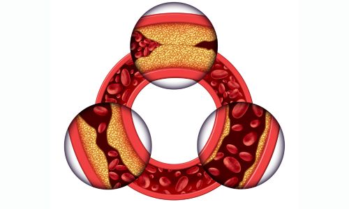
Complex coronary artery disease (complex CAD) describes advanced blockage of the heart’s arteries that cannot be treated by simple, routine techniques. Unlike a single, straightforward narrowing, complex CAD involves anatomy that is technically demanding to fix — multivessel disease (two or three major arteries affected), left main coronary artery disease, chronic total occlusions (CTOs) where an artery is completely blocked, heavily calcified lesions, and bifurcation lesions where a blockage sits at the junction of two branches.
The coronary arteries supply oxygen-rich blood to the heart muscle. When several of them are severely narrowed or blocked by atherosclerotic plaque, the heart muscle is starved of blood, causing angina, breathlessness, and a high risk of heart attack. Doctors often quantify this complexity using the SYNTAX score — a higher score means more extensive, higher-risk disease.
Because these cases carry more risk, treatment decisions are usually made by a Heart Team — an interventional cardiologist and a cardiac surgeon working together. The two main strategies are complex percutaneous coronary intervention (complex PCI), often supported by imaging (IVUS/OCT) and plaque-modifying tools such as atherectomy, and coronary artery bypass grafting (CABG). Choosing correctly can dramatically improve survival and quality of life.
2. Key Facts at a Glance
| Fact | Detail |
|---|---|
| Also known as | Complex CAD, high-risk CAD, multivessel/left-main coronary disease |
| Body system affected | Cardiovascular system (coronary arteries and heart muscle) |
| Common in | Adults over 60, people with diabetes, prior heart disease |
| Severity range | Serious to life-threatening; graded by SYNTAX score |
| Key treatments | Complex PCI (with IVUS, atherectomy), CABG, optimal medical therapy |
| Outlook | Good with timely revascularisation; depends on heart function and comorbidities |
3. Alternative Names and Medical Terminology
- Complex CAD — the common shorthand.
- Multivessel coronary artery disease — two or more major arteries diseased.
- Left main disease — narrowing of the left main stem, which supplies most of the heart.
- Chronic total occlusion (CTO) — a completely blocked artery present for more than three months.
- High SYNTAX score disease — anatomically complex disease.
- Advanced / diffuse / calcified atherosclerotic heart disease — descriptive terms used interchangeably.
Related abbreviations: PCI (percutaneous coronary intervention), CABG (coronary artery bypass grafting), IVUS/OCT (intravascular imaging).
4. Relevant Heart, Lung or Vascular Anatomy
The heart is fed by two main coronary arteries arising from the aorta. The left main coronary artery quickly divides into the left anterior descending (LAD) artery, which supplies the front wall, and the circumflex artery, which supplies the side and back. The right coronary artery (RCA) supplies the bottom of the heart and, in most people, the electrical node that sets the heartbeat.
In complex CAD, disease affects several of these vessels at once, or strikes critical junctions such as the left main bifurcation or the LAD/diagonal branch point. The internal thoracic (mammary) arteries and the saphenous veins in the leg are important because they are used as bypass grafts during surgery.
5. How the Disease Affects the Body
Complex CAD reduces blood flow to large or multiple territories of heart muscle simultaneously. When plaque narrows an artery, the muscle downstream can still function at rest but becomes ischaemic (oxygen-starved) during exertion, producing angina. When several arteries are involved, even light activity can trigger symptoms, and larger areas of muscle are at risk.
Heavily calcified and diffuse lesions stiffen the artery wall, so it resists stretching by a balloon or stent — this is why atherectomy (drilling or shaving calcium) is sometimes needed. Chronic total occlusions deprive a region entirely, though the heart may grow tiny natural bypasses called collaterals. Left main disease is especially dangerous because a single blockage there threatens most of the left ventricle.
Over time, repeated or severe ischaemia can weaken the heart muscle, causing ischaemic cardiomyopathy and heart failure. An unstable plaque can rupture and form a clot, causing a myocardial infarction (heart attack). Because multiple vessels are involved, a complication in any one can be catastrophic — which is why complex CAD demands careful, expert-led treatment planning rather than a piecemeal approach.
6. Types and Classification
Complex CAD is classified by anatomy and lesion characteristics:
- By number of vessels — single-vessel complex, two-vessel, or three-vessel (triple-vessel) disease.
- Left main disease — narrowing of the left main stem, often with additional vessels involved.
- Chronic total occlusion (CTO) — long-standing complete blockage.
- Bifurcation lesions — plaque at a branch point.
- Calcified/diffuse lesions — long, rigid, calcium-laden narrowings.
- By SYNTAX score — low (≤22), intermediate (23–32), or high (≥33) anatomical complexity, guiding the PCI-versus-CABG decision.
7. Causes of the Disease
The underlying cause is atherosclerosis — a gradual build-up of cholesterol, fatty material, inflammatory cells and calcium within the artery wall, forming plaques. In complex CAD, this process is unusually extensive, advanced, or aggressive, affecting many segments.
Contributing drivers include:
- High LDL (“bad”) cholesterol and abnormal blood fats.
- Diabetes, which accelerates and diffusely worsens atherosclerosis.
- High blood pressure damaging the artery lining over years.
- Smoking, which injures vessels and promotes clotting.
- Chronic inflammation and, in older patients, extensive vascular calcification.
8. How the Disease Develops
Atherosclerosis begins silently, often decades before symptoms. It starts with injury to the delicate inner lining (endothelium) of a coronary artery from high cholesterol, high blood pressure, tobacco or high blood sugar. LDL cholesterol seeps into the wall, is taken up by immune cells, and forms a fatty streak. Over years this matures into a fibrous plaque with a lipid core.
In complex CAD, this process is diffuse — many segments and vessels are affected rather than one. Plaques enlarge, narrowing the channel, and often become calcified, making the artery rigid. Some arteries close completely, forming a chronic total occlusion; the heart responds by developing small collateral vessels that partly maintain flow.
Two patterns cause trouble. Stable, progressive narrowing produces predictable exertional angina as the lumen shrinks. Plaque rupture — when the fibrous cap tears — triggers sudden clot formation and a heart attack. Because disease is widespread, patients may have both stable narrowings and vulnerable plaques at once. Diabetes, chronic kidney disease and continued smoking accelerate every stage, which is why complex CAD is common in these groups.
9. Risk Factors
Modifiable risk factors:
- Smoking and tobacco use.
- High LDL cholesterol and low HDL.
- High blood pressure.
- Diabetes and insulin resistance (a major driver of diffuse, complex disease).
- Obesity, physical inactivity, and an unhealthy diet.
Non-modifiable risk factors:
- Increasing age (especially over 60).
- Male sex, and post-menopausal women.
- Family history of premature heart disease.
- Chronic kidney disease and prior coronary events.
The more risk factors present, the more diffuse and complex the disease tends to be.
10. Genetic and Family-History Factors
A strong family history of early coronary disease — a parent or sibling affected before age 55 (men) or 65 (women) — meaningfully raises risk. Some people inherit familial hypercholesterolaemia, causing very high LDL cholesterol from young adulthood and aggressive, complex disease. Genetic tendencies toward diabetes, high blood pressure and lipoprotein(a) elevation also contribute.
While no single gene “causes” complex CAD, inherited susceptibility combined with lifestyle produces the extensive, calcified, multivessel patterns seen in these patients. Knowing your family history helps guide earlier screening and more intensive prevention.
11. Who Is Most at Risk?
- People with diabetes, who often develop diffuse, multivessel, calcified disease.
- Older adults, especially over 65, with years of plaque accumulation.
- Patients with chronic kidney disease, which promotes heavy calcification.
- Long-term smokers and those with untreated high cholesterol or blood pressure.
- People with prior heart attacks, stents or bypass surgery whose disease has progressed.
- Those with a strong family history of premature heart disease.
12. Prevalence and Epidemiology
Coronary artery disease is the leading cause of death worldwide. Among patients referred for coronary angiography, a substantial proportion — often roughly a third or more — have multivessel or left-main disease that qualifies as complex. Prevalence rises sharply with age and with diabetes.
Rates are increasing in low- and middle-income countries, including much of South Asia, where CAD tends to appear at younger ages. Complex, calcified disease is also more common as populations age and as more patients survive earlier heart attacks. These figures are broad approximations and vary by region, population and how “complex” is defined.
13. Signs and Symptoms
Symptoms of complex CAD reflect widespread reduced blood flow to the heart. The hallmark is angina — chest pressure, tightness, heaviness or squeezing, often triggered by exertion or stress and relieved by rest. Because multiple vessels are involved, symptoms are frequently more severe, more easily provoked, or occurring at lower activity levels than in simple single-vessel disease.
Common features include:
- Chest pain or pressure that may radiate to the arm, neck, jaw, shoulder or back.
- Breathlessness (dyspnoea) on exertion, sometimes without obvious chest pain.
- Fatigue and reduced exercise capacity.
- Palpitations or a sense of unease during activity.
- Symptoms brought on by cold weather, heavy meals or emotional stress.
In some people — particularly those with diabetes — nerve involvement blunts pain, causing silent ischaemia where the heart is starved of blood without warning symptoms. Others present for the first time with a heart attack. Any new, worsening, or rest-related chest discomfort in a person with multiple risk factors should be treated as potentially serious and evaluated promptly.
14. Early-Stage Symptoms
Early complex CAD may cause mild, exertional angina — chest tightness or breathlessness only during vigorous activity such as climbing stairs or hurrying, relieved quickly by rest. Some people notice reduced stamina or unusual fatigue rather than classic pain. In diabetics, early disease can be completely silent. These subtle signs are easy to dismiss, but in someone with several risk factors they warrant medical evaluation before disease advances.
15. Advanced-Stage Symptoms
As disease becomes more extensive, angina occurs with minimal effort or even at rest, lasts longer, and is less relieved by rest or medication (unstable angina). Patients may develop marked breathlessness, fluid retention and ankle swelling if the heart muscle weakens into heart failure. Episodes of near-fainting, severe fatigue, or recurrent chest pain signal high-risk disease that needs urgent specialist assessment and, often, revascularisation.
16. Symptoms in Women, Men and Older Adults
- Women more often have “atypical” symptoms — breathlessness, unusual fatigue, nausea, or discomfort in the jaw, neck or back rather than crushing chest pain, which can delay diagnosis.
- Men more commonly report classic central chest pressure radiating to the left arm.
- Older adults may present mainly with breathlessness, confusion, weakness or falls rather than chest pain, and often have silent disease.
- People with diabetes of any sex frequently have blunted or absent pain (silent ischaemia).
Recognising these differences helps avoid missed or late diagnoses.
17. Emergency Warning Signs
Call emergency services immediately for:
- Severe, crushing or persistent chest pain lasting more than a few minutes.
- Chest pain with sweating, nausea, or breathlessness.
- Pain spreading to the arm, jaw, neck or back at rest.
- Fainting, collapse, or a sudden severely irregular heartbeat.
These may signal a heart attack or unstable angina and require emergency care.
18. When to Seek Medical Help
See a doctor promptly if you develop new chest discomfort or breathlessness on exertion, if known angina becomes more frequent, more intense, or occurs at rest, or if your usual medication no longer controls symptoms. Anyone with multiple risk factors — especially diabetes, prior heart disease, or a strong family history — should seek evaluation for any suspicious symptoms rather than waiting.
19. Disease Stages, Grades and Severity
Complex CAD is graded in several complementary ways:
- Angina severity — the CCS (Canadian Cardiovascular Society) class I–IV, from angina with strenuous effort only, to angina at rest.
- Anatomical complexity — the SYNTAX score, categorising disease as low, intermediate or high complexity based on lesion number, location and features (calcification, CTO, bifurcation).
- Functional significance — measured by fractional flow reserve (FFR) or imaging stress tests, showing whether a narrowing actually limits blood flow.
- Heart function — left ventricular ejection fraction, since weakened muscle raises risk.
These together define severity and guide whether PCI, CABG or medical therapy is best.
20. Disease Progression
Left unaddressed, complex CAD tends to progress: plaques enlarge, more vessels become involved, and narrowings become more calcified and diffuse. Chronic total occlusions may develop. Repeated silent or symptomatic ischaemia can weaken the heart muscle, leading to ischaemic cardiomyopathy and heart failure. The pace varies — some patients remain stable for years on good medical therapy, while others deteriorate quickly, particularly with diabetes, ongoing smoking or poorly controlled cholesterol. A sudden plaque rupture can cause a heart attack at any stage.
21. Possible Complications
- Myocardial infarction (heart attack) from plaque rupture and clot.
- Heart failure from cumulative muscle damage (ischaemic cardiomyopathy).
- Dangerous arrhythmias, including ventricular tachycardia and sudden cardiac death.
- Mechanical complications after large heart attacks (e.g. ventricular septal defect, left ventricular aneurysm).
- Recurrent angina and repeat procedures.
- Procedure-related risks such as stent thrombosis, restenosis, or graft failure.
22. Related and Associated Medical Conditions
Complex CAD commonly coexists with:
- Diabetes mellitus and metabolic syndrome.
- High blood pressure and high cholesterol.
- Chronic kidney disease, which worsens calcification and procedural risk.
- Peripheral artery disease and carotid disease (atherosclerosis elsewhere).
- Heart valve disease, sometimes requiring combined surgery.
- Atrial fibrillation and heart failure.
Managing these conditions is essential to overall outcome.
23. Screening and Early Detection
There is no universal screening test for coronary disease, but people at higher risk benefit from cardiovascular risk assessment: measuring blood pressure, cholesterol, blood sugar (HbA1c) and calculating overall risk. A coronary artery calcium (CAC) score on CT can detect early, silent atherosclerosis. Those with symptoms or high risk may be referred for stress testing or CT coronary angiography. Early detection allows aggressive prevention before disease becomes complex. Learn more via the disease information hub.
24. How the Disease Is Diagnosed
Diagnosis of complex CAD combines symptom assessment, non-invasive testing and, crucially, detailed imaging of the coronary arteries. The pathway usually begins with a clinical history, examination and an ECG, followed by blood tests and often a stress test (exercise or pharmacological) or CT coronary angiography to confirm ischaemia and estimate disease burden.
The definitive test is invasive coronary angiography, in which contrast dye is injected into the coronary arteries via a catheter and X-ray images reveal the number, location and severity of blockages. In complex cases, additional tools sharpen the picture:
- Fractional flow reserve (FFR) or instantaneous wave-free ratio (iFR) to judge whether a narrowing limits flow.
- Intravascular ultrasound (IVUS) or optical coherence tomography (OCT) to assess plaque, calcium and vessel size from inside the artery.
- Echocardiography or cardiac MRI to measure heart function and scar.
From these, doctors calculate the SYNTAX score and assess overall risk. Because the disease is anatomically demanding, the findings are typically reviewed by a Heart Team who decide jointly between complex PCI and CABG. See related procedures and surgery options.
25. Physical Examination and Medical History
The doctor reviews symptoms, risk factors, and family and treatment history (previous stents, bypass, heart attacks). Physical examination may be normal between episodes but can reveal high blood pressure, signs of high cholesterol, heart murmurs, irregular pulse, or signs of heart failure such as ankle swelling and lung crackles. Evidence of disease elsewhere — weak leg pulses or carotid bruits — supports widespread atherosclerosis. History and examination guide which tests are needed.
26. Diagnostic Tests and Imaging
- Electrocardiogram (ECG) — detects ischaemia, prior heart attack, or arrhythmia.
- Echocardiogram — assesses heart muscle function and wall motion.
- Stress testing — exercise or pharmacological, with ECG, echo or nuclear imaging to reveal ischaemia.
- CT coronary angiography and calcium scoring — non-invasive imaging of the arteries.
- Invasive coronary angiography — the reference standard, mapping all blockages.
- IVUS / OCT — intravascular imaging to guide complex PCI.
- Cardiac MRI — assesses viability, scar and function.
27. Blood Tests, Biomarkers and Genetic Testing
Blood tests support diagnosis and risk assessment rather than diagnosing CAD directly:
- Lipid profile — LDL, HDL, triglycerides, and sometimes lipoprotein(a).
- HbA1c and glucose — to detect diabetes.
- Cardiac troponin — raised during a heart attack.
- Kidney function — important before contrast procedures.
- Full blood count and inflammatory markers.
Genetic testing is not routine but may be considered when familial hypercholesterolaemia or very early disease is suspected.
28. Understanding Test Results
Test results build a picture of how much heart muscle is at risk and how complex the anatomy is. A stress test showing widespread ischaemia, an angiogram revealing left main or triple-vessel disease, or a high SYNTAX score all point to complex disease needing revascularisation. FFR/iFR values below the threshold mean a narrowing is significant. A reduced ejection fraction indicates weakened muscle. Your cardiologist will explain what each result means for you and how it shapes the treatment recommendation.
29. Differential Diagnosis
Chest pain and breathlessness have many causes that doctors must distinguish from complex CAD:
- Aortic valve disease or hypertrophic cardiomyopathy causing exertional symptoms.
- Pericarditis or myocarditis.
- Pulmonary conditions — pulmonary embolism, pneumonia, pleurisy.
- Gastro-oesophageal reflux or musculoskeletal chest pain.
- Anxiety and panic attacks.
- Microvascular angina with normal large arteries.
Careful testing separates these from true multivessel coronary disease.
30. Specialist and Multidisciplinary Evaluation
Complex CAD is best managed by a Heart Team — an interventional cardiologist and a cardiac surgeon, often with imaging specialists, anaesthetists, diabetologists and heart-failure experts. Together they weigh the anatomy (SYNTAX score), heart function, comorbidities and patient preference to recommend complex PCI, CABG, or medical therapy. This collaborative approach, endorsed by international guidelines, improves outcomes and ensures patients receive a balanced, unbiased recommendation. Find experienced doctors and hospitals through our directory.
31. Treatment Goals
The aims of treatment are to:
- Relieve symptoms (angina, breathlessness) and improve quality of life.
- Reduce the risk of heart attack and death, especially in left main and multivessel disease.
- Restore blood flow to threatened heart muscle (revascularisation).
- Preserve or improve heart function.
- Control underlying risk factors to slow further disease.
32. When Is Treatment Required?
Treatment intensifies when there is significant left main or multivessel disease, ongoing angina despite medication, evidence of a large area of ischaemia, or reduced heart function. Revascularisation (PCI or CABG) is recommended to improve prognosis in high-risk anatomy and to relieve symptoms not controlled by drugs. Every patient receives optimal medical therapy; the decision about invasive treatment depends on anatomy, symptoms, heart function and overall risk, made with the Heart Team.
33. Active Monitoring and Watchful Waiting
Some patients with complex anatomy but mild symptoms, preserved heart function and low-risk features can be managed initially with optimal medical therapy and close monitoring rather than immediate intervention. This involves aggressive risk-factor control, regular review, and repeat testing if symptoms change. However, watchful waiting is not appropriate for high-risk disease such as significant left main narrowing, where revascularisation clearly improves survival. The strategy is individualised and revisited regularly.
34. Medications
Medical therapy is the foundation of care and continues even after PCI or CABG:
- Antiplatelets — aspirin, plus clopidogrel, ticagrelor or prasugrel (dual antiplatelet therapy after stents).
- Statins (and sometimes ezetimibe or PCSK9 inhibitors) to lower LDL aggressively.
- Beta-blockers to reduce angina and protect the heart.
- ACE inhibitors / ARBs, especially with diabetes, hypertension or reduced function.
- Nitrates and calcium-channel blockers for angina relief.
- Diabetes and blood-pressure medications to control risk factors.
35. Minimally Invasive Treatments
For selected patients, less invasive surgical approaches can avoid a full breastbone incision. Minimally invasive direct coronary artery bypass (MIDCAB) grafts the LAD through a small left-chest incision, and hybrid coronary revascularisation combines a minimally invasive mammary graft to the LAD with stenting of other vessels. Off-pump CABG performs bypass on a beating heart without the heart-lung machine. These options suit specific anatomy and are offered at experienced centres — see minimally invasive cardiac surgery.
36. Catheter-Based and Endovascular Treatments
Complex PCI uses catheters passed from the wrist or groin to open blocked arteries with balloons and drug-eluting stents. In complex CAD it is enhanced by specialised tools:
- Intravascular imaging (IVUS/OCT) to size vessels and guide stent placement.
- Rotational, orbital or laser atherectomy to modify heavily calcified plaque.
- CTO techniques (antegrade and retrograde) to reopen chronically blocked arteries.
- Bifurcation stenting strategies and mechanical circulatory support for high-risk cases.
These allow many complex lesions once treatable only by surgery to be tackled percutaneously. See angioplasty and endovascular stenting.
37. Surgical Treatment Options
Coronary artery bypass grafting (CABG) is often the preferred treatment for complex, multivessel and left-main disease, particularly in patients with diabetes, reduced heart function, or a high SYNTAX score. Surgeons create new routes around blockages using grafts — most importantly the left internal thoracic (mammary) artery to the LAD, which provides outstanding long-term durability, along with the right mammary, radial artery, or saphenous vein grafts to other vessels.
CABG can be performed:
- On-pump — using the heart-lung machine with the heart stopped, the traditional approach giving a still, bloodless field.
- Off-pump (beating-heart) — avoiding the machine, useful for certain patients such as those with a heavily calcified aorta.
- Minimally invasive or hybrid — smaller incisions for selected anatomy.
CABG offers excellent, complete revascularisation and durable relief of angina, and improves survival in high-risk anatomy. It requires a hospital stay of several days and a recovery of weeks, but long-term outcomes — especially with arterial grafts — are among the best in cardiac medicine. Explore coronary artery bypass grafting and general cardiac surgery.
38. Advanced and Emerging Treatments
Emerging options expand what is possible in complex CAD:
- Intravascular lithotripsy (shockwave) to fracture severe calcium safely.
- Mechanical circulatory support (e.g. temporary pumps) enabling high-risk complex PCI.
- Improved bioresorbable and drug-eluting technologies.
- Total arterial and robotic-assisted bypass surgery.
- Refined CTO techniques and physiology-guided complete revascularisation.
- Research into gene and stem-cell therapies and advanced lipid-lowering agents.
Availability varies by centre and country. See hybrid cardiac procedures.
39. Treatment Options Compared
The core choice is between complex PCI and CABG, on top of medical therapy:
- Complex PCI — less invasive, faster recovery, no chest incision; suits lower-to-intermediate SYNTAX scores and patients at high surgical risk. May need repeat procedures and long dual antiplatelet therapy.
- CABG — more invasive with longer recovery, but offers the most complete and durable revascularisation and better survival in diabetes, reduced heart function and high SYNTAX scores.
- Medical therapy alone — appropriate for lower-risk anatomy or when procedures are unsuitable.
The Heart Team weighs these against each patient’s anatomy and priorities.
40. How Doctors Choose the Right Treatment
Key factors in the decision include:
- Anatomical complexity — the SYNTAX score and lesion features.
- Presence of diabetes — favours CABG in multivessel disease.
- Left ventricular function — reduced function often favours surgery.
- Left main involvement and its complexity.
- Comorbidities and surgical risk (frailty, kidney or lung disease).
- Patient preferences and values.
Guidelines recommend a Heart Team discussion for complex or left-main disease to reach a balanced, individualised plan.
41. Benefits and Risks of Treatment
Benefits: relief of angina, improved exercise capacity and quality of life, reduced risk of heart attack, and — in high-risk anatomy — improved survival.
Risks of PCI: bleeding, vessel injury, stent thrombosis, restenosis, contrast-related kidney injury, and rarely stroke or heart attack.
Risks of CABG: the usual surgical risks of bleeding, infection, arrhythmia (especially atrial fibrillation), stroke, kidney injury, and a longer recovery. Both procedures are performed safely every day at experienced centres, and the Heart Team tailors the choice to minimise individual risk.
42. What Happens If the Disease Is Left Untreated?
Untreated complex CAD carries a high risk of heart attack, heart failure and premature death, particularly with left main or triple-vessel disease. Symptoms typically worsen, limiting daily life, and the heart muscle can progressively weaken. A vulnerable plaque may rupture at any time, causing a sudden, potentially fatal event. While some lower-risk patients remain stable on medication, high-risk anatomy left unrevascularised has a markedly worse outlook — which is why timely evaluation matters.
43. Treatment Success and Expected Outcomes
Outcomes are generally very good at experienced centres. Most patients experience substantial or complete relief of angina and improved quality of life after revascularisation. CABG with arterial grafts provides durable results lasting many years, and modern drug-eluting stents have greatly improved PCI durability. Survival improves in high-risk anatomy after appropriate revascularisation. Results depend on completeness of revascularisation, heart function, comorbidities, and adherence to medication and lifestyle changes. Exact success rates vary by patient and centre.
44. Prognosis and Long-Term Outlook
The long-term outlook for complex CAD has improved dramatically with modern therapy. With appropriate revascularisation, aggressive risk-factor control and good medication adherence, many patients live active lives for years or decades. Prognosis depends heavily on heart muscle function — those with preserved ejection fraction do best — and on comorbidities such as diabetes and kidney disease.
CABG, particularly with the internal mammary artery to the LAD, offers excellent long-term protection and durable freedom from angina; grafts, especially arterial ones, often function for many years. Complex PCI results have improved with drug-eluting stents and imaging guidance, though repeat procedures are sometimes needed. Continued attention to cholesterol, blood pressure, diabetes control, not smoking, and cardiac rehabilitation is essential — the underlying atherosclerosis persists and can progress even after successful treatment. Regular follow-up detects and addresses new problems early. Overall, a patient who engages fully with treatment and secondary prevention has a genuinely favourable outlook.
45. Recovery and Rehabilitation
Recovery differs by treatment. After complex PCI, most patients go home within a day or two and resume normal activity within about a week, continuing dual antiplatelet therapy. After CABG, hospital stay is typically several days, with breastbone healing over about six to eight weeks and full recovery over two to three months. Cardiac rehabilitation — supervised exercise, education and risk-factor counselling — is strongly recommended for all patients and improves outcomes. Gradual return to work, driving and activity is guided by the care team.
46. Follow-Up Tests and Long-Term Monitoring
Long-term follow-up includes regular clinical review, blood pressure, cholesterol and diabetes checks, and symptom assessment. ECGs and echocardiograms monitor heart function; stress testing or repeat angiography is used if symptoms recur. After stenting, adherence to antiplatelet therapy is monitored. Lifelong surveillance detects disease progression or graft/stent problems early.
47. Managing Recurrence or Disease Progression
Because atherosclerosis is a lifelong condition, new or recurrent narrowings can develop even after successful treatment — sometimes in a stented segment (restenosis), a failing graft, or a new vessel. Management includes intensifying medical therapy, and, if significant ischaemia returns, repeat PCI or, occasionally, redo CABG. Recurrent or repeat disease is discussed in detail on our recurrent coronary artery disease page. Aggressive secondary prevention is the best way to slow progression.
48. Living with the Disease
Living well with complex CAD centres on secondary prevention and consistency: taking medications reliably, attending follow-up, and sustaining a heart-healthy lifestyle. Most people return to work, travel, hobbies and, in time, intimacy, guided by their team. Understanding your medications, recognising warning symptoms, and staying engaged with cardiac rehabilitation give the best long-term results. Support from family, and from other patients, makes the adjustment easier.
49. Diet and Nutrition Guidelines
- Follow a Mediterranean-style diet rich in vegetables, fruit, whole grains, legumes, nuts and olive oil.
- Choose oily fish and lean proteins; limit red and processed meat.
- Reduce saturated and trans fats, salt, and added sugars.
- Control portions and weight; limit alcohol.
- For diabetics, manage carbohydrates to keep blood sugar well controlled.
A dietitian can personalise a plan, especially with diabetes or kidney disease.
50. Exercise and Physical-Activity Guidelines
Regular activity is protective. After treatment, most patients begin with supervised cardiac rehabilitation, then build to about 150 minutes of moderate aerobic activity per week (walking, cycling, swimming) plus light resistance work, as advised. Activity should be increased gradually and any chest pain, undue breathlessness or dizziness reported. Following heart surgery, avoid heavy lifting and upper-body strain until the breastbone heals. Your team will tailor recommendations to your heart function.
51. Medications, Activities and Habits to Avoid
- Do not stop antiplatelet medication (e.g. after a stent) without cardiology advice — this risks stent thrombosis.
- Avoid all tobacco and vaping.
- Limit alcohol and avoid recreational stimulants (e.g. cocaine), which strain the heart.
- Avoid sudden extreme exertion when unconditioned, and heavy lifting after surgery until cleared.
- Use NSAIDs cautiously and only with medical advice.
- Do not ignore new or worsening symptoms.
52. Preventing the Disease or Reducing Its Risks
Prevention targets the drivers of atherosclerosis:
- Do not smoke; avoid second-hand smoke.
- Keep LDL cholesterol low with diet and statins as advised.
- Control blood pressure and diabetes tightly.
- Maintain a healthy weight and stay physically active.
- Eat a heart-healthy diet and limit alcohol.
- Attend regular check-ups if you are at higher risk.
These measures prevent both new disease and progression after treatment.
53. Pregnancy and the Disease
Complex CAD is uncommon in women of childbearing age but can occur with familial hypercholesterolaemia, diabetes or other risk factors. Pregnancy increases the heart’s workload, so any woman with known coronary disease should have pre-pregnancy counselling and specialist (cardio-obstetric) care. Some medications, including statins and certain others, must be reviewed or stopped before conception. Management is highly individualised and should be planned with a cardiologist and obstetrician together.
54. Disease in Children and Young Adults
Complex CAD is rare in the young. When it appears early, it usually reflects familial hypercholesterolaemia, other genetic lipid disorders, diabetes, or (rarely) inflammatory conditions such as Kawasaki disease. Young adults with premature multivessel disease need intensive risk-factor management, genetic assessment and family screening. Recognising and treating these conditions early prevents progression to complex, extensive disease later in life.
55. Disease in Older Adults
Older patients frequently have the most diffuse, calcified and multivessel disease, alongside comorbidities such as kidney disease, frailty and other vascular problems. Treatment decisions carefully balance benefit against procedural risk; complex PCI with calcium-modification tools or off-pump CABG may be chosen to reduce risk. Symptoms are often atypical (breathlessness, fatigue). A geriatric-aware, individualised approach and realistic goals focused on quality of life are important.
56. Emotional Health and Patient Support
A diagnosis of complex heart disease — and the prospect of PCI or surgery — commonly causes anxiety, low mood or fear. These feelings are normal and treatable. Cardiac rehabilitation addresses emotional wellbeing alongside physical recovery, and counselling, stress-reduction techniques and support groups help. Depression can worsen heart outcomes, so it should be recognised and treated. Involving family and connecting with others who have had similar treatment provides valuable reassurance and motivation.
57. Preparing for Your Specialist Appointment
- Bring a list of symptoms, when they occur, and what triggers or relieves them.
- List all medications, allergies and past procedures (stents, bypass, heart attacks).
- Note your risk factors and family history.
- Bring previous test results, angiogram images or reports if available.
- Write down your questions and bring a family member for support.
- Be ready to discuss your goals and preferences for treatment.
58. Questions to Ask Your Doctor
- How extensive and complex is my coronary disease?
- What is my SYNTAX score and what does it mean?
- Do I need revascularisation, and is PCI or CABG better for me?
- Has my case been discussed by a Heart Team?
- What are the benefits and risks of each option in my case?
- How will my diabetes or other conditions affect the decision?
- What is my heart function (ejection fraction)?
- What recovery and follow-up should I expect?
- What medications will I need and for how long?
- What can I do to prevent my disease from progressing?
59. Cost of Diagnosis and Treatment
Costs vary widely by country, hospital and case complexity. The figures below are broad approximations in US dollars for guidance only.
| Region | Complex PCI (approx.) | CABG (approx.) |
|---|---|---|
| United States | $30,000–$90,000+ | $70,000–$200,000+ |
| United Kingdom (private) | $20,000–$45,000 | $30,000–$70,000 |
| India | $5,000–$12,000 | $6,000–$12,000 |
| Turkey | $6,000–$15,000 | $10,000–$20,000 |
| Thailand | $8,000–$18,000 | $15,000–$28,000 |
| Singapore | $18,000–$40,000 | $25,000–$60,000 |
Medical-tourism destinations such as India, Turkey and Thailand often cost roughly 50–90% less than the US or UK. Explore destinations.
60. Factors Affecting Treatment Cost
- Procedure type — CABG versus complex PCI; number of vessels and grafts/stents.
- Special tools — atherectomy, IVUS/OCT, mechanical support add cost.
- Number of stents and use of drug-eluting devices.
- Hospital type, accreditation and location.
- Surgeon/cardiologist experience and reputation.
- Length of stay, ICU time and complications.
- Comorbidities requiring extra care.
- For international patients: travel, accommodation and follow-up.
61. Choosing the Right Specialist
Look for an interventional cardiologist and cardiac surgeon with specific, high-volume experience in complex and left-main disease — complex PCI (CTO, calcium modification, imaging guidance) and CABG. Confirm they work within a Heart Team, review their credentials, board certification and outcome data, and ask how many similar cases they perform. A specialist who explains options clearly and respects your preferences is invaluable. Browse experienced doctors.
62. Choosing the Right Hospital or Treatment Centre
Choose a centre with:
- International accreditation (e.g. JCI) and strong governance.
- High procedure volumes and published outcome data for complex cases.
- Full cardiac surgery and interventional cardiology capability, plus advanced imaging and mechanical support.
- On-site ICU, cardiac rehabilitation and emergency backup.
- Good international-patient services if travelling.
Compare accredited hospitals.
63. Getting a Second Medical Opinion
Because the PCI-versus-CABG decision is nuanced, a second opinion is highly valuable in complex CAD — indeed a Heart Team review is itself a form of built-in second opinion. Seek one if you are unsure, if only one option has been offered, or before major surgery. Bring your angiogram images, reports and test results. A fresh expert review can confirm the plan or reveal alternatives, giving you confidence. Request one through our contact page.
64. Treatment Abroad and Medical-Travel Considerations
Many patients travel for high-quality, affordable complex-CAD treatment. Consider:
- Choosing a JCI-accredited hospital with experienced Heart Teams.
- Arranging a remote review of your angiogram before travelling.
- Planning for length of stay and recovery (longer for CABG) before flying home.
- Air-travel timing after a procedure, agreed with your doctors.
- Coordinating follow-up and medication with doctors at home.
- Understanding costs, language support and travel insurance.
Popular destinations include India, Turkey, Thailand and Singapore — see destinations.
65. Frequently Asked Questions
What makes coronary artery disease “complex”? Complexity comes from the anatomy — multiple vessels, left main involvement, complete blockages (CTOs), heavy calcium, or branch-point lesions — reflected in a high SYNTAX score.
Is CABG or stenting better for me? It depends on your anatomy, diabetes, heart function and preferences. CABG is often better for multivessel or left-main disease, especially with diabetes; complex PCI suits many other cases. The Heart Team decides with you.
Is complex PCI safe? Yes, at experienced centres, though it carries more risk than simple stenting. Tools like IVUS, atherectomy and circulatory support improve safety.
Will I need to take medication for life? Yes — antiplatelets, statins and other drugs are essential to protect grafts/stents and slow disease, even after successful treatment.
Can complex CAD come back after treatment? The underlying atherosclerosis persists, so new narrowings can develop. Secondary prevention and follow-up reduce this risk.
How long is recovery? About a week after complex PCI; roughly two to three months after CABG, with cardiac rehabilitation.
Can I travel abroad for treatment? Yes — many accredited international centres offer excellent complex-CAD care at lower cost, provided follow-up is coordinated.
66. Patient Stories and Treatment Experiences
The following are representative, anonymised examples for illustration only.
Rajan, India: Diagnosed with triple-vessel disease and diabetes, Rajan’s Heart Team recommended CABG using arterial grafts. Two years on, he is free of angina and back to daily walks and work.
Elena, Spain: Elena had a heavily calcified left main lesion. Her cardiologists used intravascular imaging and atherectomy during complex PCI, and she recovered within a week with lasting symptom relief.
Michael, UK: Uncertain between stenting and surgery, Michael sought a second opinion abroad. A JCI-accredited Heart Team confirmed CABG was best for his anatomy, and he travelled for treatment at a fraction of the private cost at home.
67. Latest Research and Clinical Trials
Research in complex CAD focuses on refining the PCI-versus-CABG decision through anatomy and physiology, and on improving both approaches. Advances include intravascular lithotripsy for calcified lesions, better imaging-guided (IVUS/OCT) complete revascularisation, improved CTO techniques, next-generation drug-eluting stents, and robotic and total-arterial bypass surgery. Landmark trials from major cardiology bodies continue to shape guidelines. Patients interested in clinical trials should ask their specialist or check reputable registries. (Described in general terms; consult current guidelines for specifics.)
68. Related Diseases and Conditions
- Isolated Coronary Artery Disease
- Recurrent Coronary Artery Disease
- Combined Valvular & Coronary Artery Disease
- Left Ventricular Aneurysm
- Ischemic Cardiomyopathy
- Post-Infarction Ventricular Septal Defect
69. Related Treatments and Procedures
- Coronary Artery Bypass Grafting (CABG)
- Angioplasty
- Endovascular Stenting
- Minimally Invasive Cardiac Surgery
- Hybrid Cardiac Procedures
70. Medical Glossary
- Atherosclerosis — build-up of plaque in artery walls.
- CABG — coronary artery bypass grafting; surgery to route blood around blockages.
- PCI — percutaneous coronary intervention; stenting via catheter.
- Complex PCI — advanced stenting for difficult anatomy.
- SYNTAX score — a measure of coronary disease complexity.
- Left main disease — narrowing of the main left coronary artery.
- Chronic total occlusion (CTO) — a completely blocked artery for over three months.
- Bifurcation lesion — plaque at a branch point.
- Atherectomy — removing or modifying calcified plaque.
- IVUS / OCT — imaging from inside the artery.
- FFR / iFR — measures of how much a narrowing limits blood flow.
- Drug-eluting stent — a stent that releases medication to prevent re-narrowing.
- Internal thoracic (mammary) artery — a durable graft used in CABG.
- Ejection fraction — the heart’s pumping strength.
- Ischaemia — reduced blood/oxygen supply to tissue.
71. Medical Review, Editorial Policy and Last Updated Date
Last updated: 11 July 2026.
This article is written and reviewed by qualified medical professionals and edited for clarity and accuracy. Our editorial policy aligns content with recognised guidelines from bodies such as the ACC/AHA, ESC, NHS, STS and WHO, and we review pages periodically to reflect current practice.
Disclaimer: This content is for general education only and is not a substitute for professional medical advice, diagnosis or treatment. Always consult a qualified healthcare provider about your individual situation.
72. Clinical Guidelines and Medical References
General references and guideline bodies include:
- American College of Cardiology / American Heart Association (ACC/AHA) — coronary revascularisation and chronic coronary disease guidelines.
- European Society of Cardiology (ESC) — myocardial revascularisation guidelines.
- Society of Thoracic Surgeons (STS) — surgical outcome standards.
- National Institute for Health and Care Excellence (NICE) and NHS patient resources.
- World Health Organization (WHO) — cardiovascular disease data.
Refer to the latest versions of these guidelines for detailed, current recommendations.
73. Book an Appointment or Request a Second Opinion
If you or a loved one has complex coronary artery disease, expert Heart Team assessment can make a decisive difference. Our network connects you with accredited hospitals and experienced cardiologists and cardiac surgeons worldwide.
- Book an appointment: Get started now
- Request a second opinion or ask a question: visit our contact page
Explore hospitals, doctors and destinations to plan your care with confidence.