1. Disease Overview
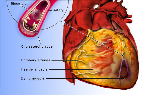
Ischemic cardiomyopathy (IC) is a condition in which the heart muscle becomes weakened and enlarged because of long-standing reduced blood supply from coronary artery disease (CAD), usually after one or more heart attacks (myocardial infarctions). When coronary arteries narrow or block, areas of heart muscle are starved of oxygen. Some muscle dies and is replaced by scar tissue; other regions survive but stop contracting effectively — so the heart’s main pumping chamber, the left ventricle, dilates and loses strength.
The result is heart failure: the heart can no longer pump enough blood to meet the body’s needs. Ischemic cardiomyopathy is the single most common cause of heart failure with reduced ejection fraction in adults worldwide. Patients typically experience breathlessness, fatigue, fluid retention, and reduced exercise tolerance.
What makes IC distinctive — and hopeful — is that a portion of the underperforming muscle may be “hibernating”: alive but dormant because of poor blood flow. If blood supply is restored through revascularization (bypass surgery or stenting), that hibernating muscle can recover function. This is why viability testing and revascularization sit at the centre of treatment, alongside guideline-directed medications, device therapy, and, in advanced cases, mechanical support or transplant. With modern care, many patients live for years with a good quality of life.
2. Key Facts at a Glance
| Fact | Detail |
|---|---|
| Also known as | Ischemic heart failure, coronary cardiomyopathy, ischemic dilated cardiomyopathy |
| Body system affected | Cardiovascular system — heart muscle (left ventricle) and coronary arteries |
| Common in | Adults over 50, more often men and those with prior heart attack, diabetes, or CAD risk factors |
| Severity range | Mild (asymptomatic LV dysfunction) to severe (advanced, end-stage heart failure) |
| Key treatments | Guideline-directed medications, revascularization (CABG/PCI), ICD/CRT devices, LVAD, transplant |
| Outlook | Variable; markedly improved with early revascularization, medical therapy, and lifestyle change |
3. Alternative Names and Medical Terminology
Ischemic cardiomyopathy is also called ischemic heart failure, ischemic dilated cardiomyopathy, or coronary cardiomyopathy. Related terms you may encounter include:
- HFrEF — heart failure with reduced ejection fraction (ejection fraction 40% or below)
- LV dysfunction — reduced left-ventricular contraction
- Post-infarction cardiomyopathy — weakening after a heart attack
- Hibernating myocardium — living but dormant muscle from chronic low blood flow
- Myocardial stunning — temporary dysfunction after a brief ischemic event
4. Relevant Heart, Lung or Vascular Anatomy
The heart has four chambers; the left ventricle is the main pumping chamber that drives oxygen-rich blood into the aorta and around the body. It is fed by the coronary arteries — chiefly the left anterior descending (LAD), left circumflex, and right coronary artery. These vessels supply the heart muscle (myocardium) itself.
In ischemic cardiomyopathy, narrowing or blockage in one or more coronary arteries reduces oxygen delivery to the territory each vessel supplies. The mitral valve, which sits between the left atrium and ventricle, often leaks (functional mitral regurgitation) as the ventricle dilates. The lungs are affected downstream: when the weakened left ventricle cannot clear blood, pressure backs up into the pulmonary circulation, causing fluid congestion and breathlessness.
5. How the Disease Affects the Body
Ischemic cardiomyopathy is fundamentally a supply-and-demand failure. Atherosclerotic plaque narrows the coronary arteries, and when a vessel closes acutely, a heart attack destroys muscle in that territory. Dead muscle is replaced by non-contracting scar (fibrosis). Surviving muscle in poorly perfused zones may enter a protective “hibernating” state, contracting weakly to conserve energy.
As functioning muscle diminishes, the left ventricle enlarges and changes shape — a process called adverse remodeling. The heart tries to compensate by beating faster and retaining fluid, but these responses eventually worsen the strain. Neurohormonal systems (the sympathetic nervous system and the renin-angiotensin-aldosterone axis) switch on, raising blood pressure and fluid load and accelerating muscle injury.
The consequences ripple outward: reduced cardiac output causes fatigue and low exertion tolerance; fluid backup into the lungs causes breathlessness; congestion in the veins causes swollen legs and abdomen. The enlarged, scarred ventricle is also electrically unstable, raising the risk of dangerous arrhythmias and sudden cardiac death. Understanding this cascade explains why treatment targets both blood supply (revascularization) and the neurohormonal drivers of remodeling (medications).
6. Types and Classification
Ischemic cardiomyopathy is one form of dilated cardiomyopathy — the ischemic subtype, caused by coronary disease rather than genetic, viral, or toxic factors. Classifications used in practice include:
- By ejection fraction: HFrEF (EF ≤40%), HFmrEF (mildly reduced, 41–49%), or preserved (rare in IC)
- By NYHA functional class: I (no symptoms) to IV (symptoms at rest)
- By viability: predominantly scarred (non-viable) versus significant hibernating/viable muscle — a key distinction for revascularization
- By extent of coronary disease: single-, double-, or triple-vessel disease, and presence of left main disease
7. Causes of the Disease
The root cause is coronary artery disease producing chronic or acute reduction in blood supply to the heart muscle. Specific mechanisms include:
- Prior myocardial infarction(s) — the most common cause, leaving scar and weakened muscle
- Chronic multivessel coronary narrowing causing hibernating myocardium without a discrete heart attack
- Repeated silent ischemia, especially in people with diabetes
- Coronary microvascular disease in some cases
- Prior coronary events left untreated or incompletely revascularized
8. How the Disease Develops
Ischemic cardiomyopathy usually develops over years. It begins with atherosclerosis — a slow buildup of cholesterol-rich plaque in the coronary arteries driven by high cholesterol, high blood pressure, smoking, and diabetes. As plaques grow, blood flow to the myocardium falls, particularly under exertion.
At some point a plaque may rupture and form a clot, causing a heart attack that kills a region of muscle within hours. Even without a full infarction, chronically underperfused muscle can shift into hibernation to survive on limited oxygen. The heart compensates for lost pumping power by enlarging and thickening, but this remodeling is ultimately maladaptive: the ventricle dilates, its geometry distorts, the mitral valve begins to leak, and neurohormonal activation drives further fibrosis.
Over months to years, a self-reinforcing cycle sets in — more remodeling, more valve leakage, more fluid retention, and progressively worse pumping. Crucially, revascularizing hibernating muscle early can interrupt this cycle and partially reverse the damage, which is why timely diagnosis matters so much.
9. Risk Factors
Modifiable risk factors:
- Smoking and tobacco use
- High LDL cholesterol and abnormal blood lipids
- High blood pressure
- Diabetes and insulin resistance
- Obesity and physical inactivity
- Poor diet high in saturated fat and salt
- Excess alcohol
Non-modifiable risk factors:
- Older age
- Male sex (though risk in women rises after menopause)
- Family history of coronary disease or early heart attack
- Prior heart attack or established coronary artery disease
10. Genetic and Family-History Factors
Ischemic cardiomyopathy itself is driven by acquired coronary disease rather than a single inherited gene. However, genetics strongly influence the underlying coronary artery disease. A family history of premature heart attack — a first-degree relative affected before roughly age 55 (men) or 65 (women) — meaningfully raises risk.
Inherited conditions such as familial hypercholesterolemia cause very high cholesterol from a young age and greatly accelerate coronary disease. Genetic tendencies toward high blood pressure, diabetes, and lipoprotein(a) elevation also contribute. If close relatives had early cardiac disease, share this with your doctor; earlier and more aggressive risk-factor management is warranted.
11. Who Is Most at Risk?
Those at highest risk include:
- People who have already had a heart attack or been diagnosed with coronary artery disease
- Adults over 50, especially men and post-menopausal women
- People with diabetes, who often have silent, diffuse coronary disease
- Long-term smokers
- Those with untreated high blood pressure or high cholesterol
- People with chronic kidney disease or metabolic syndrome
- Anyone with a strong family history of early coronary disease
12. Prevalence and Epidemiology
Ischemic cardiomyopathy is the leading cause of heart failure with reduced ejection fraction in high- and middle-income countries. Because coronary artery disease is the world’s most common cause of death, the pool of people at risk of IC is very large. Heart failure as a whole affects tens of millions of people globally, and roughly half to two-thirds of reduced-EF heart failure cases are ischemic in origin.
Prevalence rises steeply with age and is increasing in many countries as more people survive heart attacks and live longer with chronic heart disease. It is more common in men and in populations with high rates of smoking, diabetes, and hypertension. These figures are approximate and vary by region and healthcare access.
13. Signs and Symptoms
Symptoms of ischemic cardiomyopathy reflect both reduced pumping power and fluid congestion. It is possible to have early disease with no symptoms at all. As the heart weakens, common features include:
- Breathlessness on exertion, and later at rest or when lying flat
- Extreme fatigue and reduced exercise tolerance
- Swelling (edema) in the legs, ankles, and abdomen
- Heart palpitations or irregular heartbeats
- Cough or congestion from fluid in the lungs
- Chest pain or pressure (angina), particularly with exertion
- Difficulty sleeping flat, or waking breathless at night
- Dizziness or lightheadedness
- Unexpected weight gain from fluid retention
Because IC often coexists with active coronary disease, angina may accompany heart-failure symptoms. Any new or worsening symptoms should prompt medical review.
14. Early-Stage Symptoms
In early ischemic cardiomyopathy, the heart compensates well and symptoms may be subtle or absent. When present, early signs include mild breathlessness with heavier exertion (climbing stairs, brisk walking), unusual tiredness, and occasional palpitations. Some people notice reduced stamina or mild ankle swelling by the end of the day. Because these can be mistaken for aging or being out of shape, early disease is often missed — one reason regular check-ups matter for anyone with known coronary disease.
15. Advanced-Stage Symptoms
In advanced ischemic cardiomyopathy, symptoms become frequent and limiting:
- Breathlessness at rest or with minimal activity
- Orthopnea (needing to prop up on pillows to breathe) and waking gasping at night
- Marked swelling of legs, abdomen, and sometimes the whole body
- Persistent fatigue and inability to carry out daily tasks
- Poor appetite, nausea, and weight loss (cardiac cachexia)
- Confusion or reduced alertness from low output
- Frequent hospital admissions for fluid overload
- Dangerous arrhythmias, cold extremities, and low blood pressure
16. Symptoms in Women, Men and Older Adults
Men more often present with classic exertional chest pain and breathlessness following a recognized heart attack. Women are more likely to have atypical or silent coronary disease — presenting with fatigue, breathlessness, nausea, or jaw and back discomfort rather than crushing chest pain, which can delay diagnosis. Older adults frequently show non-specific features such as confusion, falls, reduced appetite, and profound fatigue rather than obvious cardiac symptoms, and often have coexisting kidney disease or frailty. People with diabetes of any sex may have blunted pain perception and silent ischemia. Awareness of these differences helps avoid missed or late diagnoses.
17. Emergency Warning Signs
Call emergency services (or your local equivalent of 911/999/112) immediately for:
- Crushing or persistent chest pain or pressure, possibly spreading to the arm, neck, or jaw
- Severe breathlessness or gasping for air, especially with pink frothy sputum
- Fainting or collapse
- A very fast, irregular, or pounding heartbeat with dizziness
- Sudden confusion, cold sweat, or grey/blue lips
These may signal a heart attack, acute heart failure, or a life-threatening arrhythmia.
18. When to Seek Medical Help
See your doctor promptly — without waiting for an emergency — if you notice new or worsening breathlessness, increasing leg or abdominal swelling, sudden weight gain of two to three kilograms over a few days, reduced exercise tolerance, or new palpitations. If you have known coronary disease or a prior heart attack, report any change in symptoms early. Prompt evaluation allows timely revascularization and medication adjustment before the heart weakens further.
19. Disease Stages, Grades and Severity
Two systems are widely used. The ACC/AHA stages describe the disease continuum:
- Stage A: at risk (coronary disease, diabetes) but no structural change
- Stage B: structural heart change (reduced EF, prior infarct) but no symptoms
- Stage C: structural disease with current or past heart-failure symptoms
- Stage D: advanced, refractory heart failure needing specialized therapy
The NYHA functional classification grades symptom severity from Class I (no limitation) to Class IV (symptoms at rest). Ejection fraction further quantifies pump strength. Severity is best judged by combining these with viability and coronary anatomy.
20. Disease Progression
Left unmanaged, ischemic cardiomyopathy tends to progress as the ventricle remodels and further coronary events occur. Repeated heart attacks or ongoing ischemia destroy more muscle; the ventricle dilates, mitral leakage worsens, and neurohormonal activation drives fibrosis. Progression is not inevitable, however: revascularization of viable muscle, guideline-directed medications, and risk-factor control can slow, halt, or partially reverse decline. Some patients remain stable for many years; others deteriorate toward advanced heart failure requiring devices, mechanical support, or transplant. The trajectory depends heavily on how much viable muscle can be rescued and how well risk factors are controlled.
21. Possible Complications
- Progressive heart failure with recurrent hospital admissions
- Arrhythmias — atrial fibrillation and life-threatening ventricular arrhythmias
- Sudden cardiac death from ventricular fibrillation
- Functional mitral regurgitation from ventricular dilation
- Blood clots and stroke from stagnant blood in a poorly contracting ventricle
- Left ventricular aneurysm or thrombus after large infarcts
- Cardiogenic shock in acute decompensation
- Kidney and liver dysfunction from congestion and low output
22. Related and Associated Medical Conditions
Ischemic cardiomyopathy rarely occurs alone. Closely associated conditions include coronary artery disease (the underlying cause), high blood pressure, diabetes, high cholesterol, and chronic kidney disease. Many patients also have atrial fibrillation, peripheral artery disease, carotid disease, or sleep apnea. Anemia, thyroid disorders, and depression frequently coexist and can worsen symptoms. Managing these comorbidities is an essential part of treatment, since each can accelerate heart-failure progression.
23. Screening and Early Detection
There is no population-wide screening test for ischemic cardiomyopathy, but people at risk should be screened for coronary disease and its risk factors. This includes regular blood-pressure checks, lipid profiles, and diabetes screening. Anyone with known coronary disease or a prior heart attack should have periodic assessment of left-ventricular function by echocardiogram, since silent decline in ejection fraction can be caught before symptoms appear. Detecting reduced EF early allows protective medications and, where indicated, revascularization to begin sooner. Learn more about assessment at accredited hospitals.
24. How the Disease Is Diagnosed
Diagnosis combines confirming reduced heart function with proving coronary disease is the cause. The pathway typically runs as follows:
- History and examination — symptoms of heart failure plus risk factors or prior heart attack.
- Electrocardiogram (ECG) — may show old infarction, Q waves, or arrhythmia.
- Echocardiogram — the key first test, measuring ejection fraction, chamber size, wall-motion abnormalities, and valve function.
- Blood tests — including natriuretic peptides (BNP/NT-proBNP) and markers of kidney and thyroid function.
- Coronary angiography — cardiac catheterization to map coronary blockages; this is what distinguishes ischemic from non-ischemic cardiomyopathy.
- Viability and imaging tests — cardiac MRI, stress echo, PET, or nuclear scans to identify scar versus hibernating (recoverable) muscle.
The diagnosis of ischemic cardiomyopathy is made when significant coronary disease (or prior infarction) explains a weakened, dilated left ventricle. Establishing how much viable muscle remains is central, because it determines whether revascularization is likely to help. A multidisciplinary heart team reviews the findings before choosing treatment.
25. Physical Examination and Medical History
The doctor asks about chest pain, breathlessness, exercise tolerance, prior heart attacks, and cardiovascular risk factors. On examination they look for signs of heart failure and fluid overload: raised jugular venous pressure, crackles in the lungs, leg and abdominal swelling, an enlarged displaced heart apex, a third heart sound (gallop rhythm), and a murmur of mitral regurgitation. Blood pressure, heart rate, and rhythm are checked. Cold, poorly perfused extremities may indicate low cardiac output. This bedside assessment guides which tests to prioritize.
26. Diagnostic Tests and Imaging
- Echocardiogram — measures ejection fraction, wall motion, chamber size, and valve leakage; the cornerstone test.
- Coronary angiography — the definitive map of coronary blockages, distinguishing ischemic disease.
- Cardiac MRI — precisely assesses function and, with late gadolinium enhancement, shows scar versus viable muscle.
- Nuclear perfusion imaging / PET — assesses blood flow and hibernating myocardium viability.
- Stress testing (exercise or pharmacologic) — reveals inducible ischemia.
- Cardiac CT / CT coronary angiography — non-invasive assessment of coronary anatomy.
- Chest X-ray — shows an enlarged heart and lung congestion.
- Holter monitoring — detects arrhythmias.
27. Blood Tests, Biomarkers and Genetic Testing
Blood tests support diagnosis and monitoring. Natriuretic peptides (BNP, NT-proBNP) rise with heart-muscle strain and help confirm heart failure and gauge severity. A cholesterol and triglyceride profile guides lipid therapy. Troponin may be checked during suspected heart attacks. Additional tests assess kidney function, electrolytes, blood count (for anemia), thyroid function, HbA1c (diabetes), and iron studies. Genetic testing is not routine for ischemic cardiomyopathy, but may be considered when an inherited lipid disorder such as familial hypercholesterolemia is suspected.
28. Understanding Test Results
Your ejection fraction (EF) is the headline number: a normal EF is roughly 55–70%; in ischemic cardiomyopathy it is usually 40% or below, and often much lower. A low EF means weaker pumping but does not by itself dictate prognosis. Viability imaging results matter greatly: substantial hibernating muscle suggests revascularization may recover function, whereas mostly scar suggests limited benefit. Angiography shows which arteries are blocked and whether they can be bypassed or stented. Natriuretic peptide levels track congestion over time. Always review results with your cardiologist, who interprets them together rather than in isolation.
29. Differential Diagnosis
Several other conditions can cause a weakened, dilated heart and must be distinguished from ischemic cardiomyopathy:
- Non-ischemic dilated cardiomyopathy — genetic, viral, alcohol-related, or idiopathic (see dilated cardiomyopathy)
- Restrictive cardiomyopathy — stiff heart muscle (see restrictive cardiomyopathy)
- Valvular heart disease as a primary cause of heart failure
- Hypertensive heart disease
- Myocarditis and infiltrative diseases (amyloidosis, sarcoidosis)
- Tachycardia-induced cardiomyopathy
Coronary angiography is the decisive test that confirms the ischemic cause.
30. Specialist and Multidisciplinary Evaluation
Optimal care involves a heart team: a cardiologist (often a heart-failure specialist), an interventional cardiologist, a cardiac surgeon, and cardiac imaging experts. For advanced disease, transplant and mechanical-support specialists, electrophysiologists, and nurse coordinators, dietitians, and rehabilitation therapists join in. The team weighs coronary anatomy, viability, symptoms, and comorbidities to decide between medication alone, revascularization, device therapy, or advanced options. You can find experienced doctors and specialist hospitals through this site.
31. Treatment Goals
The goals of treating ischemic cardiomyopathy are to:
- Relieve symptoms — breathlessness, fatigue, and fluid overload
- Restore blood supply to viable muscle through revascularization
- Slow or reverse remodeling with guideline-directed medications
- Prevent sudden cardiac death and dangerous arrhythmias
- Reduce hospitalizations and improve quality of life
- Prolong survival
- Manage risk factors to prevent further coronary events
32. When Is Treatment Required?
Treatment begins as soon as ischemic cardiomyopathy — or even asymptomatic reduced ejection fraction after a heart attack — is diagnosed. Guideline-directed medical therapy is recommended for essentially all patients with reduced EF, including those without symptoms, because it slows progression and improves survival. Revascularization is considered when significant coronary blockages supply viable muscle. Device therapy (ICD/CRT) is offered when EF stays low despite medication. Advanced options are reserved for those who deteriorate despite optimal therapy. In short, treatment is almost always required, though its intensity varies.
33. Active Monitoring and Watchful Waiting
Pure “watchful waiting” is uncommon in ischemic cardiomyopathy because early medical therapy improves outcomes. However, once patients are on optimized medication, a period of active monitoring is appropriate before deciding on devices or surgery — for example, waiting three months to see whether ejection fraction recovers on medical therapy before implanting an ICD. Stable patients with mild disease are monitored with periodic echocardiograms, symptom review, and blood tests, escalating treatment if function declines. Monitoring is active and structured, not passive neglect.
34. Medications
Medications are the backbone of care and improve both symptoms and survival. Core drug classes (“the four pillars” of modern heart-failure therapy) include:
- ACE inhibitors / ARBs / ARNI (sacubitril-valsartan) — reduce strain and reverse remodeling
- Beta-blockers — slow the heart, protect against arrhythmia, improve function
- Mineralocorticoid receptor antagonists (spironolactone, eplerenone) — reduce fibrosis and fluid
- SGLT2 inhibitors (dapagliflozin, empagliflozin) — reduce hospitalization and death
Supportive medications include diuretics for fluid overload, statins and antiplatelet agents for the underlying coronary disease, anticoagulants if atrial fibrillation or clot is present, and anti-anginal drugs. Doses are titrated gradually. Learn more under treatments.
35. Minimally Invasive Treatments
Beyond medication, several less-invasive options help selected patients. Percutaneous coronary intervention (PCI/angioplasty with stents) reopens blocked arteries without open surgery. Transcatheter mitral valve repair (edge-to-edge clip) can reduce functional mitral leakage in suitable patients with persistent symptoms. Cardiac resynchronization therapy (CRT) and implantable defibrillators (ICDs) are placed through small incisions under local or light anesthesia. Minimally invasive and hybrid cardiac approaches may be used for revascularization in appropriate cases, reducing recovery time compared with full open surgery.
36. Catheter-Based and Endovascular Treatments
Catheter-based (percutaneous) treatments are performed through a small puncture in the wrist or groin:
- Coronary angioplasty and stenting (PCI) — reopens narrowed or blocked coronary arteries to restore blood flow to hibernating muscle
- Transcatheter mitral valve repair — clip-based reduction of functional mitral regurgitation
- Device implantation — ICD and CRT leads placed via veins
- Percutaneous mechanical support — temporary pumps during high-risk procedures or shock
These approaches avoid open-chest surgery and often allow quicker recovery, though the choice between PCI and surgery depends on coronary anatomy and viability. See related procedures.
37. Surgical Treatment Options
Surgery plays a central role in ischemic cardiomyopathy because it can restore blood supply to large areas of viable muscle. The principal options are:
- Coronary artery bypass grafting (CABG) — the most established surgical treatment. Grafts (from the internal mammary artery or leg/arm vessels) bypass blocked coronary arteries. In patients with significant viable, hibernating muscle, CABG can improve pumping function, reduce symptoms, and improve survival compared with medication alone. It is often preferred over stenting for complex multivessel or left-main disease.
- Surgical ventricular reconstruction / aneurysm repair — reshaping a dilated or aneurysmal ventricle in selected cases.
- Mitral valve repair or replacement — correcting significant functional mitral regurgitation, sometimes at the same time as bypass.
- Left ventricular assist device (LVAD) — an implanted mechanical pump for advanced heart failure, used as a bridge to transplant or as long-term (“destination”) therapy.
- Heart transplantation — the definitive option for end-stage disease unresponsive to all other treatment.
The heart team’s decision hinges on coronary anatomy, viability, ventricular function, valve involvement, and overall fitness for surgery. Explore surgery options and specialist hospitals.
38. Advanced and Emerging Treatments
Research is expanding options for ischemic cardiomyopathy. Emerging and advanced approaches include regenerative therapies (stem-cell and progenitor-cell injections aiming to regrow muscle — still experimental), gene therapy targeting heart-muscle function, newer LVAD designs with better durability, and improved risk-stratification imaging to better identify who benefits from revascularization. Refinements in transcatheter valve repair, wearable and implantable remote-monitoring devices that detect early congestion, and next-generation heart-failure medications continue to enter practice. Many of these are available through clinical trials at leading centres.
39. Treatment Options Compared
- Medication alone — suits mild disease, non-viable muscle, or those unfit for procedures; improves symptoms and survival but does not restore blood flow.
- PCI (stenting) — less invasive, quicker recovery; best for suitable anatomy and can relieve ischemia, though evidence for survival benefit in cardiomyopathy is stronger for surgery in complex disease.
- CABG — more invasive with longer recovery, but offers durable revascularization and survival benefit in patients with viable muscle and multivessel disease.
- Devices (ICD/CRT) — reduce sudden death and improve function; used alongside other treatments, not instead of them.
- LVAD / transplant — reserved for advanced, end-stage disease.
Most patients receive a combination tailored to their anatomy and viability.
40. How Doctors Choose the Right Treatment
Treatment selection weighs several factors: the amount of viable, hibernating muscle (from imaging), the coronary anatomy (number and location of blockages, left-main involvement), the ejection fraction and symptoms, valve function, and the patient’s overall health, age, and surgical risk. A heart team reviews these together. For example, extensive viable muscle with complex multivessel disease favors CABG; limited viability with simpler anatomy may favor PCI or medication; persistent low EF despite therapy favors device implantation; and refractory advanced failure points toward LVAD or transplant. Patient preference and goals are integral to the decision.
41. Benefits and Risks of Treatment
Benefits: relief of breathlessness and fatigue, recovery of pumping function when viable muscle is revascularized, fewer hospitalizations, protection from sudden death (devices), and longer survival.
Risks: all revascularization carries some risk — bleeding, infection, stroke, kidney injury, and, with surgery, the risks of general anesthesia and a longer recovery. PCI carries risks of vessel injury and stent problems. Devices can have lead or infection complications. Medications may cause low blood pressure, kidney or electrolyte changes, and other side effects. For most appropriately selected patients, the benefits outweigh the risks, but the balance is individual and discussed carefully with the heart team.
42. What Happens If the Disease Is Left Untreated?
Untreated ischemic cardiomyopathy tends to worsen. Ongoing ischemia and remodeling weaken the ventricle further, symptoms intensify, and hospital admissions for fluid overload become frequent. The risk of dangerous arrhythmias and sudden cardiac death rises, as does the risk of stroke from blood clots. Hibernating muscle that could have recovered with timely revascularization may die and become permanent scar, closing the window for functional improvement. Without treatment, quality of life declines and life expectancy is significantly shortened. Early, sustained treatment changes this trajectory dramatically.
43. Treatment Success and Expected Outcomes
Outcomes vary with disease severity, how much viable muscle is present, and how well risk factors are controlled. Many patients experience meaningful symptom relief and improved exercise capacity with medication and revascularization, and some see their ejection fraction rise substantially when hibernating muscle recovers. Modern “four-pillar” medical therapy markedly reduces hospitalizations and death. Devices lower the risk of sudden cardiac death. Even advanced patients can gain years of good-quality life with LVAD or transplant. Outcomes are best when treatment starts early and viable muscle is revascularized before it becomes scar. These expectations are general and individual results differ.
44. Prognosis and Long-Term Outlook
The prognosis of ischemic cardiomyopathy depends on ejection fraction, symptom severity, extent of viable muscle, completeness of revascularization, and control of coronary risk factors. Historically, ischemic cardiomyopathy carried a worse outlook than non-ischemic forms because the underlying coronary disease continues to threaten the heart. However, the outlook has improved substantially with contemporary care: guideline-directed medications, timely revascularization of viable muscle, ICD/CRT devices, cardiac rehabilitation, and aggressive risk-factor management all extend survival and improve quality of life.
Some patients stabilize and live for many years with mild limitation; others progress to advanced heart failure despite optimal treatment. Key favorable factors include preserved viable muscle, successful revascularization, good adherence to medication, smoking cessation, and well-controlled blood pressure, cholesterol, and diabetes. Regular follow-up allows early detection of decline and timely escalation of therapy. While ischemic cardiomyopathy is a serious chronic condition, an active, informed partnership with an experienced heart team offers many patients a good and lengthening outlook.
45. Recovery and Rehabilitation
Recovery depends on the treatment received. After CABG, hospital stays are typically several days to a week, with a gradual return to normal activity over six to twelve weeks; after PCI or device implantation, recovery is quicker, often days. Cardiac rehabilitation — a supervised program of monitored exercise, education, and risk-factor counselling — is strongly recommended and improves fitness, symptoms, and survival. Recovery also involves optimizing medications, learning to monitor weight and symptoms, and adopting heart-healthy habits. Emotional recovery is important too; many rehab programs include psychological support.
46. Follow-Up Tests and Long-Term Monitoring
Long-term follow-up is essential. Typical monitoring includes periodic echocardiograms to track ejection fraction, blood tests for kidney function, electrolytes, and natriuretic peptides, ECGs and sometimes device checks, and review of symptoms and weight. Medication doses are adjusted over time. Patients are taught daily weight monitoring to catch fluid retention early. Follow-up intervals depend on stability — more frequent after a procedure or medication change, less frequent when stable. Ongoing control of cholesterol, blood pressure, and diabetes is monitored at each visit.
47. Managing Recurrence or Disease Progression
Because the underlying coronary disease persists, patients may develop new blockages, further heart attacks, or worsening heart failure over time. Management focuses on aggressive secondary prevention (statins, antiplatelet therapy, blood-pressure and diabetes control, smoking cessation), prompt evaluation of recurrent symptoms, and escalation of therapy when function declines — adding or up-titrating medications, considering repeat revascularization, upgrading devices, or, in advanced cases, referral for LVAD or transplant. Recurrent or complex coronary disease may need specialist review; see complex coronary artery disease and recurrent coronary artery disease.
48. Living with the Disease
Living well with ischemic cardiomyopathy means building sustainable habits: taking medications reliably, monitoring weight and symptoms daily, following a low-salt heart-healthy diet, staying gently active, and attending follow-up. Many people continue working and enjoying life with adjustments. Practical strategies include planning activity to conserve energy, limiting fluid when advised, avoiding smoking and excess alcohol, and getting flu and pneumonia vaccinations. A strong support network — family, heart-failure nurses, and patient groups — makes daily management easier and less isolating.
49. Diet and Nutrition Guidelines
Diet is a powerful tool. General guidance includes:
- Reduce salt (sodium) to limit fluid retention — a common target is under about 2 grams of sodium daily
- Limit fluid intake if advised by your doctor, especially in advanced disease
- Follow a heart-healthy, Mediterranean-style diet rich in vegetables, fruit, whole grains, legumes, fish, and healthy fats
- Limit saturated fat, processed foods, and added sugar to control cholesterol and weight
- Moderate or avoid alcohol
- Maintain a healthy weight and watch for sudden weight gain from fluid
A dietitian can personalize these targets, especially alongside diabetes or kidney disease.
50. Exercise and Physical-Activity Guidelines
Regular, appropriate exercise is beneficial, not dangerous, for most stable patients — but it should be guided. Cardiac rehabilitation provides a safe, supervised starting point. General principles: aim for regular moderate aerobic activity such as walking or cycling as tolerated, build up gradually, include light strength work when cleared, and stop if you develop chest pain, severe breathlessness, dizziness, or palpitations. Avoid sudden strenuous exertion in unstable phases. Your cardiologist will tailor recommendations to your ejection fraction, symptoms, and any recent procedures.
51. Medications, Activities and Habits to Avoid
- Do not smoke — smoking accelerates coronary disease and undermines every treatment
- Avoid excess salt and fluid if you have fluid retention
- Limit or avoid alcohol and any recreational drugs (cocaine and stimulants are especially harmful)
- Avoid NSAID painkillers (like ibuprofen) where possible — they cause fluid retention and can worsen heart failure
- Be cautious with some herbal supplements and decongestants; check with your doctor
- Do not stop heart-failure medications abruptly without medical advice
- Avoid sudden extreme exertion during unstable periods
52. Preventing the Disease or Reducing Its Risks
Because ischemic cardiomyopathy stems from coronary artery disease, prevention centres on protecting the coronary arteries:
- Don’t smoke, and avoid secondhand smoke
- Keep cholesterol low with diet and statins when indicated
- Control blood pressure and diabetes
- Stay physically active and maintain a healthy weight
- Eat a heart-healthy diet low in salt and saturated fat
- Treat coronary disease early and take secondary-prevention medications after any heart attack
- Attend regular check-ups if you have risk factors
53. Pregnancy and the Disease
Ischemic cardiomyopathy is uncommon in women of childbearing age but does occur, and pregnancy places major extra demands on an already weakened heart. Women with known ischemic cardiomyopathy who are considering pregnancy should have pre-conception counselling with a cardiologist and obstetrician experienced in high-risk pregnancy, because a low ejection fraction significantly raises maternal risk. Some heart-failure medications (such as ACE inhibitors, ARBs, ARNI, and SGLT2 inhibitors) are unsafe in pregnancy and must be reviewed beforehand. Any pregnancy in this setting requires specialist, multidisciplinary care.
54. Disease in Children and Young Adults
Classic ischemic cardiomyopathy from atherosclerotic coronary disease is rare in children and young adults. When heart-muscle weakness occurs in younger people, other causes are far more likely — genetic dilated cardiomyopathy, myocarditis, or congenital coronary anomalies. Rare young-adult cases of true ischemic cardiomyopathy may follow familial hypercholesterolemia, premature atherosclerosis, cocaine use, or a coronary artery abnormality. Young patients with unexplained heart failure need careful evaluation to identify the cause; see also congenital heart disease.
55. Disease in Older Adults
Ischemic cardiomyopathy is most common in older adults, who often have multiple coexisting conditions — kidney disease, diabetes, atrial fibrillation, and frailty. Treatment must be individualized: medication doses may need gentler titration, and the risks and benefits of surgery weighed against overall health and life expectancy and the patient’s own goals. Older patients still benefit greatly from guideline-directed therapy, cardiac rehabilitation, and appropriate revascularization or devices. Attention to quality of life, medication burden, and support at home is especially important in this group.
56. Emotional Health and Patient Support
A diagnosis of ischemic cardiomyopathy can bring anxiety, low mood, and fear about the future — this is common and treatable. Depression and anxiety are frequent in heart failure and can worsen outcomes if unaddressed. Support strategies include talking openly with your care team, joining heart-failure or cardiac-rehab support groups, involving family, and seeking professional counselling or, when needed, treatment for depression. Many cardiac rehabilitation programs incorporate psychological support. You are not alone; emotional wellbeing is a genuine part of recovery.
57. Preparing for Your Specialist Appointment
To make the most of your appointment:
- Write down your symptoms, when they occur, and how they limit you
- Bring a complete medication list, including supplements
- Gather previous test results (echocardiograms, angiograms, blood tests)
- Note your medical history, prior heart attacks, and family history
- Track your weight and any recent changes
- Prepare your questions in advance (see next section)
- Consider bringing a family member to help remember information
58. Questions to Ask Your Doctor
- How severe is my ischemic cardiomyopathy, and what is my ejection fraction?
- How much of my heart muscle is viable and could recover with treatment?
- Would I benefit from bypass surgery (CABG) or a stent (PCI)?
- Which medications should I take, and what are their side effects?
- Do I need an ICD or CRT device to protect against sudden death?
- What symptoms should prompt me to seek urgent care?
- How much salt and fluid should I have each day?
- What kind of exercise is safe for me?
- What is my long-term outlook, and how often will I need follow-up?
- Should I consider treatment at a specialist or overseas centre?
59. Cost of Diagnosis and Treatment
Costs vary widely by country, hospital, and complexity. The figures below are approximate ranges for reference only.
| Treatment | US / UK / Western Europe (approx.) | India / Turkey / Thailand (approx.) |
|---|---|---|
| Diagnostic workup (echo, angiogram, imaging) | $3,000–$10,000 | $600–$2,500 |
| PCI / angioplasty with stents | $20,000–$45,000 | $4,000–$10,000 |
| Coronary artery bypass (CABG) | $70,000–$150,000+ | $6,000–$15,000 |
| ICD / CRT device implantation | $30,000–$80,000 | $8,000–$20,000 |
| LVAD implantation | $150,000–$400,000+ | $50,000–$120,000 |
| Heart transplant | $500,000–$1,400,000+ | $70,000–$180,000 |
Medical-tourism destinations such as India, Turkey, Thailand, and Singapore often deliver comparable quality at roughly 50–90% lower cost. Explore destinations.
60. Factors Affecting Treatment Cost
Cost depends on:
- Type of treatment (medication vs. PCI vs. CABG vs. LVAD/transplant)
- Complexity of coronary disease and number of grafts or stents
- Hospital type and accreditation (JCI-accredited centres may cost more but offer quality assurance)
- Country and city of treatment
- Length of hospital stay and intensive-care needs
- Surgeon and cardiologist experience
- Devices and implants used
- Complications and follow-up care
- Rehabilitation and medication costs over time
61. Choosing the Right Specialist
Look for a cardiologist or cardiac surgeon with specific experience in heart failure and ischemic cardiomyopathy. Consider their volume of revascularization and heart-failure cases, board certification, and outcomes. For advanced disease, seek a centre with a dedicated heart-failure and transplant program. Good communication, willingness to explain options, and a genuine multidisciplinary team approach matter. Browse experienced doctors and, if you wish, request a second opinion before major surgery.
62. Choosing the Right Hospital or Treatment Centre
Choose a hospital with:
- International accreditation such as JCI (Joint Commission International)
- A high volume of cardiac surgery and interventional procedures
- Published or transparent outcome data
- A full multidisciplinary heart team and modern imaging
- Cardiac rehabilitation and heart-failure clinic services
- Advanced capabilities (LVAD, transplant) if you may need them
- Good aftercare and clear communication, especially for international patients
Compare accredited hospitals and destinations through this site.
63. Getting a Second Medical Opinion
A second opinion is valuable before major decisions such as bypass surgery, device implantation, or transplant evaluation — especially when treatment recommendations differ or viability is uncertain. Another experienced heart team may confirm the plan, suggest alternatives, or clarify whether revascularization is likely to help. Seeking a second opinion is a normal and reasonable step; a good doctor will support it. You can request a second opinion through this site.
64. Treatment Abroad and Medical-Travel Considerations
Many patients travel abroad for high-quality, more affordable cardiac care. Leading destinations — India, Turkey, Thailand, Singapore, and others — offer JCI-accredited hospitals, experienced surgeons, and modern technology at a fraction of Western prices. When planning treatment abroad, consider: the hospital’s accreditation and surgical volume, the surgeon’s experience, clear cost estimates, language and communication support, arrangements for travel and recovery time (heart surgery requires several weeks before flying home), and coordination of follow-up with your doctor at home. Fitness to fly and clot-prevention on long flights should be discussed. Explore options under destinations and hospitals.
65. Frequently Asked Questions
Is ischemic cardiomyopathy the same as a heart attack? No. A heart attack is a sudden event; ischemic cardiomyopathy is the chronic weakening of the heart muscle that can result from one or more heart attacks or long-standing coronary disease.
Can ischemic cardiomyopathy be reversed? It cannot always be reversed, but if significant hibernating (viable) muscle is present, restoring blood flow through bypass or stenting — plus medications — can partly recover heart function.
Will I need surgery? Not everyone does. Many patients do well on medications and devices. Surgery (CABG) is offered when there is viable muscle supplied by blocked arteries suitable for bypass.
What is ejection fraction and why does it matter? It is the percentage of blood the left ventricle pumps out with each beat. A low value indicates weaker pumping and helps guide treatment, though it is only one part of the picture.
Can I exercise? Yes — supervised, gradual exercise through cardiac rehabilitation is beneficial for most stable patients. Your doctor will tailor a safe plan.
Is ischemic cardiomyopathy inherited? The disease itself is acquired from coronary artery disease, but the tendency to develop coronary disease can run in families.
How long can I live with ischemic cardiomyopathy? Outlook varies widely. With early revascularization, modern medications, devices, and risk-factor control, many people live for many years with good quality of life.
66. Patient Stories and Treatment Experiences
The following stories are representative and anonymized to illustrate typical experiences.
Rajesh, India — After two heart attacks left his ejection fraction very low, viability imaging showed substantial hibernating muscle. Following bypass surgery and four-pillar medication, his breathlessness eased and his heart function improved over several months. He now walks daily and has returned to part-time work.
Margaret, United Kingdom — Diagnosed with ischemic cardiomyopathy and persistent low EF despite medication, Margaret received an ICD and cardiac resynchronization device. She reports fewer symptoms, reassurance about sudden-death risk, and an active social life supported by a local heart-failure group.
Ahmed, UAE — Facing long waits and high costs at home, Ahmed travelled to a JCI-accredited hospital abroad for bypass surgery at a fraction of the price. He valued the coordinated care and clear follow-up plan shared with his cardiologist back home.
67. Latest Research and Clinical Trials
Research in ischemic cardiomyopathy is active and encouraging. Major directions include better viability imaging to identify who truly benefits from revascularization, refinements in the timing and choice of CABG versus PCI, and the rapid uptake of SGLT2 inhibitors and ARNI as standard heart-failure therapy. Regenerative medicine (stem-cell and gene therapies) is being studied to regrow or protect heart muscle, though it remains experimental. Newer LVAD designs and improved transplant management are extending options for advanced disease, and remote monitoring devices aim to catch decompensation early. Patients interested in trials should ask their heart team or specialist centres about eligibility. (These are general research directions, not specific study citations.)
68. Related Diseases and Conditions
- Coronary artery disease — the underlying cause of ischemic cardiomyopathy
- Complex coronary artery disease
- Left ventricular aneurysm
- Dilated cardiomyopathy
- Restrictive cardiomyopathy
- Mitral valve disease
69. Related Treatments and Procedures
- Coronary artery bypass grafting
- Angioplasty
- Minimally invasive cardiac surgery
- Electrophysiological procedures (for devices such as ICD/CRT)
- Hybrid cardiac procedures
70. Medical Glossary
- Ischemia — reduced blood (and oxygen) supply to tissue
- Cardiomyopathy — disease of the heart muscle
- Myocardial infarction — heart attack; death of heart muscle from blocked blood flow
- Ejection fraction (EF) — percentage of blood pumped from the left ventricle per beat
- Hibernating myocardium — living but dormant muscle from chronic low blood flow that may recover with revascularization
- Revascularization — restoring blood flow via bypass surgery or stenting
- CABG — coronary artery bypass grafting
- PCI — percutaneous coronary intervention (angioplasty and stenting)
- HFrEF — heart failure with reduced ejection fraction
- Remodeling — change in the heart’s size and shape after injury
- ICD — implantable cardioverter-defibrillator, which treats dangerous rhythms
- CRT — cardiac resynchronization therapy, which coordinates ventricular contraction
- LVAD — left ventricular assist device, a mechanical pump
- Natriuretic peptides (BNP/NT-proBNP) — blood markers of heart strain
71. Medical Review, Editorial Policy and Last Updated Date
Last updated: 11 July 2026.
This article is reviewed for accuracy against recognized cardiology guidance from bodies such as the ACC/AHA, the European Society of Cardiology, and the NHS. Our editorial policy is to present clear, balanced, evidence-aligned information using qualified ranges rather than invented statistics, and to update content periodically as guidelines evolve.
Disclaimer: This content is for general education only and is not a substitute for professional medical advice, diagnosis, or treatment. Always consult a qualified cardiologist or physician about your individual condition. Never disregard or delay seeking medical advice because of something you read here.
72. Clinical Guidelines and Medical References
This article draws on general, widely accepted medical knowledge and the published guidance of reputable bodies, including:
- American College of Cardiology / American Heart Association (ACC/AHA) heart-failure and coronary revascularization guidelines
- European Society of Cardiology (ESC) heart-failure guidelines
- NHS and other national health-service patient information
- Society of Thoracic Surgeons (STS) resources on cardiac surgery
- Standard cardiology textbooks and peer-reviewed literature
These are cited as general reference sources; always confirm current recommendations with your treating specialist.
73. Book an Appointment or Request a Second Opinion
If you or a loved one has ischemic cardiomyopathy, expert help is available. Our network connects patients with experienced cardiologists, cardiac surgeons, and accredited hospitals worldwide — including affordable, high-quality destinations for medical travel.
- Book an appointment: Get started here
- Request a second opinion or ask a question: Contact us
Take the next step toward expert cardiac care and a stronger, healthier heart.