1. Disease Overview
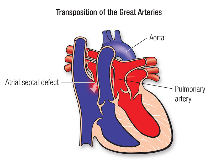
Transposition of the great arteries (TGA) is a serious congenital heart defect present at birth in which the two main arteries leaving the heart are connected to the wrong pumping chambers. In the most common and most critical form, dextro-transposition (d-TGA), the aorta arises from the right ventricle and the pulmonary artery arises from the left ventricle — the reverse of normal. This creates two separate, parallel circulations instead of one connected loop.
Normally, oxygen-poor (“blue”) blood returns to the right side of the heart, is pumped to the lungs, picks up oxygen, returns to the left side, and is then pumped out to the body. In d-TGA, oxygen-poor blood returning from the body is pumped straight back out to the body, while oxygenated blood from the lungs is pumped straight back to the lungs. The body is therefore starved of oxygen unless there is a connection (a “mixing” point) between the two circuits.
Without such mixing — through a hole between the chambers or an open ductus — d-TGA is rapidly fatal. Because of this, it is one of the true neonatal cardiac emergencies, usually presenting with cyanosis (blue discolouration) within hours or days of birth. Modern treatment, chiefly the arterial switch operation, has transformed TGA from an almost uniformly fatal condition into one with excellent survival and quality of life. Learn more about the broader group of congenital heart conditions.
2. Key Facts at a Glance
| Key Fact | Detail |
|---|---|
| Also known as | d-TGA, dextro-TGA, complete transposition, TGA |
| Body system affected | Heart and great vessels (cardiovascular system) |
| Common in | Newborns; slightly more common in male infants |
| Severity range | Life-threatening at birth; excellent long-term outlook after surgery |
| Key treatments | Prostaglandin, balloon atrial septostomy, arterial switch operation |
| Outlook | Very good after successful arterial switch in early infancy |
3. Alternative Names and Medical Terminology
- d-TGA / dextro-TGA — complete transposition (the emergency neonatal form).
- l-TGA / levo-TGA — congenitally corrected transposition (ccTGA), a rarer form in which the ventricles are also swapped, so circulation is functionally corrected but the right ventricle supports the body.
- TGA — general abbreviation for transposition of the great arteries.
- Ventriculo-arterial (VA) discordance — the precise anatomical term meaning the arteries arise from the wrong ventricles.
- Related descriptors: “transposition with intact ventricular septum”, “transposition with VSD”.
4. Relevant Heart, Lung or Vascular Anatomy
The heart has four chambers: two upper atria and two lower ventricles. The right ventricle normally pumps deoxygenated blood into the pulmonary artery toward the lungs; the left ventricle pumps oxygenated blood into the aorta toward the body. Four valves control flow, and the coronary arteries — which supply the heart muscle itself — normally branch from the base of the aorta.
In d-TGA this arrangement is reversed at the arterial level. The aorta connects to the right ventricle and the pulmonary artery connects to the left ventricle. The atrioventricular connections are normal (right atrium to right ventricle, left atrium to left ventricle). The coronary artery pattern, and any associated ventricular septal defect (VSD), atrial septal defect (ASD) or patent ductus arteriosus (PDA), are anatomically critical because they determine both the child’s survival before surgery and the complexity of the repair.
5. How the Disease Affects the Body
In normal circulation, the systemic and pulmonary loops run in series — every drop of blood alternates between body and lungs. In d-TGA, the loops run in parallel: deoxygenated blood circulates around the body over and over without reaching the lungs, while oxygenated blood circulates repeatedly through the lungs without reaching the body.
The only thing keeping a newborn alive is mixing between the two circuits. This mixing occurs through a patent foramen ovale/ASD (at atrial level), a VSD (at ventricular level), or a patent ductus arteriosus (PDA) (between the great arteries). As the ductus naturally closes in the first days of life and any atrial communication is small, oxygen levels fall further, producing profound cyanosis and, if untreated, metabolic acidosis, organ dysfunction, and death.
Even with adequate mixing, the tissues receive blood with a lower oxygen content than normal, so the infant works harder to compensate — breathing faster and with a higher heart rate. This is why the immediate goals of care are to keep the ductus open (with prostaglandin) and to improve mixing (with balloon atrial septostomy) until definitive surgery can restore normal series circulation.
6. Types and Classification
- d-TGA with intact ventricular septum (IVS) — the classic emergency form; depends heavily on the ductus and atrial mixing.
- d-TGA with ventricular septal defect (VSD) — a hole allows more mixing, so cyanosis may be milder but heart failure can develop.
- d-TGA with VSD and left ventricular outflow tract obstruction (LVOTO) — pulmonary stenosis complicates repair and may require different operations.
- Congenitally corrected TGA (l-TGA / ccTGA) — both the ventricles and great arteries are swapped, so blood flow is physiologically “corrected”, but the morphological right ventricle pumps to the body and tends to fail over decades.
7. Causes of the Disease
TGA arises from abnormal development of the heart and great vessels during the first eight weeks of pregnancy, when the single outflow tract normally divides and spirals to connect each ventricle to the correct artery. In transposition, this spiralling (conotruncal) process is disturbed, so the arteries end up connected to the wrong chambers.
In most cases no single cause is identified and the defect is not the result of anything a parent did or did not do. It results from a combination of genetic and environmental influences affecting early cardiac looping and outflow-tract septation. It is generally considered a sporadic (multifactorial) malformation rather than a strongly inherited one.
8. How the Disease Develops
TGA is a structural defect that forms before birth, not a condition that develops later in life. During early embryonic development, the truncus arteriosus (the common outflow trunk) normally divides into the aorta and pulmonary artery via a spiral septum, aligning each artery with the correct ventricle. In transposition, this septum forms straight rather than spiralled, or the conal (infundibular) tissue develops abnormally, so the aorta connects to the right ventricle and the pulmonary artery to the left.
Because the abnormality is established in the womb, the fetus is usually unaffected in utero: the placenta provides oxygen, and fetal shunts (the ductus arteriosus and foramen ovale) allow blood to bypass the lungs. Problems appear only after birth, when the baby must rely on its own lungs. As the ductus arteriosus and foramen ovale begin to close over the first hours to days of life, the parallel circulation can no longer mix adequately, and cyanosis rapidly worsens. The clinical “development” of TGA is therefore really the unmasking of a fixed defect as the normal newborn transition takes place.
9. Risk Factors
- Maternal diabetes, particularly poorly controlled, is associated with a higher risk of conotruncal defects.
- Maternal viral illness or rubella during pregnancy.
- Poor maternal nutrition and certain medication or alcohol exposures in early pregnancy.
- Male sex of the infant (d-TGA is somewhat more common in boys).
- Family history of congenital heart disease (a modest increase in risk).
Most babies with TGA, however, have no identifiable risk factor.
10. Genetic and Family-History Factors
Isolated d-TGA is usually sporadic and only rarely part of a recognised genetic syndrome — a contrast with several other congenital heart defects. The recurrence risk in a future sibling is generally low (in the low single-digit percentages). Occasionally TGA is associated with laterality (heterotaxy) disorders and abnormalities of left–right patterning genes. Congenitally corrected TGA (l-TGA) similarly is usually sporadic. Genetic counselling and fetal echocardiography are reasonable for families with a prior affected child or other congenital heart disease.
11. Who Is Most at Risk?
- Newborns of mothers with diabetes or with viral illness in early pregnancy.
- Male infants, who show a modest predominance.
- Babies with a family history of congenital heart disease.
- Infants with other conotruncal defects identified on fetal scanning.
Because most cases are sporadic, any newborn can be affected, which is why routine newborn pulse-oximetry screening is valuable for early detection.
12. Prevalence and Epidemiology
TGA is one of the more common cyanotic congenital heart defects, occurring in roughly 2 to 3 of every 10,000 live births and accounting for a meaningful share of babies presenting with cyanosis in the newborn period. It is more common in boys than girls. It occurs worldwide across all ethnic and geographic groups. Congenitally corrected TGA (l-TGA) is considerably rarer. These figures are approximate and vary between registries and regions.
13. Signs and Symptoms
The hallmark of d-TGA is cyanosis appearing within the first hours to days of life. Signs and symptoms include:
- Blue or dusky discolouration of the lips, tongue, skin and nail beds (central cyanosis) that does not improve with oxygen.
- Rapid or laboured breathing (tachypnoea).
- Poor feeding and tiring during feeds.
- Weak pulses and cool extremities if circulation is failing.
- A heart murmur — often absent in simple d-TGA, or present if there is a VSD or pulmonary stenosis.
In d-TGA with a large VSD, cyanosis may be less obvious at first, and the baby may instead present later with signs of heart failure — breathlessness, sweating with feeds and poor weight gain — over the first weeks of life. Because cyanosis can be subtle in some lighting and skin tones, low oxygen saturation on pulse oximetry is often the first objective clue.
14. Early-Stage Symptoms
In simple d-TGA the “early stage” is essentially the first hours of life. The baby may look well at delivery, then develop progressive blueness and fast breathing as the ductus arteriosus starts to close. Oxygen saturations are low and typically do not rise substantially with supplemental oxygen, a distinguishing feature. Feeding may become difficult as the baby tires.
15. Advanced-Stage Symptoms
Untreated or inadequately mixed d-TGA progresses to deep cyanosis, lethargy, poor feeding, and metabolic acidosis as tissues are starved of oxygen. The baby may become floppy and unresponsive, with signs of shock and multi-organ compromise. In d-TGA with a large VSD, “advanced” disease more often means congestive heart failure and, over months if unrepaired, the risk of irreversible pulmonary vascular disease. These are late findings that modern early surgery aims to prevent entirely.
16. Symptoms in Women, Men and Older Adults
TGA presents in newborns, so classic adult sex differences do not apply. There is a modest male predominance at birth. In adults, the relevant population is those who had surgery in childhood: patients repaired with the older atrial switch (Mustard or Senning) operations may develop right-ventricular failure, arrhythmias and baffle problems in adulthood, whereas those who had the arterial switch generally do well but need lifelong follow-up for coronary and pulmonary-artery issues. Congenitally corrected l-TGA may not cause symptoms until adulthood, when the systemic right ventricle begins to fail.
17. Emergency Warning Signs
Seek emergency care immediately for a newborn or infant with:
- Deep or worsening blue colour of lips, tongue or body.
- Very fast, laboured or grunting breathing.
- Limpness, unresponsiveness or extreme sleepiness.
- Refusal to feed with a mottled, cold or greyish appearance.
These indicate critically low oxygen and require urgent hospital assessment.
18. When to Seek Medical Help
Any cyanosis, fast breathing, or poor feeding in a newborn warrants immediate medical attention. Because many babies with TGA are now detected before or at birth through fetal echocardiography and newborn pulse-oximetry screening, prompt referral to a paediatric cardiology centre is essential the moment transposition is suspected.
19. Disease Stages, Grades and Severity
TGA is not “staged” like a progressive disease; severity is defined by anatomy and physiology:
- Adequacy of mixing — the single most important early factor; poor mixing means severe cyanosis.
- Associated defects — presence and size of a VSD, ASD, PDA, or LVOTO.
- Coronary artery pattern — some patterns make the arterial switch more technically demanding.
- Timing of diagnosis and treatment — earlier intervention correlates with better outcomes.
20. Disease Progression
Untreated, d-TGA progresses rapidly and fatally over the first days to weeks of life as ductal and atrial mixing declines. With modern care, the “progression” is instead through a planned pathway: stabilise with prostaglandin, improve mixing with balloon atrial septostomy if needed, then perform the arterial switch operation in the first two weeks of life. After successful repair, most children follow a normal developmental trajectory with periodic cardiac surveillance rather than ongoing disease progression.
21. Possible Complications
- Severe hypoxia and acidosis before surgery if mixing is inadequate.
- Coronary artery problems after arterial switch (kinking or narrowing of the reimplanted coronaries).
- Supravalvar pulmonary stenosis or neo-aortic valve regurgitation and root dilatation over time after arterial switch.
- Baffle obstruction, baffle leaks and arrhythmias after older atrial switch operations, with eventual systemic right-ventricular failure.
- Neurodevelopmental effects in a minority, related to pre-operative hypoxia and complex surgery.
22. Related and Associated Medical Conditions
TGA frequently coexists with ventricular septal defect (VSD), atrial septal defect (ASD), patent ductus arteriosus (PDA) and pulmonary (left ventricular outflow tract) stenosis. It shares the conotruncal family of defects with conditions such as tetralogy of Fallot. Some patients have coronary artery anomalies that influence surgery. Long-term, associated concerns include arrhythmias and, for atrial-switch survivors, heart failure.
23. Screening and Early Detection
- Fetal echocardiography — increasingly detects TGA before birth, allowing delivery at or near a cardiac surgical centre.
- Routine newborn pulse-oximetry screening — flags the low oxygen saturation of TGA even when cyanosis is subtle.
- Prenatal ultrasound anomaly scan — may raise suspicion (though the outflow-tract views can be difficult).
Antenatal detection markedly improves the newborn’s condition at the time of treatment.
24. How the Disease Is Diagnosed
Diagnosis begins with clinical suspicion in a cyanotic newborn whose oxygen levels do not respond to supplemental oxygen (a “failed hyperoxia test”). The definitive diagnostic test is echocardiography (heart ultrasound), which demonstrates the aorta arising from the right ventricle and the pulmonary artery from the left, defines any associated ASD, VSD, PDA or outflow obstruction, and maps the coronary artery pattern essential for surgical planning.
Supporting investigations include a chest X-ray, which may show the classic “egg-on-a-string” cardiac silhouette with a narrow upper mediastinum, and an electrocardiogram (ECG). Pulse oximetry confirms low saturations. Cardiac catheterisation is now used less for diagnosis and more for the therapeutic balloon atrial septostomy, though it can clarify coronary anatomy when echocardiography is inconclusive. Cardiac CT or MRI may be used in complex or older patients to define anatomy and ventricular function. When TGA is diagnosed antenatally, the delivery is planned so that neonatal stabilisation and surgery can proceed without delay. Prompt, accurate diagnosis is the foundation of the excellent outcomes now achievable.
25. Physical Examination and Medical History
Examination of a newborn with d-TGA typically reveals central cyanosis, tachypnoea, and normal or only slightly increased work of breathing initially. The second heart sound is often single and loud. A murmur may be absent in simple TGA, or present with a VSD or pulmonary stenosis. History focuses on timing and progression of cyanosis, feeding, any antenatal diagnosis, maternal diabetes or illness, and family history of congenital heart disease.
26. Diagnostic Tests and Imaging
- Echocardiography — the primary diagnostic tool; defines the transposed arteries, associated defects, and coronary pattern.
- Chest X-ray — may show the “egg-on-a-string” heart and increased pulmonary vascular markings.
- ECG — often normal for a newborn early on; may show right-ventricular dominance.
- Pulse oximetry — documents low, oxygen-resistant saturations.
- Cardiac catheterisation — mainly therapeutic (balloon septostomy); clarifies coronary anatomy if needed.
- Cardiac MRI / CT — reserved for complex anatomy and long-term follow-up.
27. Blood Tests, Biomarkers and Genetic Testing
There is no blood test that diagnoses TGA. Arterial blood gas shows low oxygen and may reveal metabolic acidosis if circulation is compromised. Lactate helps gauge tissue oxygen delivery. Routine pre-operative bloods (full blood count, clotting, electrolytes, kidney and liver function, blood group and cross-match) prepare for surgery. Genetic testing is not routinely needed for isolated d-TGA but may be considered when there are additional anomalies or a heterotaxy/laterality disorder.
28. Understanding Test Results
On echocardiography, the key finding is ventriculo-arterial discordance — the aorta (which gives off the coronary and head/neck vessels) arising from the right ventricle, and the pulmonary artery (which branches to the lungs) from the left. Reports will also describe the atrial and ventricular septum (any ASD or VSD), the ductus arteriosus, any outflow obstruction, and the coronary artery pattern, which is graded by standardised descriptions because it guides the surgeon. Low oxygen saturations that do not correct with oxygen point strongly to a mixing problem such as TGA. Your child’s cardiologist will interpret these findings in context and explain the surgical plan.
29. Differential Diagnosis
Other causes of cyanosis in a newborn that must be distinguished include:
- Tetralogy of Fallot and other cyanotic conotruncal defects.
- Total anomalous pulmonary venous connection (TAPVC).
- Tricuspid atresia and single-ventricle conditions.
- Truncus arteriosus and pulmonary atresia.
- Persistent pulmonary hypertension of the newborn and respiratory/lung disease.
Echocardiography reliably separates TGA from these.
30. Specialist and Multidisciplinary Evaluation
Care of TGA requires a paediatric cardiac team: a paediatric cardiologist, a congenital cardiac surgeon, a neonatologist, a cardiac anaesthetist and intensivist, specialist nurses, and perfusionists for cardiopulmonary bypass. Fetal cardiology, genetics, neurodevelopmental and social support services contribute across the pathway. Families benefit from choosing an experienced congenital cardiac centre with a dedicated team. Explore experienced paediatric heart specialists and treatment destinations.
31. Treatment Goals
- Immediate: ensure adequate mixing and oxygen delivery to keep the baby stable (prostaglandin, balloon septostomy).
- Definitive: restore normal series circulation by connecting each ventricle to the correct artery — the arterial switch operation.
- Long-term: preserve ventricular function, prevent complications, and support normal growth, development and quality of life.
32. When Is Treatment Required?
Treatment is always required, and urgently, in d-TGA — it is a neonatal emergency. Prostaglandin is started as soon as TGA is suspected to keep the ductus open. Balloon atrial septostomy is performed within hours if mixing is inadequate. The arterial switch operation is ideally carried out within the first one to two weeks of life, while the left ventricle is still “trained” by pumping against the higher resistance of the fetal/neonatal lungs. Congenitally corrected l-TGA may be managed more conservatively until problems arise.
33. Active Monitoring and Watchful Waiting
For d-TGA there is no role for watchful waiting — timely surgery is essential. “Monitoring” applies to the congenitally corrected (l-TGA) form, where a well-functioning heart may be observed with regular echocardiograms and clinical review, with intervention reserved for systemic right-ventricular dysfunction, significant tricuspid regurgitation, or heart block. All repaired patients enter lifelong surveillance rather than watchful waiting of an untreated defect.
34. Medications
- Prostaglandin E1 (alprostadil) — the critical first drug, given by infusion to keep the ductus arteriosus open and maintain mixing until surgery.
- Inotropes and vasoactive drugs — to support blood pressure and cardiac output around surgery.
- Diuretics — for heart failure, especially with a large VSD.
- Post-operative medications — pain relief, and sometimes anti-arrhythmics.
- Medication alone cannot correct TGA; drugs stabilise the baby for surgery and support recovery.
35. Minimally Invasive Treatments
The chief minimally invasive, catheter-based intervention in TGA is the balloon atrial septostomy (Rashkind procedure), performed via a vein without open surgery to enlarge the atrial communication and improve mixing. In older, previously repaired patients, some later problems — such as pulmonary artery narrowing or baffle stenosis after atrial switch — can be treated with catheter balloon dilation and stenting rather than repeat open surgery. Fuller definitive correction, however, requires open-heart surgery.
36. Catheter-Based and Endovascular Treatments
- Balloon atrial septostomy — emergency catheter procedure to create or enlarge an atrial opening for mixing.
- Balloon angioplasty and stenting — for pulmonary-artery stenosis or baffle obstruction seen in later follow-up.
- Device closure — occasionally for residual shunts or baffle leaks in atrial-switch survivors.
These procedures are performed in the cardiac catheterisation laboratory. See related endovascular stenting techniques.
37. Surgical Treatment Options
The arterial switch operation (ASO, or Jatene procedure) is the modern operation of choice for d-TGA. Performed on cardiopulmonary bypass in the first days to weeks of life, the surgeon divides both great arteries above the valves and reconnects them to the correct ventricles, so the aorta arises from the left ventricle and the pulmonary artery from the right. The technically demanding, defining step is transferring the coronary arteries from the original aortic root to the new aortic root; success depends heavily on the coronary anatomy. Any VSD is closed at the same operation.
Before the arterial switch became standard, the atrial switch operations — the Mustard and Senning procedures — redirected blood at the atrial level using a baffle, so oxygenated and deoxygenated blood reached the correct arteries even though the arteries were left transposed. These operations gave good early survival but left the right ventricle pumping to the body, leading to late right-ventricular failure and arrhythmias; they are now largely historic but remain relevant to many adults living with earlier repairs. For d-TGA with VSD and severe pulmonary stenosis, the Rastelli operation (or its variants) routes the left ventricle to the aorta through the VSD and connects the right ventricle to the pulmonary artery with a conduit. Congenitally corrected l-TGA may be managed with a “double switch” in selected cases. All are specialised congenital heart procedures performed at experienced surgical centres.
38. Advanced and Emerging Treatments
Advances focus on refining the arterial switch (coronary transfer techniques for unusual patterns), improving neuroprotection during neonatal bypass, and better fetal diagnosis and delivery planning. For adults with failing systemic right ventricles after atrial switch, research explores heart failure therapies, resynchronisation, mechanical support and, ultimately, transplantation. Tissue-engineered conduits and valves are being studied to reduce the need for repeat operations in Rastelli-type repairs. Growing use of 3D imaging and printing aids surgical planning in complex anatomy.
39. Treatment Options Compared
- Arterial switch (ASO) — anatomically corrects TGA and preserves the left ventricle as the systemic pump; the preferred modern operation, best done in the neonatal period.
- Atrial switch (Mustard/Senning) — physiologically corrects circulation but leaves the right ventricle supporting the body; largely historic due to late complications.
- Rastelli / double switch — reserved for specific anatomy (TGA with VSD and pulmonary stenosis, or l-TGA); more complex, may need conduit replacement.
- Catheter septostomy — a temporising measure, not definitive correction.
40. How Doctors Choose the Right Treatment
The choice depends on the exact anatomy: presence and size of a VSD, degree of any pulmonary stenosis, the coronary artery pattern, ventricular function, and the baby’s age and left-ventricular “training”. Simple d-TGA and d-TGA with VSD are corrected by arterial switch; TGA with VSD and severe LVOTO by Rastelli-type repair; and l-TGA is individualised. Timing matters greatly — the arterial switch is best performed before the left ventricle “detrains”. Decisions are made by the multidisciplinary team with the family.
41. Benefits and Risks of Treatment
Benefits: the arterial switch anatomically cures d-TGA, restores normal circulation, and offers excellent long-term survival and quality of life with a systemic left ventricle.
Risks: as major neonatal open-heart surgery, it carries risks of bleeding, infection, arrhythmia, and — specific to this operation — coronary artery problems, later pulmonary artery narrowing, and neo-aortic valve leak or root dilatation. Atrial-switch survivors face arrhythmias, baffle issues and right-ventricular failure. In experienced centres, operative mortality is now low and the benefits greatly outweigh the risks.
42. What Happens If the Disease Is Left Untreated?
Untreated d-TGA is almost always fatal in early infancy — the majority of babies would die within the first weeks to months of life as mixing fails. Those with a large VSD may survive a little longer but develop heart failure and, over months, irreversible pulmonary vascular disease (Eisenmenger physiology), which precludes later repair. This grim natural history is precisely why urgent stabilisation and early surgery are non-negotiable.
43. Treatment Success and Expected Outcomes
In experienced congenital cardiac centres, the arterial switch operation has high survival, with most children leaving hospital and growing normally. Long-term studies show that the great majority of arterial-switch survivors reach adulthood with good exercise capacity and a systemic left ventricle that functions well. Outcomes are best with early diagnosis, favourable coronary anatomy, and high-volume surgical experience. Some patients need later catheter or surgical intervention for pulmonary artery narrowing or neo-aortic valve issues, but overall the outlook is very good. These are general expectations; individual results vary.
44. Prognosis and Long-Term Outlook
The long-term outlook after a successful arterial switch is excellent — the operation anatomically corrects the defect and preserves the left ventricle as the body’s pump. Most survivors enjoy normal or near-normal daily life, schooling, work and physical activity, with lifelong cardiology follow-up to watch for the recognised late issues: coronary artery narrowing, supravalvar pulmonary stenosis, and neo-aortic root dilatation or valve regurgitation.
Adults who had the older atrial switch (Mustard/Senning) operations have a more guarded outlook because the right ventricle supports the systemic circulation and may fail over the decades, along with a tendency to atrial arrhythmias and baffle problems; they need close specialist care and, occasionally, advanced heart-failure treatment or transplantation. Congenitally corrected l-TGA patients may remain well for years but are at risk of systemic right-ventricular failure, tricuspid regurgitation and heart block in adulthood. Overall, transposition has been transformed from a fatal condition into one compatible with a full life when treated in a modern programme, provided patients remain engaged with lifelong congenital-heart follow-up.
45. Recovery and Rehabilitation
After the arterial switch, the baby recovers in a cardiac intensive care unit, initially ventilated and supported with medications, then progressively weaned. Most infants are ready to go home within one to a few weeks if recovery is uncomplicated. At home, parents monitor feeding, weight gain, breathing and the wound. Growth and development are generally normal, and early developmental support is offered where needed. Older children and adults undergoing later procedures follow standard cardiac recovery and, where appropriate, cardiac rehabilitation.
46. Follow-Up Tests and Long-Term Monitoring
All patients need lifelong follow-up with a congenital cardiologist. Surveillance typically includes periodic clinical review, ECG, echocardiography, and — as patients grow — exercise testing and sometimes cardiac MRI/CT to assess the coronary arteries, pulmonary arteries, neo-aortic root and ventricular function. Holter monitoring may be used to detect arrhythmias, particularly in atrial-switch survivors. The frequency is individualised.
47. Managing Recurrence or Disease Progression
TGA does not “recur”, but late complications can develop and progress. Management includes catheter dilation or stenting for pulmonary artery or baffle narrowing, surgery for significant neo-aortic valve regurgitation or root enlargement, anti-arrhythmic drugs, ablation or pacemakers for rhythm problems, and heart-failure therapy for a failing systemic right ventricle in atrial-switch or l-TGA patients. Regular follow-up allows problems to be caught and treated early.
48. Living with the Disease
Most children and adults who had an arterial switch live essentially normal lives — attending school, working, exercising and, for many, participating in sport. Practical considerations include staying in lifelong cardiac follow-up, carrying a summary of the cardiac history, antibiotic advice for some dental/surgical procedures where indicated, and planning around pregnancy, occupation and insurance. Patients with atrial-switch repairs or l-TGA may need more activity guidance depending on ventricular function and rhythm.
49. Diet and Nutrition Guidelines
For infants, the focus is adequate nutrition and weight gain, sometimes with fortified feeds or extra calories before and after surgery; feeding support is provided if the baby tires. For older children and adults, a balanced, heart-healthy diet — plenty of fruit and vegetables, whole grains, limited salt and processed food — supports overall cardiovascular health. Those with heart failure may need fluid and salt guidance from their team. There is no special “TGA diet”.
50. Exercise and Physical-Activity Guidelines
After a successful arterial switch with good heart function, most patients can participate in normal activity and sport, guided by periodic exercise testing. Recommendations are individualised according to ventricular function, any residual pulmonary artery narrowing, coronary findings and arrhythmia risk. Patients with atrial-switch repairs, systemic right-ventricular dysfunction, or l-TGA may be advised to avoid intense competitive or isometric exertion. Always follow your congenital cardiologist’s specific advice.
51. Medications, Activities and Habits to Avoid
- Never stop prescribed heart medications without medical advice.
- Adults should avoid smoking, excess alcohol and recreational drugs, which stress the heart.
- Discuss strenuous competitive or contact sport with your cardiologist before participating.
- Seek advice on antibiotic prophylaxis for certain dental/surgical procedures if you have residual defects or prosthetic material.
- Women should obtain pre-conception cardiology counselling before pregnancy.
52. Preventing the Disease or Reducing Its Risks
TGA cannot reliably be prevented, as it forms early in fetal development from largely unknown causes. General measures that support healthy pregnancy and may reduce congenital heart defect risk include good control of maternal diabetes, taking folic acid, avoiding alcohol, smoking and unnecessary medications in pregnancy, keeping vaccinations (including rubella) up to date, and attending antenatal care. Fetal echocardiography does not prevent TGA but enables early detection and safer delivery planning.
53. Pregnancy and the Disease
Two contexts matter. First, maternal factors (diabetes, illness, exposures) modestly influence the risk of a baby having TGA. Second, women who themselves had TGA repaired can often have successful pregnancies but should have pre-pregnancy and specialist cardiac-obstetric care, particularly if they have a systemic right ventricle (atrial switch or l-TGA), arrhythmias or reduced heart function, which raise pregnancy risk. There is also a small increased chance of congenital heart disease in the baby, so fetal echocardiography is advised.
54. Disease in Children and Young Adults
TGA is fundamentally a condition of the newborn, corrected in infancy. Children who had an arterial switch usually grow and develop normally with routine cardiology follow-up, though a minority need attention for pulmonary artery narrowing or neo-aortic valve issues. Young adults transition to adult congenital heart disease (ACHD) services and should remain engaged with specialist care, especially those with atrial-switch repairs or l-TGA who face higher long-term risks. Learn more about congenital heart disease across the lifespan.
55. Disease in Older Adults
Older adults with TGA are long-term survivors of childhood surgery. Those with atrial-switch (Mustard/Senning) repairs may develop systemic right-ventricular failure, atrial arrhythmias and baffle complications over the decades. Congenitally corrected l-TGA may only declare itself in later adulthood with heart failure, tricuspid regurgitation or complete heart block. These patients need lifelong ACHD follow-up and may require devices, catheter procedures, or advanced heart-failure care.
56. Emotional Health and Patient Support
A diagnosis of a serious newborn heart defect is frightening and stressful for families. Support includes clear information from the cardiac team, psychological and social support, and peer/parent networks and congenital heart charities. As children grow, counselling and transition programmes help them understand and manage their condition. Adults may face anxiety about the future, exercise, pregnancy or work; specialist ACHD nurses and support groups are valuable resources. Read real patient stories and reach out via our contact page.
57. Preparing for Your Specialist Appointment
- Bring all previous records: operative notes, echo/MRI reports, and a summary of procedures and medications.
- Note the exact type of repair (arterial switch, Mustard/Senning, Rastelli) and dates.
- Write down current symptoms, exercise tolerance and any palpitations.
- List all medications and allergies.
- Prepare your questions in advance (see the next section) and consider bringing a family member.
58. Questions to Ask Your Doctor
- What exact type of transposition and repair does my child (or do I) have?
- What operation is recommended and why, and when should it be done?
- What are the specific risks of surgery in our case, given the coronary anatomy?
- What is the expected recovery and hospital stay?
- What long-term complications should we watch for?
- How often will follow-up and imaging be needed?
- What activities, sports or work are safe?
- What should we know about pregnancy and inheritance?
- Is this the right centre and surgeon for this operation, and what is their experience?
- What are the signs that would mean we need urgent review?
59. Cost of Diagnosis and Treatment
Costs vary widely by country, hospital and complexity. The figures below are broad approximations in US dollars for the arterial switch operation and associated care.
| Region | Approximate Cost (USD) |
|---|---|
| United States | $80,000 – $250,000+ |
| United Kingdom (private) | $50,000 – $150,000 |
| Singapore | $40,000 – $90,000 |
| Thailand | $20,000 – $45,000 |
| Turkey | $18,000 – $40,000 |
| India | $8,000 – $25,000 |
Medical-tourism destinations such as India, Turkey and Thailand often provide high-quality congenital cardiac surgery at roughly 50–90% less than the US or UK. Always confirm what a quote includes. Explore treatment destinations and hospitals.
60. Factors Affecting Treatment Cost
- Complexity of anatomy (simple d-TGA vs. TGA with VSD/LVOTO or difficult coronaries).
- Type of operation and need for conduits or later reinterventions.
- Length of ICU and hospital stay and any complications.
- Hospital accreditation, surgeon experience and country.
- Pre-operative catheter procedures, imaging and follow-up.
- Travel, accommodation and interpreter services for international patients.
61. Choosing the Right Specialist
Choose a congenital (paediatric) cardiac surgeon and cardiologist who regularly perform the arterial switch and manage TGA, ideally within a high-volume programme. Ask about case numbers, outcomes, and experience with complex coronary patterns. Continuity into adult congenital heart disease services matters for lifelong care. Browse experienced heart specialists.
62. Choosing the Right Hospital or Treatment Centre
Look for a dedicated congenital cardiac centre with:
- International accreditation (e.g., JCI) and strong published outcomes.
- High surgical volume in neonatal and congenital heart surgery.
- A full multidisciplinary team and paediatric cardiac ICU.
- Catheter-lab, fetal cardiology and neurodevelopmental services.
- Clear communication and support for international families.
Compare hospitals and surgical options.
63. Getting a Second Medical Opinion
Because TGA repair is complex and high-stakes, a second opinion from another experienced congenital centre is entirely reasonable and often reassuring — especially regarding operative approach, timing and coronary anatomy. A good specialist will welcome it. Sharing echo images and reports allows a meaningful review. Request a second opinion through our team.
64. Treatment Abroad and Medical-Travel Considerations
Many families travel for congenital cardiac surgery to access experienced surgeons and lower costs. Key considerations:
- Confirm the centre’s accreditation, volume and TGA-specific outcomes.
- Plan for a prolonged stay covering surgery, recovery and initial follow-up.
- Arrange records transfer, imaging sharing and clear communication.
- Consider travel logistics for a fragile newborn and post-operative flying advice.
- Establish follow-up arrangements back home for lifelong care.
Explore medical-travel destinations and contact us for guidance.
65. Frequently Asked Questions
Is TGA an emergency? Yes. d-TGA is a neonatal emergency; babies need prostaglandin and often balloon septostomy urgently, followed by early surgery.
Can TGA be cured? The arterial switch operation anatomically corrects d-TGA, and most children go on to live normal lives with lifelong follow-up.
When is the surgery done? The arterial switch is ideally performed in the first one to two weeks of life, while the left ventricle is still conditioned to pump to the body.
What is balloon atrial septostomy? A catheter procedure that enlarges the opening between the atria to improve mixing of oxygenated and deoxygenated blood before surgery.
What is the difference between d-TGA and l-TGA? d-TGA is the classic emergency form; l-TGA (congenitally corrected TGA) has both ventricles and arteries swapped, so circulation works but the right ventricle supports the body and may fail later.
Will my child be able to play sport? Usually yes after a successful arterial switch with good heart function, guided by periodic exercise testing and their cardiologist.
Does TGA run in families? Isolated d-TGA is usually sporadic with a low recurrence risk; genetic counselling is available for concerned families.
Do adults with old atrial-switch repairs need special care? Yes — they need lifelong adult congenital heart follow-up for arrhythmias and possible right-ventricular failure.
66. Patient Stories and Treatment Experiences
The following are representative, anonymised examples for illustration only.
- Aarav, India — Diagnosed with d-TGA soon after birth, Aarav received prostaglandin and a balloon septostomy, then an arterial switch at ten days old. Now a lively school-age child, he attends yearly cardiology checks and plays football with his friends.
- Sofia, Spain — Detected antenatally, Sofia was delivered at a cardiac centre and operated on within her first two weeks. Her parents credit early diagnosis for her smooth recovery and normal development.
- James, United Kingdom — Now in his thirties, James had a Mustard operation as a baby in an earlier era. He lives a full life but sees an adult congenital heart team regularly to monitor his systemic right ventricle and heart rhythm.
67. Latest Research and Clinical Trials
Contemporary research focuses on long-term outcomes of the arterial switch — particularly the fate of the coronary arteries, neo-aortic root and pulmonary arteries into adulthood — and on neuroprotection during neonatal surgery to optimise developmental outcomes. Investigators are studying advanced heart-failure therapies and mechanical support for adults with failing systemic right ventricles, tissue-engineered valves and conduits to reduce reoperations, and improvements in fetal diagnosis and delivery planning. Families interested in trials should ask their congenital cardiac centre about current registries and studies; reputable information is available through major congenital heart programmes and specialist societies.
68. Related Diseases and Conditions
- Congenital Heart Disease (overview)
- Tetralogy of Fallot
- Ventricular Septal Defects
- Atrial Septal Defects
- Patent Ductus Arteriosus
- Single Ventricle Physiological Conditions
69. Related Treatments and Procedures
- Congenital Heart Procedures
- Hybrid Cardiac Procedures
- Endovascular Stenting
- Electrophysiological Procedures
- Minimally Invasive Cardiac Surgery
70. Medical Glossary
- d-TGA (dextro-transposition) — the common form where the aorta arises from the right ventricle and the pulmonary artery from the left.
- l-TGA / congenitally corrected TGA — both ventricles and arteries are swapped, so circulation functions but the right ventricle supports the body.
- Ventriculo-arterial discordance — arteries connected to the wrong ventricles.
- Parallel circulation — the two circuits run separately rather than in series, requiring mixing to survive.
- Ductus arteriosus (PDA) — a fetal vessel between aorta and pulmonary artery; kept open by prostaglandin to allow mixing.
- Prostaglandin E1 (alprostadil) — the medication used to keep the ductus open.
- Balloon atrial septostomy (Rashkind) — catheter procedure to enlarge the atrial opening for mixing.
- Arterial switch operation (Jatene / ASO) — the operation that reconnects the great arteries and coronaries to the correct chambers.
- Atrial switch (Mustard/Senning) — older baffle operations that redirect blood at atrial level.
- Rastelli operation — repair for TGA with VSD and pulmonary stenosis using a conduit.
- Coronary transfer — reimplantation of the coronary arteries during the arterial switch.
- Cyanosis — bluish discolouration from low blood oxygen.
- Neo-aortic valve/root — the original pulmonary valve/root that becomes the systemic (aortic) valve after the switch.
- Systemic right ventricle — a right ventricle pumping to the body, prone to late failure (in atrial switch and l-TGA).
71. Medical Review, Editorial Policy and Last Updated Date
Last updated: 11 July 2026.
This article is written and reviewed following BestHeartSurgery.com’s editorial policy, aligning content with recognised guidance from bodies such as the ACC/AHA, ESC, NHS and specialist congenital cardiac societies, and with standard paediatric cardiology and cardiac surgery references. Content is intended to be accurate, balanced and up to date, and is reviewed by qualified medical professionals.
Disclaimer: This information is provided for educational purposes only and is not a substitute for professional medical advice, diagnosis or treatment. Always seek the guidance of a qualified paediatric cardiologist or congenital cardiac surgeon regarding your child’s or your own condition.
72. Clinical Guidelines and Medical References
This content draws on general, publicly available guidance and standard textbook knowledge, including:
- American College of Cardiology / American Heart Association (ACC/AHA) guidelines on congenital and adult congenital heart disease.
- European Society of Cardiology (ESC) guidelines for the management of adult congenital heart disease.
- NHS and other national health-service patient information on transposition of the great arteries.
- Society of Thoracic Surgeons (STS) congenital heart surgery resources.
- Standard paediatric cardiology and congenital cardiac surgery textbooks.
Please consult the current versions of these guidelines and your care team for detailed, personalised recommendations.
73. Book an Appointment or Request a Second Opinion
If your child has been diagnosed with transposition of the great arteries, or you are an adult living with a previous repair, our network can connect you with experienced congenital cardiac specialists and accredited hospitals worldwide.
- Book an appointment: /contact/
- Request a second opinion or ask a question: Contact us
Explore top hospitals, specialist doctors, and treatment destinations to make an informed, confident decision about care.