1. Disease Overview
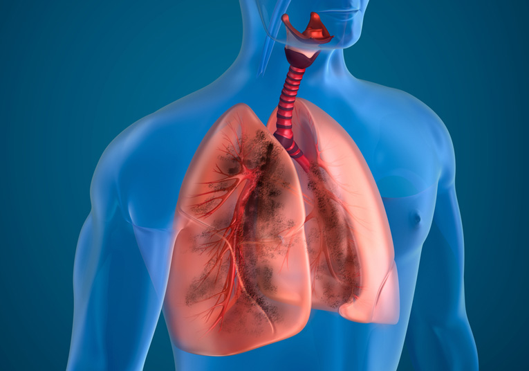
Lung disease refers to any condition that stops the lungs from working normally. On a heart-surgery portal, the focus is on surgically relevant pulmonary conditions — those a cardiothoracic (thoracic) surgeon may treat, or that directly influence the heart. These include lung cancer and lung tumours, bronchiectasis, emphysema and giant bullae, pleural disease (pleural effusions, empyema, pneumothorax, mesothelioma) and pulmonary vascular disease such as pulmonary hypertension, which can strain the right side of the heart and lead to cor pulmonale.
The lungs and heart share the chest cavity and are functionally inseparable: the right ventricle pumps blood through the lungs to collect oxygen, and the left heart depends on that oxygenated return. When lung tissue, airways or pulmonary vessels are diseased, the heart must work harder. Many patients therefore see both a pulmonologist and a cardiothoracic surgeon.
Modern thoracic surgery has shifted from large open incisions toward minimally invasive video-assisted thoracoscopic surgery (VATS) and robotic surgery, allowing lung resections with smaller wounds, less pain and faster recovery. This page explains causes, symptoms, diagnosis, surgical and non-surgical treatment, costs, and how to seek care abroad. It is educational and does not replace a consultation with a qualified doctor.
2. Key Facts at a Glance
| Fact | Detail |
|---|---|
| Also known as | Pulmonary disease, respiratory disease, thoracic disease |
| Body system affected | Respiratory system (lungs, airways, pleura, pulmonary vessels) — closely linked to the heart |
| Common in | Smokers, older adults, people with occupational or environmental exposure |
| Severity range | Mild and manageable to life-threatening (advanced cancer, severe pulmonary hypertension) |
| Key treatments | Medications, oxygen, VATS/robotic resection, lobectomy, pleural procedures, transplant |
| Outlook | Highly variable — excellent for early-stage resectable disease, guarded for advanced disease |
3. Alternative Names and Medical Terminology
Lung disease is broadly termed pulmonary disease or respiratory disease. Common surgically relevant subtypes and their terms include:
- Lung cancer — bronchogenic carcinoma; NSCLC (non-small-cell lung cancer), SCLC (small-cell lung cancer)
- COPD — chronic obstructive pulmonary disease; includes emphysema and chronic bronchitis
- Bronchiectasis — permanent airway dilatation
- Bulla / bullae — large air-filled spaces; giant bullous emphysema
- Pleural disease — effusion, empyema, pneumothorax, mesothelioma
- Pulmonary hypertension (PH / PAH) and cor pulmonale (right-heart strain from lung disease)
- VATS — video-assisted thoracoscopic surgery
4. Relevant Heart, Lung or Vascular Anatomy
The lungs sit inside the thorax (chest), one on each side of the heart. Air enters via the trachea, which divides into the right and left main bronchi, then into smaller bronchioles ending in alveoli — tiny air sacs where oxygen enters the blood and carbon dioxide leaves.
Each lung is divided into lobes — three on the right (upper, middle, lower) and two on the left (upper, lower) — the anatomical basis for surgical lobectomy. The lungs are wrapped in the pleura, a double membrane with a thin fluid layer; the space between the layers is the pleural cavity.
Crucially for a heart-surgery site: the pulmonary arteries carry deoxygenated blood from the right ventricle to the lungs, and the pulmonary veins return oxygenated blood to the left atrium. Diseased lungs raise resistance in these vessels, forcing the right heart to pump against higher pressure.
5. How the Disease Affects the Body
Lung disease disrupts gas exchange — the movement of oxygen into and carbon dioxide out of the blood. The mechanism depends on the type:
- Obstructive disease (COPD, emphysema, bronchiectasis, asthma) narrows or damages airways, trapping air and making exhalation difficult. Emphysema destroys alveolar walls, reducing the surface area for gas exchange.
- Restrictive disease (pulmonary fibrosis, some pleural disease) stiffens lung tissue or limits chest expansion, so the lungs cannot fill fully.
- Tumours occupy space, block airways, invade nearby structures and can spread (metastasise).
- Pleural disease compresses lung tissue with fluid, air or thickened membranes.
- Pulmonary vascular disease raises pressure in the lung circulation.
Reduced oxygen (hypoxaemia) makes tissues work harder and can cause breathlessness, fatigue and, over time, strain on the right ventricle. Chronic lung disease that raises pulmonary pressures leads to cor pulmonale — enlargement and eventual failure of the right heart. This heart-lung interdependence is why cardiothoracic teams manage many of these patients jointly.
6. Types and Classification
Surgically relevant lung disease is grouped as:
- Airway diseases — asthma, chronic bronchitis, emphysema/COPD, bronchiectasis
- Lung tissue (parenchymal) diseases — pulmonary fibrosis, sarcoidosis, infections
- Lung tumours — lung cancer (NSCLC, SCLC), benign tumours, metastases, and tumours of the heart and lung
- Pleural diseases — effusion, empyema, pneumothorax, mesothelioma
- Pulmonary vascular diseases — pulmonary hypertension, pulmonary embolism, cor pulmonale
Lung cancer uses the TNM staging system (tumour, nodes, metastasis). COPD is graded by the GOLD classification. Pulmonary hypertension is classified into five WHO groups by underlying cause.
7. Causes of the Disease
- Tobacco smoking — the leading cause of COPD and most lung cancers
- Air pollution and occupational exposure — asbestos (mesothelioma), silica, coal dust, industrial fumes
- Infections — recurrent or severe infections can cause bronchiectasis and empyema
- Genetic conditions — alpha-1 antitrypsin deficiency, cystic fibrosis
- Radon and environmental radiation
- Autoimmune and inflammatory disease — can cause fibrosis and pulmonary hypertension
- Blood clots — chronic pulmonary embolism can lead to pulmonary hypertension
Many conditions have more than one contributing cause, and smoking amplifies the effect of other exposures.
8. How the Disease Develops
Most surgically relevant lung disease develops slowly over years. In smokers, repeated inhalation of toxins inflames airways and destroys alveolar walls, gradually producing emphysema and airflow obstruction. Damaged DNA in airway lining cells can accumulate mutations that eventually form a cancerous tumour — often decades after exposure begins.
Bronchiectasis develops when infection or inflammation permanently widens and scars airways, creating pockets where mucus pools and repeated infection occurs — a self-perpetuating cycle. Emphysematous bullae form as destroyed lung tissue coalesces into large air spaces that compress healthy lung.
Pleural disease may develop acutely (a spontaneous pneumothorax) or gradually (a slowly accumulating malignant effusion). Pulmonary hypertension develops as small lung arteries thicken and stiffen, steadily raising the pressure the right ventricle must overcome. Over time the right ventricle hypertrophies, dilates and can fail. Because progression is often silent early on, many patients present only when symptoms or an incidental scan reveal advanced disease.
9. Risk Factors
Modifiable:
- Smoking and second-hand smoke
- Occupational exposure (asbestos, silica, dusts, fumes)
- Air pollution
- Recurrent untreated respiratory infections
- Obesity and physical inactivity
Non-modifiable:
- Increasing age
- Male sex (slightly higher for some cancers, though the gap is narrowing)
- Family history and inherited conditions
- Prior lung disease or chest radiotherapy
The single most important, avoidable risk factor across the group is tobacco use.
10. Genetic and Family-History Factors
Most lung disease is driven by environment and smoking, but genetics matter. Alpha-1 antitrypsin deficiency is an inherited condition that causes early emphysema, sometimes in non-smokers. Cystic fibrosis and primary ciliary dyskinesia cause bronchiectasis from childhood. A family history of lung cancer modestly raises individual risk, and certain inherited mutations influence tumour behaviour. Some forms of pulmonary arterial hypertension run in families (e.g. BMPR2 gene mutations). Genetic counselling and, where appropriate, tumour molecular profiling (see section 27) increasingly guide diagnosis and treatment.
11. Who Is Most at Risk?
- Current and former smokers, especially with long smoking histories
- Older adults, particularly those over 55–60
- Workers exposed to asbestos, silica, coal or industrial chemicals
- People with chronic or recurrent chest infections
- Those with inherited conditions (alpha-1 antitrypsin deficiency, cystic fibrosis)
- People with connective-tissue disease or unresolved blood clots (for pulmonary hypertension)
- Residents of high-pollution or high-radon areas
12. Prevalence and Epidemiology
Lung disease is among the most common causes of illness and death worldwide. COPD affects hundreds of millions of people globally and is a leading cause of death. Lung cancer is one of the most frequently diagnosed cancers and the leading cause of cancer death worldwide, largely because it is often found late. Bronchiectasis is increasingly recognised, especially in older adults. Pulmonary hypertension is far less common but seriously affects quality and length of life. Figures vary widely by country, smoking rates and air quality; the numbers here are broad approximations rather than precise statistics.
13. Signs and Symptoms
Symptoms depend on the specific condition but commonly include:
- Breathlessness (dyspnoea) — at first with exertion, later at rest
- Chronic cough — dry or productive
- Coughing up blood (haemoptysis) — always warrants urgent assessment
- Wheezing and chest tightness
- Recurrent chest infections
- Chest pain, especially with pleural disease
- Fatigue and reduced exercise tolerance
- Unexplained weight loss (a red flag for cancer)
- Ankle swelling and signs of right-heart strain in advanced pulmonary hypertension or cor pulmonale
Many people attribute early symptoms to ageing, being unfit or a “smoker’s cough”, which delays diagnosis. Any persistent respiratory symptom deserves medical review.
14. Early-Stage Symptoms
Early lung disease is frequently silent or mild. When present, early signs include a persistent cough, mild breathlessness on exertion (climbing stairs, brisk walking), occasional wheeze, and more frequent chest infections. Early lung cancer often causes no symptoms at all and is found incidentally on a scan done for another reason — a key argument for screening at-risk groups.
15. Advanced-Stage Symptoms
As disease advances, symptoms intensify: breathlessness at rest, persistent cough, haemoptysis, chest pain, marked fatigue and weight loss. Advanced lung cancer may cause hoarseness, difficulty swallowing, or symptoms from spread to bone, brain or liver. Severe COPD and pulmonary hypertension produce cyanosis (bluish lips), leg swelling, and signs of right heart failure (cor pulmonale). Advanced pleural disease causes large effusions with severe breathlessness.
16. Symptoms in Women, Men and Older Adults
Historically lung cancer and COPD were seen mainly in men, but rising female smoking has narrowed that gap; women are more likely to develop certain lung cancers (such as adenocarcinoma) and may present with subtler symptoms. Men still have higher rates of asbestos-related and occupational lung disease. Older adults often have milder or atypical symptoms, may attribute breathlessness to ageing, and frequently have coexisting heart disease that complicates the picture. Fatigue and confusion can dominate in the elderly.
17. Emergency Warning Signs
Seek emergency care immediately for:
- Severe or sudden breathlessness or inability to speak in full sentences
- Coughing up significant blood
- Sudden sharp chest pain with breathlessness (possible pneumothorax or pulmonary embolism)
- Blue lips or face (cyanosis)
- Fainting or collapse
- Confusion or drowsiness from low oxygen
18. When to Seek Medical Help
Book a non-urgent appointment for a cough lasting more than three weeks, breathlessness that is new or worsening, repeated chest infections, unexplained weight loss, or any coughing up of blood. Anyone with a significant smoking history and new respiratory symptoms should be assessed promptly, as should people with known occupational asbestos exposure.
19. Disease Stages, Grades and Severity
Severity is measured differently by condition:
- Lung cancer uses TNM staging (Stage I–IV), determining resectability and prognosis. Early stages (I–II) are often surgically curable; Stage IV means distant spread.
- COPD is graded by the GOLD system using lung-function (spirometry) and symptoms.
- Pulmonary hypertension is graded by pressure measurements and functional class (WHO Functional Class I–IV).
- Pleural disease severity relates to fluid volume, infection and lung compression.
Accurate staging drives every treatment decision and is completed before surgery is planned.
20. Disease Progression
Untreated progression varies by type. COPD tends to worsen gradually, punctuated by acute “exacerbations”. Lung cancer, if untreated, grows locally, invades adjacent structures and metastasises. Bronchiectasis follows a cycle of infection and further airway damage. Pulmonary hypertension steadily raises right-heart workload until the right ventricle fails. Progression can often be slowed or halted by stopping smoking, treating infection, resecting tumours early and managing pulmonary pressures.
21. Possible Complications
- Respiratory failure — the lungs cannot maintain adequate oxygen
- Cor pulmonale and right heart failure — the direct heart-lung link
- Recurrent pneumonia and empyema
- Pneumothorax — collapsed lung, especially with bullous disease
- Massive haemoptysis
- Metastatic spread of cancer
- Pulmonary embolism
- Malignant pleural effusion
- Reduced tolerance for other surgery, including cardiac surgery
22. Related and Associated Medical Conditions
Lung disease commonly coexists with coronary artery disease, heart failure, atrial fibrillation and other heart-rhythm disorders, because smoking and ageing damage both organs. Pulmonary hypertension links directly to left-heart and valve disease. Patients often also have diabetes, osteoporosis (worsened by steroids), depression and general deconditioning. Recognising these associations is essential when planning surgery, since coexisting heart disease affects anaesthetic and surgical risk.
23. Screening and Early Detection
Low-dose CT (LDCT) screening is recommended in many countries for high-risk people — typically older adults (roughly 50–80) with a substantial smoking history. Screening can detect lung cancer early, when it is small and curable by surgery, significantly improving survival. There is no equivalent mass-screening programme for most other lung diseases, but spirometry can detect COPD early, and at-risk individuals (e.g. asbestos-exposed workers) should have regular medical review.
24. How the Disease Is Diagnosed
Diagnosis follows a structured pathway that combines history, examination, imaging and tissue sampling. It usually begins with a chest X-ray, prompted by symptoms or an abnormal screening scan. If an abnormality is seen, a CT scan of the chest provides detailed images of the lungs, airways, pleura and mediastinum.
For suspected cancer, a PET-CT scan assesses spread, and tissue biopsy confirms the diagnosis — obtained by bronchoscopy, CT-guided needle biopsy, endobronchial ultrasound (EBUS) or, when needed, surgical biopsy via VATS. Lymph nodes are sampled to complete staging.
For airway disease, spirometry and lung-function tests measure airflow and lung volumes. For pulmonary vascular disease, an echocardiogram estimates pulmonary pressures and assesses the right heart, and right-heart catheterisation confirms pulmonary hypertension. Pleural fluid is sampled by pleural aspiration (thoracentesis) and analysed. A multidisciplinary team (MDT) reviews all results to reach a diagnosis and plan treatment. See our treatments and procedures pages for related diagnostic services.
25. Physical Examination and Medical History
The doctor asks about smoking history, occupational exposures, symptoms, their duration, and family history. Examination may reveal an abnormal breathing pattern, wheeze or crackles on listening to the chest, reduced breath sounds over an effusion or collapse, finger clubbing (associated with some lung cancers and chronic disease), cyanosis, and signs of right-heart strain such as raised neck veins and ankle swelling. Weight and general condition are noted, as they influence fitness for surgery.
26. Diagnostic Tests and Imaging
- Chest X-ray — first-line imaging
- CT chest — detailed anatomy, tumour size and location
- PET-CT — detects metabolically active tumour and spread
- Bronchoscopy / EBUS — direct airway view and node sampling
- CT-guided biopsy — tissue from peripheral lesions
- Spirometry and lung-function tests — airflow and volumes
- Echocardiogram — right-heart function and pulmonary pressures
- Right-heart catheterisation — confirms pulmonary hypertension
- VATS biopsy — surgical tissue diagnosis when others are inconclusive
27. Blood Tests, Biomarkers and Genetic Testing
Blood tests assess general health, infection, kidney and liver function, and clotting before surgery. Arterial blood gases measure oxygen and carbon dioxide levels. For lung cancer, molecular and genetic profiling of tumour tissue (e.g. EGFR, ALK, ROS1 mutations and PD-L1 expression) guides targeted therapy and immunotherapy. Alpha-1 antitrypsin levels are checked in early or non-smoking emphysema. Certain autoimmune blood markers help identify causes of fibrosis and pulmonary hypertension.
28. Understanding Test Results
Results are interpreted together, not in isolation. A CT may show a nodule (which may be benign or malignant), a mass, an effusion or emphysematous changes. Spirometry reports airflow as percentages of predicted values; low ratios indicate obstruction. TNM stage summarises how far cancer has spread. Pulmonary artery pressures from catheterisation confirm and grade pulmonary hypertension. Your specialist will explain what your specific results mean for treatment options and outlook; ask for plain-language explanations.
29. Differential Diagnosis
Because breathlessness and cough are common, doctors must distinguish lung disease from other causes: heart failure, coronary artery disease and valve disease can all cause breathlessness; anaemia causes fatigue and breathlessness; anxiety can mimic respiratory symptoms; and infections such as tuberculosis can resemble cancer on imaging. A lung mass must be distinguished between primary cancer, metastasis, infection and benign tumour — which is why tissue biopsy is essential.
30. Specialist and Multidisciplinary Evaluation
Surgically relevant lung disease is managed by a multidisciplinary team: respiratory physicians (pulmonologists), thoracic (cardiothoracic) surgeons, radiologists, pathologists, oncologists, anaesthetists and specialist nurses. For pulmonary hypertension, cardiologists are central. The MDT reviews imaging, biopsy results and fitness assessments to agree the safest, most effective plan. You can find experienced doctors and specialist hospitals through this site.
31. Treatment Goals
- Cure where possible (early-stage cancer via complete resection)
- Relieve symptoms — breathlessness, cough, pain
- Preserve and improve lung function
- Slow or halt disease progression
- Prevent complications such as infection, pneumothorax and cor pulmonale
- Protect the heart by reducing pulmonary strain
- Maintain quality of life and independence
32. When Is Treatment Required?
Treatment is required when disease causes symptoms, threatens life, or is curable if acted upon. Resectable lung cancer should be treated promptly with surgery. Symptomatic bullae or recurrent pneumothorax warrant surgical repair. Large or infected pleural effusions need drainage. Severe COPD with breathlessness needs medical therapy and sometimes lung-volume-reduction surgery. Pulmonary hypertension requires targeted drug therapy. Small, benign, asymptomatic nodules may simply be monitored (see section 33).
33. Active Monitoring and Watchful Waiting
Not every finding needs immediate treatment. Small lung nodules with a low chance of cancer are commonly followed with interval CT scans to check for growth — a widely used, evidence-based approach that avoids unnecessary surgery. Mild, stable COPD may be managed with lifestyle change and monitoring. Watchful waiting is only appropriate when the risk of the condition is genuinely low and the patient is reliably followed up.
34. Medications
Medications play a major role, often alongside or instead of surgery:
- Bronchodilators (inhalers) — open airways in COPD and asthma
- Inhaled and oral corticosteroids — reduce airway inflammation
- Antibiotics — treat and prevent chest infections and empyema
- Chemotherapy and immunotherapy — treat lung cancer, sometimes before or after surgery
- Targeted therapies — for tumours with specific mutations
- Pulmonary vasodilators — lower pressure in pulmonary hypertension
- Diuretics — relieve fluid overload in cor pulmonale
- Anticoagulants — for pulmonary embolism
35. Minimally Invasive Treatments
Minimally invasive approaches have transformed thoracic care. Video-assisted thoracoscopic surgery (VATS) and robotic-assisted surgery allow lobectomy, wedge resection, bulla removal and pleural procedures through small “keyhole” incisions rather than a large open chest opening. Benefits include less pain, shorter hospital stay, faster recovery and smaller scars. Bronchoscopic (endobronchial) treatments can place valves to reduce lung volume in emphysema or control airway tumours without open surgery. Learn more on our video-assisted thoracic surgery page.
36. Catheter-Based and Endovascular Treatments
Some lung-related problems are treated through blood vessels rather than open surgery. Pulmonary artery balloon angioplasty (BPA) can treat chronic clot-related pulmonary hypertension in selected patients. Bronchial artery embolisation stops severe or recurrent haemoptysis by blocking the bleeding vessel via a catheter. Endovascular stenting techniques may relieve compression of major vessels by tumour. These procedures are performed by interventional radiologists or cardiologists and avoid a chest incision; see our endovascular stenting page.
37. Surgical Treatment Options
Surgery is central to treating many lung conditions, especially early-stage cancer. Options include:
- Wedge resection / segmentectomy — removal of a small portion of lung, preserving healthy tissue; suitable for small tumours or limited lung reserve.
- Lobectomy — removal of an entire lobe; the standard curative operation for many early-stage lung cancers.
- Pneumonectomy — removal of a whole lung, reserved for larger or central tumours.
- Bullectomy — removal of giant emphysematous bullae to allow healthy lung to expand.
- Lung volume reduction surgery (LVRS) — removal of the most damaged emphysematous lung to improve breathing in selected severe COPD.
- Pleural procedures — pleurodesis and decortication for recurrent effusions, empyema or trapped lung; pleurectomy for mesothelioma.
- Lung transplantation — for end-stage lung disease when other options are exhausted.
Most resections today are performed by VATS or robotic techniques where feasible, reserving open thoracotomy for complex cases. A thorough assessment of lung function and heart fitness precedes surgery to ensure the patient can tolerate removal of lung tissue. Explore our surgery section for more detail.
38. Advanced and Emerging Treatments
Progress is rapid. Immunotherapy and targeted molecular therapies have improved outcomes in advanced lung cancer and are increasingly used before surgery to shrink tumours. Robotic bronchoscopy enables biopsy and treatment of very peripheral lesions. Stereotactic body radiotherapy (SBRT) treats small tumours non-invasively in patients unfit for surgery. Endobronchial valves and thermal ablation reduce lung volume or destroy tumours without open surgery. For pulmonary hypertension, new drug combinations and balloon pulmonary angioplasty continue to expand options.
39. Treatment Options Compared
- Medication / inhalers — non-invasive, manage symptoms and slow progression; do not remove tumours.
- VATS / robotic resection — minimally invasive, faster recovery; needs adequate lung reserve.
- Open thoracotomy — allows complex resections; more pain and longer recovery.
- Radiotherapy / SBRT — no incision; an alternative when surgery is too risky.
- Chemotherapy / immunotherapy — treats spread; used with or instead of surgery.
- Transplantation — for end-stage disease; limited by donor availability and lifelong immunosuppression.
The best choice balances cure potential, the patient’s lung and heart fitness, and personal preference.
40. How Doctors Choose the Right Treatment
The decision depends on the diagnosis and stage, the patient’s lung function and cardiac fitness, coexisting illnesses, age and preferences. For lung cancer, resectability and stage are decisive; for COPD, the degree of airflow obstruction and symptom burden guide therapy. Fitness testing (spirometry, exercise testing and heart assessment) determines whether a patient can safely tolerate lung resection. The multidisciplinary team weighs these factors to recommend the safest effective plan, which is then discussed with the patient.
41. Benefits and Risks of Treatment
Benefits: potential cure of early cancer, relief of breathlessness and cough, prevention of complications, and protection of the heart from pulmonary strain.
Risks: as with any major surgery — bleeding, infection, air leak, pneumonia, blood clots and anaesthetic risks. Removing lung tissue permanently reduces lung capacity, which matters most in patients with limited reserve. Medication side-effects and, for transplantation, the risks of immunosuppression must also be weighed. Minimally invasive techniques reduce many surgical risks.
42. What Happens If the Disease Is Left Untreated?
Untreated lung cancer grows, spreads and becomes incurable, greatly shortening survival. Untreated COPD worsens toward respiratory failure and cor pulmonale. Untreated bronchiectasis leads to repeated severe infections and lung destruction. Untreated pneumothorax or empyema can be life-threatening. Untreated pulmonary hypertension progressively strains the right heart until it fails. Early treatment dramatically improves outcomes, which is why timely diagnosis matters so much.
43. Treatment Success and Expected Outcomes
Outcomes vary widely. Early-stage lung cancer treated with complete surgical resection has a good chance of cure, with many patients surviving long term. Symptom relief from bullectomy, LVRS and pleural procedures is often substantial. COPD treatment cannot reverse damage but can markedly improve symptoms and reduce flare-ups. Pulmonary hypertension therapy improves function and survival. Results depend heavily on disease stage, the patient’s overall fitness and the experience of the surgical centre — a reason to choose a high-volume hospital.
44. Prognosis and Long-Term Outlook
Prognosis is highly individual. For lung cancer, outlook is closely tied to stage at diagnosis: early, resectable disease carries a far better prognosis than advanced disease — underscoring the value of screening and prompt action. For COPD, stopping smoking is the single most powerful step to improve long-term outlook and slow decline. For bronchiectasis, good infection control allows many people to live well for decades. For pulmonary hypertension and cor pulmonale, modern therapies have improved survival though the condition remains serious.
Across all lung disease, key factors improving prognosis include quitting smoking, early treatment, good nutrition, staying active, keeping vaccinations up to date, and treating coexisting heart disease. Because the lungs and heart work as a unit, protecting cardiac health is part of protecting long-term lung outcomes. Regular follow-up allows problems to be caught and addressed early, and many patients enjoy a good quality of life with appropriate care.
45. Recovery and Rehabilitation
Recovery after lung surgery depends on the procedure and approach. After VATS or robotic resection, many patients leave hospital within a few days; open surgery requires longer. Early mobilisation, breathing exercises and chest physiotherapy prevent complications. Pulmonary rehabilitation — a structured programme of exercise, education and breathing techniques — is highly valuable for COPD and after lung surgery, improving stamina and confidence. Full recovery from major resection may take several weeks to a few months.
46. Follow-Up Tests and Long-Term Monitoring
After treatment for lung cancer, patients are followed with regular CT scans and clinic reviews to detect recurrence early. COPD is monitored with periodic spirometry and symptom review. Pulmonary hypertension needs regular echocardiograms and functional assessment. Follow-up also monitors lung function, heart health and general wellbeing, and provides ongoing support for smoking cessation and rehabilitation.
47. Managing Recurrence or Disease Progression
If lung cancer recurs, further treatment — additional surgery, radiotherapy, chemotherapy or immunotherapy — may be offered depending on the situation. Progressive COPD may require step-up inhaler therapy, oxygen, or consideration of lung-volume-reduction or transplant. Worsening pulmonary hypertension may need additional drugs. Ongoing management focuses on controlling symptoms, protecting the right heart, and maintaining quality of life, with regular MDT review.
48. Living with the Disease
Living well with lung disease means stopping smoking, staying as active as possible, eating well, keeping vaccinations current (flu, pneumococcal, COVID-19), and taking medications and inhalers correctly. Pacing activity, using breathing techniques and joining pulmonary rehabilitation help manage breathlessness. Avoiding pollution, smoke and respiratory infections reduces flare-ups. Support from family, patient groups and the care team makes a real difference to daily life.
49. Diet and Nutrition Guidelines
- Eat a balanced diet rich in fruit, vegetables, whole grains and lean protein
- Maintain a healthy weight — being underweight weakens recovery, while obesity worsens breathlessness
- Ensure adequate protein to support healing after surgery
- Stay well hydrated to keep mucus thin and easier to clear
- Eat smaller, more frequent meals if a full stomach worsens breathlessness
- Limit salt if there is fluid retention or right-heart strain
Good nutrition improves surgical outcomes and general resilience.
50. Exercise and Physical-Activity Guidelines
Regular, appropriate exercise strengthens the muscles that support breathing and improves stamina. Pulmonary rehabilitation offers a supervised, tailored programme. Walking, cycling and light strength training are usually encouraged; activity should be paced and increased gradually. Even people with significant lung disease benefit from staying active. After lung surgery, exercise is reintroduced progressively under guidance. Always agree an activity plan with your care team, especially if you also have heart disease.
51. Medications, Activities and Habits to Avoid
- Do not smoke — the single most important thing to avoid
- Avoid second-hand smoke, air pollution and occupational dusts/fumes
- Avoid respiratory infections — practise good hygiene and stay vaccinated
- Avoid unprescribed sedatives that can suppress breathing
- Be cautious with high altitude and flying if oxygen levels are low — seek advice first
- Avoid strenuous unaccustomed exertion without guidance in severe disease
- Check with your doctor before starting new medications or supplements
52. Preventing the Disease or Reducing Its Risks
The most effective prevention is never smoking or quitting smoking — this dramatically lowers the risk of COPD and lung cancer. Additional measures include avoiding second-hand smoke, reducing exposure to asbestos, silica and other occupational hazards (with proper protective equipment), reducing air-pollution exposure, testing homes for radon where relevant, staying vaccinated, and treating chest infections promptly. Participating in LDCT screening if eligible allows early detection.
53. Pregnancy and the Disease
Pregnancy places extra demands on the heart and lungs. Most surgically relevant lung disease is uncommon during the reproductive years, but women with cystic fibrosis, severe asthma, or pulmonary hypertension need specialist care, as pregnancy can be high-risk — pulmonary hypertension in particular carries serious maternal risk and requires expert multidisciplinary management. Any woman with significant lung disease who is pregnant or planning pregnancy should be reviewed by both respiratory and obstetric specialists.
54. Disease in Children and Young Adults
In children and young adults, surgically relevant lung disease is usually due to congenital conditions, cystic fibrosis or bronchiectasis rather than cancer or COPD. Spontaneous pneumothorax can occur in tall, young people and may need surgical treatment. Some congenital lung malformations require resection in infancy or childhood. Care is delivered by paediatric respiratory and thoracic specialists, with attention to growth and long-term lung development.
55. Disease in Older Adults
Older adults bear most of the burden of lung cancer, COPD and pulmonary hypertension. They frequently have coexisting heart disease, kidney impairment and frailty, which raise surgical risk and influence treatment choice. Minimally invasive VATS and robotic surgery, and non-surgical options such as SBRT, are especially valuable in this group. Careful assessment of fitness, and shared decision-making that respects the patient’s priorities and quality of life, are essential.
56. Emotional Health and Patient Support
A diagnosis of serious lung disease — particularly cancer — can cause anxiety, low mood and fear. Breathlessness itself is frightening and can be distressing. Support is available through counselling, patient support groups, specialist nurses and, where needed, mental-health services. Sharing concerns with family and the care team helps. Pulmonary rehabilitation also improves confidence and mood. Emotional wellbeing is an important part of overall care, not an afterthought.
57. Preparing for Your Specialist Appointment
- Write down your symptoms, when they began and how they have changed
- List your smoking and occupational history
- Bring a list of all medications and inhalers
- Note your medical history and any family history of lung disease
- Bring previous scans, X-rays and test results if available
- Prepare your questions (see section 58)
- Consider bringing a family member for support and to help remember information
58. Questions to Ask Your Doctor
- What exactly is my diagnosis, and what type of lung disease do I have?
- What stage or severity is it, and what does that mean?
- What tests do I still need?
- What are my treatment options, including surgery?
- Am I fit enough for surgery, and which approach (VATS, robotic or open) would you use?
- What are the benefits and risks of each option?
- How will treatment affect my breathing and daily life?
- What is my expected recovery time and long-term outlook?
- How will you protect my heart during and after treatment?
- What can I do — such as stopping smoking — to improve my outcome?
59. Cost of Diagnosis and Treatment
Costs vary widely by country, hospital and the complexity of care. The figures below are broad approximations in US dollars for illustration and often exclude scans, medication and follow-up.
| Country / Region | VATS Lobectomy (approx.) | Notes |
|---|---|---|
| United States | $40,000 – $90,000+ | Highest costs |
| United Kingdom (private) | $20,000 – $45,000 | NHS free at point of care |
| Singapore | $18,000 – $40,000 | High quality, JCI-accredited |
| Thailand | $10,000 – $22,000 | Popular medical-tourism hub |
| Turkey | $8,000 – $18,000 | Growing thoracic expertise |
| India | $5,000 – $12,000 | Often 50–90% less than US/UK |
Medical tourism to India, Turkey, Thailand and Singapore can reduce costs substantially while offering internationally accredited care. Explore destinations for more.
60. Factors Affecting Treatment Cost
- Type and extent of surgery — wedge resection vs lobectomy vs pneumonectomy or transplant
- Surgical approach — robotic and VATS may cost more than open surgery upfront
- Disease stage — advanced cancer needing chemotherapy/immunotherapy costs more
- Hospital and country — accreditation, reputation and location
- Length of hospital and ICU stay
- Diagnostics — PET-CT, biopsy, genetic profiling
- Complications and rehabilitation needs
- Medications — targeted and immunotherapy drugs are expensive
61. Choosing the Right Specialist
Look for a board-certified thoracic (cardiothoracic) surgeon or respiratory physician with specific experience in your condition. Key considerations include the surgeon’s case volume, experience with minimally invasive VATS and robotic techniques, published outcomes, and access to a full multidisciplinary team. Good communication and willingness to answer questions matter too. Browse experienced doctors on this site.
62. Choosing the Right Hospital or Treatment Centre
Choose a hospital with:
- International accreditation (e.g. JCI) and strong quality standards
- High volume of thoracic surgery and good published outcomes
- A full multidisciplinary team and modern imaging
- Robotic and VATS capability
- Intensive care and pulmonary rehabilitation services
- Clear support for international patients if travelling
See our curated hospitals and destinations pages.
63. Getting a Second Medical Opinion
A second opinion is wise before major lung surgery or when a diagnosis is uncertain. It can confirm the diagnosis and stage, clarify whether surgery is the best option, and may reveal minimally invasive or non-surgical alternatives. Reputable specialists welcome second opinions. You can request a second opinion through this site to review your scans and reports with an experienced team.
64. Treatment Abroad and Medical-Travel Considerations
Many patients travel abroad for lung treatment to access high-quality care at lower cost or with shorter waits. When planning medical travel, consider hospital accreditation, the surgeon’s experience, clear cost estimates, language support, and arrangements for travel, accommodation and follow-up. Because lung surgery reduces breathing capacity, discuss the safety of flying afterward with your surgeon. Coordinate care so that your home doctor receives full records. Our destinations pages profile leading medical-tourism countries.
65. Frequently Asked Questions
Is lung disease always caused by smoking? No. Smoking is the leading cause of COPD and lung cancer, but lung disease also results from genetics, infections, occupational exposure, pollution and autoimmune conditions — and some patients have never smoked.
Can lung disease affect my heart? Yes. Lung disease raises pressure in the pulmonary circulation, which strains the right side of the heart and can cause cor pulmonale. This is why lung and heart care are closely linked.
Is keyhole (VATS) surgery as effective as open surgery? For suitable patients, minimally invasive VATS and robotic surgery achieve comparable cancer outcomes to open surgery, with less pain and faster recovery.
Will I be able to breathe normally after part of my lung is removed? Most people with adequate reserve adapt well after lobectomy or wedge resection. Fitness testing beforehand ensures you can tolerate surgery.
How is lung cancer detected early? Low-dose CT screening in high-risk people can find lung cancer when it is small and curable, before symptoms appear.
Can lung disease be cured? Early-stage lung cancer can often be cured by surgery. COPD and bronchiectasis cannot be cured but can be well controlled. Outlook depends on the specific condition.
Is it safe to fly after lung surgery? Usually yes after recovery, but timing depends on your lung function; always check with your surgeon first.
66. Patient Stories and Treatment Experiences
The following are representative, anonymised examples for illustration only.
Rajesh, India — A lifelong non-smoker, Rajesh had an early lung tumour found incidentally on a scan. He underwent a robotic-assisted lobectomy, went home within four days, and returned to work within a month.
Fatima, UAE — Fatima had severe emphysema with a large bulla compressing healthy lung. After a VATS bullectomy at an accredited centre abroad, her breathlessness improved markedly and she resumed daily walks.
David, UK — Diagnosed with a recurrent pneumothorax, David chose keyhole surgery with pleurodesis. He recovered quickly and has had no further collapses.
67. Latest Research and Clinical Trials
Research continues to improve lung-disease care. Advances include immunotherapy and targeted drugs given before surgery to shrink tumours, robotic bronchoscopy for very early diagnosis and treatment, stereotactic radiotherapy for inoperable small cancers, and new therapies for pulmonary hypertension. Trials are also refining minimally invasive and lung-preserving surgery. Leading centres and guideline bodies regularly update recommendations; ask your specialist whether a suitable clinical trial is available for your condition.
68. Related Diseases and Conditions
- Tumours of the Heart and Lung
- Heart Failure
- Coronary Artery Disease
- Infective Endocarditis
- Pulmonary Valve Disease
- Tricuspid Valve Disease
69. Related Treatments and Procedures
- Video-Assisted Thoracic Surgery (VATS)
- Endoscopic Heart Surgery
- Minimally Invasive Cardiac Surgery
- Endovascular Stenting
- Other Procedures
70. Medical Glossary
- Alveoli — tiny air sacs in the lungs where gas exchange occurs
- Bronchoscopy — examination of the airways with a thin camera
- Bulla — a large air-filled space formed by destroyed lung tissue
- Cor pulmonale — right-heart enlargement/failure caused by lung disease
- Emphysema — destruction of alveoli causing airflow obstruction
- Haemoptysis — coughing up blood
- Lobectomy — surgical removal of a lobe of the lung
- Pleura — the double membrane lining the lungs and chest cavity
- Pleurodesis — a procedure to seal the pleural space and prevent fluid or air build-up
- Pneumothorax — a collapsed lung caused by air in the pleural space
- Pulmonary hypertension — high blood pressure in the lung arteries
- Spirometry — a breathing test measuring airflow and lung volumes
- TNM staging — cancer staging by tumour, nodes and metastasis
- VATS — video-assisted thoracoscopic (keyhole) surgery
71. Medical Review, Editorial Policy and Last Updated Date
Last updated: 11 July 2026.
This article is written for patient education and reviewed against reputable sources including guidance from the American College of Chest Physicians, the European Respiratory Society, GOLD, the NHS, WHO and major thoracic-surgery bodies. Our editorial policy emphasises accuracy, clarity and balance; content is reviewed by qualified medical professionals and updated periodically.
Disclaimer: This information is educational and is not a substitute for professional medical advice, diagnosis or treatment. Always consult a qualified doctor about your individual situation.
72. Clinical Guidelines and Medical References
This content aligns with general guidance from recognised bodies, including:
- GOLD (Global Initiative for Chronic Obstructive Lung Disease)
- American College of Chest Physicians (CHEST)
- European Respiratory Society (ERS) and European Society of Cardiology (ESC)
- National Institute for Health and Care Excellence (NICE) and the NHS
- World Health Organization (WHO)
- Society of Thoracic Surgeons (STS) and national cancer guideline bodies
Refer to your specialist and current national guidelines for individualised, up-to-date recommendations.
73. Book an Appointment or Request a Second Opinion
If you or a loved one has lung disease and want expert advice or a surgical opinion, our network can help you find experienced cardiothoracic specialists and accredited hospitals worldwide.
- Book an appointment: Get started here
- Request a second opinion or ask a question: Contact us
- Explore top hospitals, doctors and destinations for lung-disease care.
Take the first step toward clearer breathing and better health today.