1. Disease Overview
Combined valvular and coronary artery disease is a situation in which a person has two important heart problems at the same time: a diseased heart valve (most often a narrowed aortic valve or a leaking mitral valve) and significant blockages in the coronary arteries that supply blood to the heart muscle. Each condition is serious on its own; together they strain the heart from two directions — the valve makes the heart work harder to pump blood, while the blocked arteries starve the heart muscle of oxygen.
This combination is common in older adults because both aortic stenosis and atherosclerosis (the fatty buildup that causes coronary disease) share the same risk factors and become more frequent with age. When both are present and severe, cardiologists and cardiac surgeons usually recommend treating them in a single combined operation — a coronary artery bypass graft (CABG) together with valve repair or replacement. This avoids leaving one problem untreated, which could undermine the benefit of fixing the other.
Combined surgery carries a somewhat higher operative risk than either procedure alone, but modern techniques, careful patient selection, and experienced heart teams make excellent long-term outcomes possible. This guide covers the anatomy, causes, symptoms, diagnosis, and full range of treatment and recovery considerations — including options for patients considering care at leading international heart centres.
2. Key Facts at a Glance
| Fact | Detail |
|---|---|
| Also known as | Concomitant valve and coronary disease; combined valve-CABG disease |
| Body system affected | Cardiovascular (heart valves + coronary arteries) |
| Common in | Older adults, typically 60+; shared with atherosclerosis risk profile |
| Severity range | Moderate to severe; both components can be life-threatening |
| Key treatments | Combined CABG + valve repair/replacement; sometimes hybrid or staged catheter approaches |
| Outlook | Generally good after successful combined surgery, though higher risk than single procedures |
3. Alternative Names and Medical Terminology
- Concomitant coronary and valvular heart disease
- Combined valve and CABG disease / “valve-plus-bypass” patients
- Coexisting CAD and valvular heart disease (VHD)
- Frequently described by the specific combination, e.g. “aortic stenosis with coronary artery disease” or “mitral regurgitation with CAD”
- Related surgical terms: AVR + CABG (aortic valve replacement plus bypass), MV repair + CABG
- CAD = coronary artery disease; VHD = valvular heart disease
4. Relevant Heart, Lung or Vascular Anatomy
The heart has four chambers and four valves (aortic, mitral, tricuspid, pulmonary) that keep blood flowing in one direction. The aortic valve guards the exit from the left ventricle into the aorta; the mitral valve sits between the left atrium and left ventricle. These left-sided valves are most often involved in this combined disease.
The heart muscle itself is fed by the coronary arteries, which branch from the base of the aorta — chiefly the left anterior descending (LAD), left circumflex, and right coronary artery (RCA). Atherosclerotic plaque narrows these vessels and reduces blood flow to the myocardium. In combined disease, a stiff or leaking valve forces the left ventricle to work harder and thicken, raising its oxygen demand — precisely when narrowed coronaries are least able to supply it. This interplay is why both problems must be considered together.
5. How the Disease Affects the Body
When a valve is diseased, the heart must compensate. In aortic stenosis, the narrowed valve makes the left ventricle pump against high resistance, so the muscle thickens (hypertrophy) and demands more oxygen. In mitral regurgitation, blood leaks backward, forcing the ventricle to handle extra volume and eventually enlarge. Either way, the heart muscle works harder and its oxygen needs rise.
At the same time, coronary artery disease limits the supply of oxygen-rich blood to that same overworked muscle. The result is a dangerous mismatch: greater demand meets reduced supply. Patients may develop angina, breathlessness, fatigue, and reduced exercise capacity, and over time the strained ventricle can weaken, leading to heart failure, rhythm disturbances, or a heart attack.
Because the two problems amplify each other, each accelerates the other’s harm — even moderate blockages can tip a heart burdened by a failing valve into failure. This mutual worsening is why simultaneous correction is so often recommended.
6. Types and Classification
Combined disease is usually classified by which valve is involved and the pattern of coronary disease:
- Aortic stenosis + CAD — the most common combination in older patients.
- Aortic regurgitation + CAD — leaking aortic valve with blockages.
- Mitral regurgitation + CAD — often ischemic mitral regurgitation, where the coronary disease itself damages the valve apparatus.
- Mitral stenosis + CAD — less common, often rheumatic in origin.
- Multi-valve disease + CAD — two or more valves plus coronary blockages.
Coronary disease is graded by the number of diseased vessels (single-, double-, or triple-vessel) and involvement of the left main artery. The severity and combination guide whether surgery, catheter treatment, or a hybrid approach is best.
7. Causes of the Disease
The two components often share underlying causes but can also arise independently:
- Age-related degeneration (calcific valve disease) — the leading cause of aortic stenosis in older adults, driven by calcium buildup.
- Atherosclerosis — fatty plaque accumulation causing coronary blockages; shares risk factors with calcific valve disease.
- Ischemic damage — a previous heart attack can weaken the muscle and stretch the mitral valve, causing ischemic mitral regurgitation.
- Rheumatic heart disease — still an important cause of valve damage in many countries.
- Congenital valve abnormalities — e.g. a bicuspid aortic valve that wears out early.
- Infective endocarditis — infection damaging a valve, occasionally alongside coronary disease.
8. How the Disease Develops
Combined valvular and coronary disease usually develops slowly over years to decades. Shared risk factors — high blood pressure, high cholesterol, diabetes, smoking, and ageing — quietly drive two parallel processes. In the coronary arteries, cholesterol-laden plaque narrows the vessels; in the valves, similar inflammatory and calcium-deposition processes stiffen the leaflets (aortic stenosis) or weaken the supporting tissues (mitral disease).
For a long time the heart compensates — the left ventricle thickens or enlarges to maintain output, and patients may feel well despite advancing disease. Eventually compensation reaches its limit: the valve becomes tight or leaky enough to cause symptoms, while narrowed coronaries can no longer meet the muscle’s rising oxygen demand.
A key accelerant is a heart attack: sudden coronary blockage can injure the muscle that anchors the mitral valve, triggering leakage almost overnight. Because both conditions progress silently at first, many patients are diagnosed only when symptoms appear or when a routine echocardiogram or angiogram reveals both problems at once.
9. Risk Factors
Modifiable risk factors:
- High blood pressure (hypertension)
- High cholesterol / abnormal lipids
- Diabetes mellitus
- Smoking and tobacco use
- Obesity and physical inactivity
- Poor diet and chronic kidney disease
Non-modifiable risk factors:
- Advancing age (the strongest driver of combined disease)
- Male sex (somewhat higher coronary risk)
- Family history of heart or valve disease
- Congenital valve abnormalities (e.g. bicuspid aortic valve)
- Prior rheumatic fever
Because these factors drive both valve and coronary disease, patients with several of them are especially likely to develop the combined condition.
10. Genetic and Family-History Factors
Genetics play a meaningful but partial role. A family history of premature coronary artery disease raises an individual’s own risk, and inherited disorders of cholesterol metabolism such as familial hypercholesterolaemia can cause early, aggressive atherosclerosis. On the valve side, a bicuspid aortic valve — the most common inherited heart abnormality — often runs in families and predisposes to early aortic stenosis or regurgitation. Certain connective-tissue conditions can also affect valve tissue. While no single gene “causes” combined disease, a strong family history of heart attacks, valve surgery, or sudden cardiac death should prompt earlier screening and closer attention to modifiable risk factors.
11. Who Is Most at Risk?
- Older adults, particularly those over 65–70, in whom calcific valve disease and atherosclerosis frequently coexist.
- People with long-standing hypertension, diabetes, or high cholesterol.
- Smokers and former smokers.
- Patients with a prior heart attack or known coronary disease who later develop valve problems (or vice versa).
- Those with a bicuspid aortic valve or prior rheumatic fever.
- People with chronic kidney disease, which accelerates both vascular and valvular calcification.
12. Prevalence and Epidemiology
Combined valvular and coronary disease is common in the surgical population. Among older patients undergoing valve surgery — especially for aortic stenosis — a substantial proportion (often estimated at roughly a third to a half) also have significant coronary artery disease requiring bypass. Because both conditions rise steeply with age, and because populations worldwide are ageing, the number of patients needing combined valve-plus-CABG surgery has grown over recent decades. Exact figures vary widely by country, referral patterns, and how “significant” coronary disease is defined, so these numbers should be viewed as approximate. What is consistent is that the combination becomes markedly more frequent after age 65.
13. Signs and Symptoms
Because both conditions burden the heart, symptoms often overlap and can be hard to attribute to one cause. Common symptoms include:
- Chest pain or tightness (angina) — from reduced coronary blood flow, and sometimes from the thickened heart muscle in aortic stenosis.
- Shortness of breath, especially on exertion or when lying flat.
- Fatigue and reduced exercise tolerance.
- Palpitations or awareness of an irregular heartbeat.
- Dizziness, light-headedness, or fainting (syncope) — classic for severe aortic stenosis.
- Swelling of the ankles and legs, or weight gain from fluid retention (signs of heart failure).
- A heart murmur heard by a doctor, reflecting turbulent flow across a diseased valve.
Because the combined condition can deteriorate quickly, any new or worsening chest pain, breathlessness, or fainting warrants prompt medical assessment.
14. Early-Stage Symptoms
In early stages, many patients have no symptoms at all, and the disease is found incidentally on a check-up or an echocardiogram. When symptoms first appear, they are usually mild and easy to dismiss:
- Feeling more breathless than usual on stairs or hills.
- Tiring easily during activities that were previously comfortable.
- Occasional mild chest tightness with exertion that eases with rest.
- A vague sense of reduced stamina.
These subtle changes are important warning signs, particularly in older adults with known risk factors, and deserve evaluation rather than being attributed simply to “getting older.”
15. Advanced-Stage Symptoms
As the disease progresses and the heart’s compensation fails, symptoms become more pronounced and limiting:
- Chest pain at rest or with minimal effort.
- Severe breathlessness, including waking at night gasping for air (paroxysmal nocturnal dyspnoea).
- Fainting or near-fainting, especially with exertion.
- Marked fatigue and inability to carry out daily tasks.
- Swelling of the legs, abdomen, or generalized fluid overload.
- Palpitations from atrial fibrillation or other rhythm disturbances.
Advanced symptoms signal that the heart is under serious strain and that treatment — usually surgery — should not be delayed.
16. Symptoms in Women, Men and Older Adults
Symptoms can differ by group. Women more often report atypical presentations of coronary disease — such as unusual fatigue, breathlessness, nausea, or jaw and back discomfort rather than classic chest pain — which can delay diagnosis. Men more commonly describe typical exertional chest pain. Older adults frequently under-report symptoms, attributing breathlessness and fatigue to age or reduced activity; they may present late, sometimes with heart failure or fainting. Because older patients form the bulk of this combined condition, clinicians maintain a high index of suspicion and often rely on objective testing rather than symptoms alone.
17. Emergency Warning Signs
Seek emergency care immediately for:
- Severe or crushing chest pain, especially if spreading to the arm, neck, or jaw (possible heart attack).
- Sudden severe breathlessness or an inability to breathe when lying down.
- Fainting or collapse.
- A very fast, very slow, or highly irregular heartbeat with dizziness.
- Signs of stroke — sudden weakness, slurred speech, or facial droop.
These can indicate a heart attack, acute heart failure, or a life-threatening rhythm and require urgent treatment.
18. When to Seek Medical Help
Arrange a prompt medical review if you notice new or worsening chest discomfort, breathlessness, unusual fatigue, ankle swelling, palpitations, or episodes of dizziness or fainting — particularly if you are older or have known heart risk factors. A newly detected heart murmur also warrants evaluation. Early assessment allows both the valve and coronary components to be measured and treated before the heart is seriously damaged. You can find experienced cardiologists through our doctors directory.
19. Disease Stages, Grades and Severity
Severity is assessed separately for each component and then together:
- Valve severity — graded as mild, moderate, or severe using echocardiography (e.g. valve area and pressure gradients for aortic stenosis; regurgitant volume for leaks).
- Coronary severity — described by the number of diseased vessels, degree of narrowing (percentage stenosis), and whether the left main artery is involved. Tools such as the SYNTAX score help quantify overall coronary complexity.
- Symptom class — the NYHA (New York Heart Association) classes I–IV grade functional limitation.
- Overall risk — surgical risk scores (e.g. STS or EuroSCORE) combine valve, coronary, and patient factors to estimate operative risk.
The combined picture, not any single measure, drives the treatment decision.
20. Disease Progression
Left unaddressed, both components tend to worsen. Aortic stenosis narrows further each year until symptoms and risk climb sharply, while coronary plaques enlarge and can rupture, causing heart attacks. As the valve tightens or leaks more and coronary flow falls, the left ventricle eventually decompensates, leading to heart failure, atrial fibrillation, and reduced survival. Progression is not always linear — a heart attack or arrhythmia can trigger rapid deterioration — which is why timely intervention is emphasized.
21. Possible Complications
- Heart failure — the most common consequence of untreated combined disease.
- Heart attack (myocardial infarction) from coronary blockage.
- Atrial fibrillation and other arrhythmias.
- Sudden cardiac death, particularly with severe aortic stenosis.
- Stroke, from clots or atrial fibrillation.
- Pulmonary hypertension and right-heart strain.
- Infective endocarditis of the diseased valve.
- Perioperative complications if surgery is delayed until the heart is severely weakened.
22. Related and Associated Medical Conditions
Patients frequently have other conditions that share risk factors or complicate treatment:
- Hypertension and high cholesterol
- Type 2 diabetes
- Peripheral arterial disease and carotid artery disease
- Chronic kidney disease
- Atrial fibrillation
- Chronic obstructive pulmonary disease (COPD)
- Anaemia and thyroid disorders
These comorbidities influence surgical risk and are carefully assessed before any operation.
23. Screening and Early Detection
There is no single population screening test, but the condition is often detected early through routine care. A heart murmur heard during a physical exam is a key clue that prompts an echocardiogram. Patients with known coronary disease should have their valves assessed, and patients with known valve disease undergo coronary evaluation before any planned surgery. Managing shared risk factors — blood pressure, cholesterol, diabetes, and smoking — and having periodic check-ups after age 60 improves the chance of early detection. Anyone with risk factors and new exertional symptoms should be evaluated promptly.
24. How the Disease Is Diagnosed
Diagnosis follows a stepwise pathway that must characterize both the valve and the coronary arteries. It usually begins when a patient reports symptoms or a doctor detects a heart murmur or abnormal ECG. The core diagnostic tool is the echocardiogram (ultrasound of the heart), which measures valve severity, chamber sizes, and pumping function. This confirms and grades the valve problem.
Because valve surgery is being contemplated, the coronary arteries must be mapped. The gold standard is coronary angiography (cardiac catheterization), in which contrast dye and X-ray images reveal the location and severity of blockages; CT coronary angiography offers a non-invasive alternative. Additional tests — ECG, chest X-ray, stress testing, and sometimes cardiac MRI — assess heart function and muscle viability, while blood tests check kidney function, blood counts, and cardiac biomarkers.
The results are reviewed together by a heart team — cardiologists and cardiac surgeons — who integrate valve severity, coronary anatomy, symptoms, and overall health to plan treatment. Because both components must be quantified precisely, diagnosis of combined disease is more involved than for either condition alone.
25. Physical Examination and Medical History
The doctor reviews symptoms, risk factors, and prior cardiac events, then examines the patient. Key findings may include a heart murmur (its timing and character suggesting which valve is affected), an abnormal pulse, signs of heart failure (raised neck veins, lung crackles, ankle swelling), and evidence of vascular disease elsewhere. A history of angina, breathlessness, fainting, or a prior heart attack is particularly relevant. The exam guides which tests to prioritize, though imaging is always needed to confirm the diagnosis.
26. Diagnostic Tests and Imaging
- Echocardiogram (transthoracic and sometimes transoesophageal) — the primary test for valve severity and heart function.
- Coronary angiography — the gold standard for mapping coronary blockages before surgery.
- CT coronary angiography — non-invasive coronary imaging.
- Electrocardiogram (ECG) — detects rhythm problems, prior heart attack, and left-ventricular strain.
- Chest X-ray — shows heart size and lung congestion.
- Cardiac MRI — assesses muscle viability, scar, and complex valve disease.
- Stress testing — evaluates functional capacity and inducible ischemia in selected patients.
27. Blood Tests, Biomarkers and Genetic Testing
Blood tests support diagnosis and surgical planning rather than making the diagnosis directly. Common tests include a full blood count, kidney and liver function, electrolytes, blood glucose and HbA1c, and a lipid profile. Cardiac troponin is measured if a heart attack is suspected, and BNP or NT-proBNP helps gauge heart-failure severity. Coagulation tests and blood typing are done before surgery. Genetic testing is not routine but may be considered when there is a strong family history of premature coronary disease, familial hypercholesterolaemia, or bicuspid aortic valve.
28. Understanding Test Results
Interpreting results means combining several measures. For the valve, echocardiography reports figures such as valve area and pressure gradient (for stenosis) or regurgitant severity (for leaks), classified as mild, moderate, or severe. For the coronaries, the angiogram describes each blockage by location and percentage narrowing, with 70% or greater usually considered significant (50% for the left main). The ejection fraction indicates how well the heart pumps. Your cardiologist explains how these numbers fit together — for example, a severe valve plus significant multi-vessel coronary disease typically points toward combined surgery. It is always reasonable to ask the team to explain your specific results in plain language.
29. Differential Diagnosis
Because symptoms overlap, doctors distinguish combined disease from conditions that can mimic it:
- Isolated coronary artery disease without significant valve involvement.
- Isolated valve disease without meaningful coronary blockages.
- Heart failure from cardiomyopathy or hypertension.
- Lung conditions such as COPD causing breathlessness.
- Arrhythmias producing palpitations or fainting.
- Anaemia or thyroid disease causing fatigue and breathlessness.
Echocardiography plus coronary imaging usually clarifies which problems are truly present and how severe each is.
30. Specialist and Multidisciplinary Evaluation
Combined disease is best managed by a heart team. This typically includes an interventional/general cardiologist, a cardiac surgeon, an imaging cardiologist, a cardiac anaesthetist, and often a structural heart specialist. For higher-risk patients, input from nephrology, pulmonology, or geriatric medicine may be added. The team reviews all imaging and test results together to decide whether combined surgery, a hybrid strategy, or a catheter-based approach offers the best balance of benefit and risk. This collaborative model is the standard of care and is available at major heart centres worldwide, including many featured in our hospitals directory.
31. Treatment Goals
- Relieve symptoms such as angina, breathlessness, and fatigue.
- Correct the valve problem to restore normal blood flow.
- Restore coronary blood supply to the heart muscle.
- Prevent complications such as heart failure, heart attack, and sudden death.
- Improve survival and quality of life.
- Address both conditions safely, ideally in a single, well-planned operation.
32. When Is Treatment Required?
Treatment — usually surgery — is generally recommended when the valve disease is severe and causing symptoms, or when severe valve disease is found alongside significant coronary blockages needing bypass. Even when valve disease is moderate, surgeons often correct it at the same time as bypass surgery, because operating on the heart twice carries more risk than doing both together. Conversely, if coronary disease is discovered during work-up for valve surgery, bypass is added to the valve operation. The precise thresholds depend on valve type, symptoms, heart function, and overall risk, and are decided by the heart team. Urgent treatment is needed for unstable symptoms or a recent heart attack.
33. Active Monitoring and Watchful Waiting
Not every patient needs immediate surgery. When valve disease is not yet severe and coronary disease is stable, a period of watchful waiting with regular follow-up is appropriate. This involves periodic echocardiograms to track valve progression, symptom review, and optimal control of risk factors and medications. The aim is to intervene at the right moment — before irreversible heart damage occurs, but without operating prematurely. Patients under surveillance should report new or worsening symptoms promptly, as these often signal that the time for intervention has arrived.
34. Medications
Medicines do not cure the valve or the blockages, but they relieve symptoms, protect the heart, and manage risk factors while awaiting or after surgery:
- Statins — lower cholesterol and stabilize coronary plaque.
- Antiplatelet drugs (e.g. aspirin) — reduce clot risk in coronary disease.
- Beta-blockers — ease angina and control heart rate.
- ACE inhibitors / ARBs — help in heart failure and hypertension (used cautiously in severe aortic stenosis).
- Diuretics — relieve fluid overload and breathlessness.
- Anticoagulants — for atrial fibrillation or mechanical valves.
- Nitrates — relieve angina (used carefully in severe aortic stenosis).
Medication choices are individualized, as some drugs must be used cautiously depending on the specific valve lesion.
35. Minimally Invasive Treatments
For selected patients, surgeons may perform valve and coronary procedures through smaller incisions rather than a full breastbone opening (sternotomy). Approaches such as minimally invasive valve surgery and minimally invasive direct coronary bypass (MIDCAB) can reduce pain, scarring, and recovery time. However, addressing both the valve and multiple coronary blockages minimally invasively is technically demanding, so full sternotomy remains the standard for most combined cases. Eligibility depends on the number and location of coronary lesions, valve type, and the surgeon’s experience. Explore related options in our minimally invasive procedures section.
36. Catheter-Based and Endovascular Treatments
Catheter techniques allow some patients to avoid or reduce open surgery. Percutaneous coronary intervention (PCI) with stents opens blocked arteries via a catheter, while transcatheter aortic valve implantation (TAVI/TAVR) replaces a diseased aortic valve without opening the chest. For higher-risk or elderly patients with combined disease, a hybrid strategy — for example, PCI to treat the coronaries followed by TAVI for the valve — is increasingly used as an alternative to combined open surgery. The heart team weighs coronary complexity, valve suitability, and overall risk when choosing between surgical and catheter-based routes. See our angioplasty and endovascular stenting pages for more.
37. Surgical Treatment Options
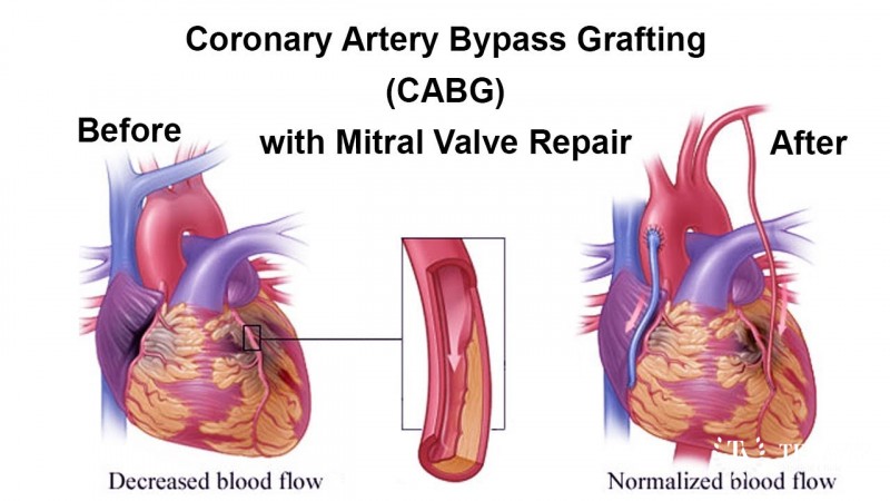
Combined open-heart surgery is the established treatment for most patients with severe valve disease and significant coronary blockages. In a single operation, the surgeon performs coronary artery bypass grafting (CABG) — using the patient’s own arteries or veins to route blood around the blockages — together with valve repair or replacement.
The usual sequence is to construct the bypass grafts first, then repair or replace the valve while the heart is stopped and supported by a heart-lung (cardiopulmonary bypass) machine. This ordering limits the time the heart is under cardiac arrest and ensures both problems are fully addressed. For the aortic valve, replacement with a mechanical or tissue (bioprosthetic) valve is typical; for the mitral valve, repair is preferred when feasible because it preserves the patient’s own valve, with replacement reserved for valves that cannot be repaired.
The mechanical-versus-tissue choice depends on age, bleeding risk, and willingness to take lifelong blood thinners. Combined surgery is more complex and carries somewhat higher operative risk than either procedure alone, but in experienced hands it offers excellent, durable results and treats both conditions definitively. Learn more on our CABG and aortic valve procedures pages.
38. Advanced and Emerging Treatments
Innovation continues to expand options for combined disease:
- Hybrid procedures combining PCI or TAVI with minimally invasive surgery in one planned strategy.
- Robotic and endoscopic techniques for valve repair and bypass through tiny incisions.
- Newer transcatheter valve devices, including transcatheter mitral and tricuspid systems.
- Sutureless and rapid-deployment valves that shorten operative time in combined cases.
- Improved myocardial-protection and off-pump strategies to reduce operative stress.
Many of these are performed at high-volume international centres. See our hybrid cardiac procedures page for details.
39. Treatment Options Compared
- Combined open surgery (CABG + valve) — the most definitive and durable option; treats both problems at once but is the most invasive, with a longer recovery and higher upfront risk.
- Hybrid approach (PCI + TAVI, or PCI + minimally invasive valve) — less invasive, favored for older or higher-risk patients, but may be less durable for the coronary component and not suitable for all anatomies.
- Staged procedures — treating one problem then the other in separate sessions; occasionally chosen but generally exposes the patient to two interventions.
- Medical therapy alone — reserved for patients unfit for any procedure; controls symptoms but does not correct the underlying disease.
The right choice depends on anatomy, risk, and patient preference.
40. How Doctors Choose the Right Treatment
The heart team weighs many factors:
- Severity and type of valve disease and complexity of coronary blockages.
- The patient’s age, frailty, and other medical conditions.
- Heart function (ejection fraction) and symptom burden.
- Surgical risk scores (STS, EuroSCORE) and anatomical suitability for catheter options.
- Patient preferences, including willingness to take lifelong anticoagulation.
Younger, fitter patients often do best with combined open surgery, while older, higher-risk patients may be better served by hybrid or transcatheter strategies. Shared decision-making ensures the plan fits the individual.
41. Benefits and Risks of Treatment
Benefits:
- Relief of angina, breathlessness, and fatigue.
- Correction of both problems, often in a single operation.
- Reduced risk of heart failure, heart attack, and sudden death.
- Improved survival and quality of life.
Risks:
- Higher operative risk than single procedures — including bleeding, stroke, infection, kidney injury, and arrhythmias.
- Risks of anaesthesia and the heart-lung machine.
- Possible need for a pacemaker or reoperation.
- Valve-related issues over time (wear of tissue valves; anticoagulation needs for mechanical valves).
Overall, for patients with severe combined disease, the benefits of well-timed treatment substantially outweigh the risks.
42. What Happens If the Disease Is Left Untreated?
If severe symptomatic combined disease is left untreated, the outlook is poor. The overburdened heart progressively fails, causing worsening breathlessness, repeated hospital admissions, and declining quality of life. Untreated severe aortic stenosis carries a high risk of sudden death once symptoms appear, and untreated coronary blockages raise the risk of heart attack, while complications such as atrial fibrillation and stroke accumulate. Because the two conditions worsen each other, deterioration can be faster than with either alone — timely treatment dramatically changes this trajectory.
43. Treatment Success and Expected Outcomes
Modern combined valve-and-bypass surgery has good success rates in experienced centres. Most patients experience marked relief of symptoms, improved exercise capacity, and better long-term survival. Operative risk is higher than for single procedures but remains acceptable, particularly in patients without severe frailty or advanced organ damage. Long-term outcomes depend on heart function before surgery, the durability of the chosen valve, control of risk factors afterward, and adherence to medications and follow-up. Patients treated before the heart is severely weakened generally do best. Outcomes are described here in general terms; your surgical team can provide individualized estimates based on your specific findings.
44. Prognosis and Long-Term Outlook
The long-term outlook after successful treatment is generally favourable, especially when surgery is performed before irreversible heart damage occurs. Many patients return to active, comfortable lives and enjoy substantially improved survival compared with leaving the disease untreated.
Prognosis depends on the strength of the heart muscle before surgery, the completeness of coronary revascularization, the durability of the valve, and how well risk factors (blood pressure, cholesterol, diabetes, smoking) are controlled afterward. Patients with well-preserved heart function tend to do very well over many years.
Ongoing care matters: lifelong attention to medications, healthy lifestyle, and regular follow-up — including periodic echocardiograms — helps sustain the benefit of surgery. Tissue valves may eventually wear out, while mechanical valves need lifelong anticoagulation. Frailty, advanced age, severe pre-existing heart failure, or significant kidney disease can moderate the outlook, which is why individualized assessment matters.
45. Recovery and Rehabilitation
Recovery is gradual. Most patients spend 1–2 days in intensive care and around 5–8 days in hospital, though this varies. Early days focus on breathing exercises, gentle mobilization, pain control, and wound care. Full recovery of energy and return to normal activities typically takes 6–12 weeks, longer than for single procedures because the operation is more extensive. A structured cardiac rehabilitation programme significantly improves recovery, confidence, and long-term outcomes and is strongly recommended.
46. Follow-Up Tests and Long-Term Monitoring
Lifelong follow-up is essential. This usually includes periodic echocardiograms to check valve function and heart pumping, ECGs to monitor rhythm, and blood tests. Patients on anticoagulation for a mechanical valve or atrial fibrillation need regular blood-clotting monitoring (INR). Coronary status is reviewed if new symptoms arise. Follow-up also tracks control of cholesterol, blood pressure, and diabetes. The frequency is tailored to the individual — often more frequent in the first year, then annually if stable.
47. Managing Recurrence or Disease Progression
Even after successful treatment, the disease process can continue. Coronary disease may recur in native vessels or grafts, and tissue valves may degenerate over years. Management centres on aggressive control of risk factors, adherence to statins and other medications, and prompt evaluation of new symptoms. If recurrence occurs, options range from medication adjustment to repeat catheter procedures (PCI or valve-in-valve TAVI) or, less commonly, redo surgery. Regular monitoring ensures problems are caught early, when treatment is safer and more effective.
48. Living with the Disease
Living with combined valvular and coronary disease means partnering actively in your care. Most patients can lead full lives after treatment by taking medications reliably, attending follow-up appointments, and maintaining heart-healthy habits. Practical steps include monitoring symptoms, avoiding smoking, staying physically active within advised limits, eating well, and managing stress. Patients on anticoagulation should learn about interactions with food and other medicines and carry appropriate medical information. With good self-management and support, quality of life after treatment is generally very good.
49. Diet and Nutrition Guidelines
A heart-healthy diet supports both the valve and coronary components:
- Emphasize vegetables, fruits, whole grains, legumes, and fish.
- Choose healthy fats (olive oil, nuts) over saturated and trans fats.
- Limit salt to help control blood pressure and fluid retention.
- Reduce added sugars and refined carbohydrates, especially with diabetes.
- Limit alcohol and avoid processed, high-sodium foods.
- If on warfarin, keep vitamin-K intake (leafy greens) consistent rather than fluctuating.
A dietitian can tailor advice to your medications and other conditions.
50. Exercise and Physical-Activity Guidelines
Regular, appropriate exercise strengthens the heart and aids recovery. Before treatment, activity should be guided by symptoms and the severity of valve disease — strenuous exertion may be limited in severe aortic stenosis. After surgery, most patients build up gradually through a cardiac rehabilitation programme, progressing from gentle walking to more sustained aerobic activity. General goals include regular moderate aerobic exercise most days once cleared, plus light strengthening. Patients should avoid heavy lifting until the breastbone heals (about 6–8 weeks after sternotomy) and always follow their team’s individualized advice.
51. Medications, Activities and Habits to Avoid
- Do not smoke — tobacco accelerates both coronary and valve disease.
- Avoid stopping prescribed medicines (especially antiplatelets or anticoagulants) without medical advice.
- Use caution with nitrates and strong blood-pressure–lowering drugs in severe aortic stenosis.
- Avoid excessive alcohol and high-salt, high-fat processed foods.
- Avoid strenuous or competitive exertion with untreated severe valve disease.
- After surgery, avoid heavy lifting and driving until cleared by your surgeon.
- Check for drug and food interactions if taking warfarin.
52. Preventing the Disease or Reducing Its Risks
While age-related valve disease cannot be fully prevented, the coronary component and overall progression can be substantially reduced by:
- Not smoking and avoiding tobacco.
- Keeping blood pressure, cholesterol, and blood sugar well controlled.
- Eating a heart-healthy diet and staying physically active.
- Maintaining a healthy weight.
- Treating conditions like diabetes and kidney disease.
- Attending regular check-ups so that both valve and coronary problems are detected early.
53. Pregnancy and the Disease
Combined valvular and coronary disease is uncommon during pregnancy because it mostly affects older adults. However, younger women with significant valve disease (e.g. from a bicuspid valve or rheumatic disease) who also have risk factors need specialist care. Pregnancy increases the heart’s workload and can unmask or worsen valve disease. Any woman with known heart or valve disease planning pregnancy should have pre-conception counselling with a cardiologist and obstetric team, and those on anticoagulation require careful medication management, as some blood thinners are unsafe in pregnancy.
54. Disease in Children and Young Adults
The combined condition is rare in the young. When valve disease occurs in children or young adults, it is usually congenital (such as a bicuspid aortic valve) or rheumatic, and significant coronary disease is uncommon at these ages — though inherited cholesterol disorders can cause early coronary disease. Young patients are managed by specialists in congenital and structural heart disease, with emphasis on valve preservation and long-term planning. Explore related conditions in our congenital heart disease section.
55. Disease in Older Adults
Older adults represent the large majority of patients with combined disease, as both calcific valve disease and atherosclerosis rise steeply with age. Elderly patients more often have other medical conditions, frailty, and reduced kidney function, which raise surgical risk. For this group, less invasive options such as TAVI combined with PCI are frequently favored. Careful, individualized assessment — including frailty and quality-of-life considerations — guides whether open surgery, a hybrid approach, or conservative management is most appropriate.
56. Emotional Health and Patient Support
Facing two serious heart conditions and major surgery can cause anxiety, low mood, or fear. These feelings are normal and treatable. Support from family, patient groups, and the care team helps, as does clear information about what to expect. Cardiac rehabilitation programmes address emotional as well as physical recovery, and counselling or professional mental-health support should be sought if anxiety or depression persists. Addressing emotional health improves both recovery and long-term wellbeing.
57. Preparing for Your Specialist Appointment
To make the most of your appointment:
- List your symptoms, when they occur, and what triggers them.
- Bring a complete list of medications and doses.
- Note your medical history, prior heart events, and family history.
- Bring previous test results (echocardiograms, angiograms) if available.
- Write down your questions in advance.
- Consider bringing a family member to help remember information.
You can request an appointment or a second opinion through our contact page.
58. Questions to Ask Your Doctor
- How severe is my valve disease, and how severe are my coronary blockages?
- Do I need surgery, and if so, how urgently?
- Would a combined operation or a hybrid/catheter approach be better for me?
- What type of valve (mechanical or tissue) do you recommend, and why?
- What are the specific risks of surgery in my case?
- Will I need lifelong blood thinners?
- How long is recovery, and when can I return to normal activities?
- What are my options if I choose not to have surgery?
- How will we monitor my heart afterward?
- What can I do to improve my outcome before and after treatment?
59. Cost of Diagnosis and Treatment
Costs vary widely by country, hospital, and complexity. The figures below are approximate ranges for combined valve-plus-bypass surgery and should be confirmed with individual centres. Medical-travel destinations often offer major savings.
| Region | Approx. cost of combined valve + CABG surgery (USD) |
|---|---|
| United States | $70,000 – $200,000+ |
| United Kingdom / Western Europe | $40,000 – $90,000 |
| Singapore | $30,000 – $60,000 |
| Thailand | $18,000 – $40,000 |
| Turkey | $15,000 – $35,000 |
| India | $8,000 – $20,000 |
Diagnostic tests (echocardiogram, angiography) add several hundred to a few thousand dollars. India, Turkey, and Thailand often cost roughly 50–90% less than the US or UK for comparable quality at accredited centres.
60. Factors Affecting Treatment Cost
- Country and hospital chosen, and its accreditation and reputation.
- Type of procedure (open combined surgery vs. hybrid/TAVI + PCI).
- Valve type (mechanical, tissue, or transcatheter device).
- Number of bypass grafts and overall complexity.
- Length of hospital and ICU stay and any complications.
- Surgeon and team experience.
- Additional costs — diagnostics, medications, rehabilitation, and for international patients, travel and accommodation.
61. Choosing the Right Specialist
Look for a cardiac surgeon and cardiology team experienced in combined valve-and-bypass procedures. Helpful criteria include:
- High case volume in combined and valve surgery.
- Board certification and specialist training.
- Access to a multidisciplinary heart team.
- Transparent, favourable outcome data.
- Good communication and willingness to answer questions.
Browse experienced cardiac specialists in our doctors directory.
62. Choosing the Right Hospital or Treatment Centre
Choose a centre with:
- International accreditation (e.g. JCI) for quality and safety.
- High volume of cardiac and combined valve surgeries.
- A full heart team and modern imaging and surgical facilities.
- Strong outcome and infection-control records.
- Good post-operative and rehabilitation support, and services for international patients.
Explore leading facilities in our hospitals directory and popular destinations.
63. Getting a Second Medical Opinion
Because combined disease involves complex, high-stakes decisions, a second opinion is valuable and encouraged. Another expert may confirm the diagnosis, suggest an alternative approach (for example, a hybrid strategy instead of open surgery), or reassure you about the recommended plan. Bring your echocardiogram, angiogram, and test reports so the reviewing specialist has full information. Seeking a second opinion is a normal, responsible step — you can request one through our contact page.
64. Treatment Abroad and Medical-Travel Considerations
Many patients travel abroad for combined heart surgery to access high-quality care at lower cost. Leading centres in India, Turkey, Thailand, and Singapore offer experienced heart teams and JCI-accredited facilities. When planning, consider:
- Accreditation and surgical volume of the hospital.
- Clear cost estimates and what they include.
- Travel fitness — long flights soon after heart surgery require medical clearance.
- Language support and international patient services.
- Follow-up arrangements on return home, coordinated with your local doctor.
See our destinations and hospitals pages to compare options.
65. Frequently Asked Questions
Q: Can both the valve and the blocked arteries be fixed in one operation? A: Yes. Combined CABG plus valve repair/replacement in a single operation is the standard approach for most patients with severe combined disease.
Q: Is combined surgery riskier than a single procedure? A: It carries a somewhat higher operative risk, but in experienced centres the results are very good and it avoids the greater risk of operating twice.
Q: Should I choose a mechanical or tissue valve? A: Mechanical valves last longer but require lifelong blood thinners; tissue valves avoid long-term anticoagulation but may wear out. Your age and preferences guide the choice.
Q: Can I avoid open surgery? A: Some patients, especially older or higher-risk ones, may be candidates for a hybrid approach using stents (PCI) and a transcatheter valve (TAVI).
Q: How long will I be in hospital? A: Typically around 5–8 days, with 1–2 days in intensive care, though this varies.
Q: How long until I feel normal again? A: Most people return to normal activities within 6–12 weeks, aided by cardiac rehabilitation.
Q: Will the disease come back? A: Coronary disease can progress and tissue valves can wear over time, so lifelong follow-up and risk-factor control are important.
Q: Is treatment abroad safe? A: At accredited, high-volume international centres, outcomes can be excellent. Choose carefully and plan follow-up.
66. Patient Stories and Treatment Experiences
The following stories are representative and anonymized to illustrate typical experiences.
Robert, United Kingdom: In his early 70s, Robert developed breathlessness and chest tightness. Tests showed severe aortic stenosis with two blocked arteries. He underwent combined valve replacement and bypass surgery and, after cardiac rehabilitation, returned to gardening and daily walks within a few months.
Anita, India: Anita, 66, was found to have a leaking mitral valve after a small heart attack. Her heart team repaired the valve and bypassed the blocked artery in one operation. She describes feeling far more energetic a few months later.
James, Canada: Considered higher-risk due to age and kidney disease, James was treated with a hybrid approach — stents for his coronaries followed by a transcatheter valve. He avoided open surgery and recovered quickly.
67. Latest Research and Clinical Trials
Research continues to refine care for combined disease. Active areas include transcatheter valve technologies (including newer mitral and tricuspid devices) that expand less-invasive options, improved hybrid strategies combining PCI and TAVI, and better risk-prediction tools to guide who benefits most from combined open surgery versus catheter approaches. Studies also explore valve durability, optimal anticoagulation strategies, and enhanced myocardial protection during surgery. Guideline bodies such as the ACC/AHA and ESC/EACTS regularly update recommendations as evidence evolves. Patients interested in trials should ask their heart team about opportunities at major academic centres.
68. Related Diseases and Conditions
- Isolated Coronary Artery Disease
- Complex Coronary Artery Disease
- Aortic Valve Disease
- Mitral Valve Disease
- Combined Multi-Valvular Disease
- Ischemic Cardiomyopathy
69. Related Treatments and Procedures
- Coronary Artery Bypass Grafting (CABG)
- Aortic Valve Procedures
- Mitral Valve Procedures
- Hybrid Cardiac Procedures
- Angioplasty
70. Medical Glossary
- Aortic stenosis — narrowing of the aortic valve, restricting blood flow out of the heart.
- Mitral regurgitation — leakage of the mitral valve, allowing blood to flow backward.
- CABG — coronary artery bypass grafting; surgery to route blood around blocked arteries.
- Ischemic mitral regurgitation — valve leakage caused by coronary disease damaging the heart muscle.
- Atherosclerosis — plaque buildup that narrows arteries.
- Ejection fraction — the percentage of blood the left ventricle pumps out with each beat.
- TAVI/TAVR — transcatheter aortic valve implantation; valve replacement via a catheter.
- PCI — percutaneous coronary intervention; opening arteries with stents via catheter.
- Bioprosthetic valve — a tissue valve made from animal or human tissue.
- Mechanical valve — a durable artificial valve requiring lifelong anticoagulation.
- Sternotomy — surgical opening of the breastbone to access the heart.
- Cardiopulmonary bypass — the heart-lung machine that supports circulation during surgery.
- NYHA class — a scale (I–IV) grading heart-failure symptoms.
- STS/EuroSCORE — tools estimating surgical risk.
71. Medical Review, Editorial Policy and Last Updated Date
Last updated: 11 July 2026. This article is written for patient education and reviewed against current cardiology and cardiac-surgery guidance from bodies such as the ACC/AHA, ESC/EACTS, STS, and NHS. Our editorial process emphasizes accuracy, clarity, and balance, and content is periodically updated as guidelines evolve. This information is educational only and is not a substitute for professional medical advice, diagnosis, or treatment. Always consult a qualified cardiologist or cardiac surgeon about your individual situation.
72. Clinical Guidelines and Medical References
This content aligns with general recommendations from leading authorities, including:
- American College of Cardiology / American Heart Association (ACC/AHA) valvular and coronary disease guidelines.
- European Society of Cardiology / European Association for Cardio-Thoracic Surgery (ESC/EACTS) guidelines on valvular heart disease and myocardial revascularization.
- Society of Thoracic Surgeons (STS) practice standards and risk models.
- National Health Service (NHS) and World Health Organization (WHO) patient-information resources.
These sources are cited in general terms; consult the latest published guidelines and your care team for specific clinical guidance.
73. Book an Appointment or Request a Second Opinion
If you or a loved one has combined valvular and coronary artery disease, expert evaluation can clarify the best treatment path. Book an appointment or get started here, or reach our team through our contact page to request a second opinion. You can also explore experienced doctors, accredited hospitals, and leading destinations for cardiac care worldwide.