1. Disease Overview
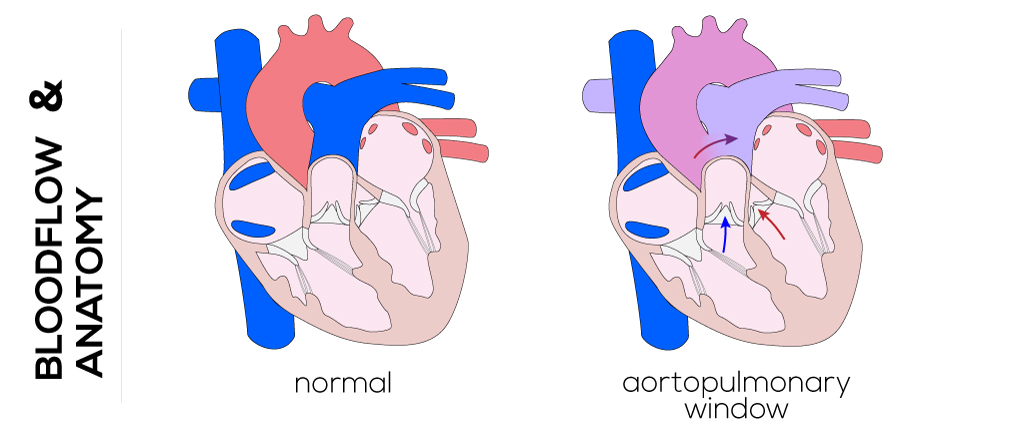
Aortopulmonary window (APW) is a rare congenital heart defect in which there is an abnormal opening, or communication, between the ascending aorta and the main pulmonary artery, while the two arterial valves (the aortic and pulmonary valves) remain separate and normally formed. This distinguishes it from truncus arteriosus, in which a single valve and vessel arise from the heart.
Because the aorta carries blood at much higher pressure than the pulmonary artery, blood shunts from left to right through the window, sending too much blood into the lungs. This large left-to-right shunt typically causes early heart failure and pulmonary overcirculation within the first weeks to months of life. Over time, if untreated, the high lung blood flow damages the pulmonary arteries and leads to pulmonary hypertension, which can become irreversible.
APW accounts for only a small fraction of all congenital heart defects, but it is important because symptoms appear early and the condition is almost always treated with surgical patch closure in infancy. With prompt diagnosis and repair before permanent lung damage develops, most children go on to lead normal, active lives. This page explains APW in plain language, from anatomy and symptoms through diagnosis, surgery, recovery and long-term outlook, with information for families considering treatment abroad.
2. Key Facts at a Glance
| Feature | Detail |
|---|---|
| Also known as | Aortopulmonary septal defect, AP window, aortopulmonary fenestration |
| Body system affected | Heart and great arteries (cardiovascular and pulmonary circulation) |
| Common in | Newborns and young infants; a rare congenital defect |
| Severity range | Serious; large shunt causes early heart failure and pulmonary hypertension if untreated |
| Key treatments | Surgical patch closure in infancy; occasionally transcatheter device closure in selected cases |
| Outlook | Excellent with early surgical repair before irreversible lung damage |
3. Alternative Names and Medical Terminology
- Aortopulmonary window (APW) — the most common name
- Aortopulmonary septal defect (APSD)
- Aorto-pulmonary window / AP window / AP fenestration
- Aortopulmonary communication (a broader descriptive term)
It should not be confused with patent ductus arteriosus (PDA), a persistent fetal vessel, or with truncus arteriosus, where a single valve serves both circulations. In APW there are two normal semilunar valves and a direct side-to-side connection between the great vessels.
4. Relevant Heart, Lung or Vascular Anatomy
Normally, the aorta and the main pulmonary artery arise side by side from the heart and are completely separated by a wall (the aortopulmonary septum) that forms during early fetal development. The aorta carries oxygen-rich blood to the body at high pressure; the pulmonary artery carries oxygen-poor blood to the lungs at lower pressure. Each vessel has its own valve — the aortic valve and the pulmonary valve.
In APW, a portion of that dividing wall fails to form, leaving a window between the ascending aorta and the main pulmonary artery, usually just above the valves. The size, position and length of this window vary. Because the coronary arteries and the origin of the right pulmonary artery lie nearby, their relationship to the defect is important surgically. Associated anomalies of the arch, coronary arteries or other structures may coexist.
5. How the Disease Affects the Body
The core problem in APW is an abnormal mixing of the two circulations through the window. Because the aorta operates at systemic (high) pressure and the pulmonary artery at low pressure, blood is driven from the aorta into the pulmonary artery — a left-to-right shunt. This means a large volume of already-oxygenated blood is recirculated back through the lungs instead of going out to the body.
Two consequences follow. First, the lungs receive far too much blood flow (pulmonary overcirculation), which makes the lungs congested and stiff, causing fast, labored breathing and feeding difficulty. Second, the left side of the heart must pump a greatly increased volume, leading to volume overload and congestive heart failure — often within the first few weeks of life. The heart enlarges and works harder to compensate.
If the shunt is not corrected, the small pulmonary arteries respond to the high flow and pressure by thickening and narrowing. Over months to a few years this produces pulmonary arterial hypertension and, eventually, irreversible pulmonary vascular disease (Eisenmenger physiology), at which point the shunt may reverse and cause low oxygen levels (cyanosis). This is why early diagnosis and repair are essential.
6. Types and Classification
APW is commonly classified by the location and extent of the defect. A widely used scheme (Mori/Richardson types) describes:
- Type I (proximal): the window lies just above the semilunar valves, near the sinuses.
- Type II (distal): the window is located more distally on the ascending aorta.
- Type III (total defect): a large or near-complete absence of the aortopulmonary septum.
An additional category includes defects with anomalous origin of the right pulmonary artery from the aorta. APW is also described as simple (isolated) or complex when combined with other lesions such as interrupted aortic arch, ventricular septal defect, coarctation or coronary anomalies. Classification guides the surgical approach.
7. Causes of the Disease
APW results from a failure of normal fetal development — specifically, incomplete formation and fusion of the aortopulmonary septum that should divide the common arterial trunk into a separate aorta and pulmonary artery during the first weeks of pregnancy. The exact trigger is usually unknown.
- It is a congenital (present-at-birth) structural defect, not caused by anything the child does.
- It is generally sporadic, arising by chance during early cardiac development.
- No single environmental cause has been proven, though disturbances in the migration and development of certain embryonic cells are thought to play a role.
8. How the Disease Develops
During the fifth to eighth week of pregnancy, spiral ridges of tissue grow and fuse to divide the single embryonic outflow trunk into the aorta and pulmonary artery. In APW, a segment of this septum fails to complete, leaving a persistent opening between the two great vessels.
Before birth, the defect causes few problems because pressures in the aorta and pulmonary artery are similar and the lungs are not yet the main route of blood flow. After birth, the natural fall in pulmonary artery pressure creates a pressure difference across the window, and left-to-right shunting begins to increase over the first days and weeks of life. As pulmonary resistance drops further, shunt flow rises, producing pulmonary overcirculation and heart failure. If untreated, the pulmonary arteries gradually remodel and stiffen, so the natural history moves from high-flow heart failure toward fixed pulmonary vascular disease.
9. Risk Factors
APW is largely a chance event, and no strong, well-established risk factors are recognized. Factors that generally increase the likelihood of congenital heart defects overall may play a background role:
- Maternal health during pregnancy — poorly controlled diabetes, certain infections, or exposure to some medications, alcohol or toxins (general congenital-heart risk factors)
- Family history of congenital heart disease (uncommon for APW specifically)
- Coexisting congenital anomalies, as APW is sometimes part of a broader pattern of defects
Because most cases are sporadic, the absence of any of these factors does not prevent APW.
10. Genetic and Family-History Factors
APW is usually sporadic and is not typically inherited in a simple pattern. It occurs less often as part of recognized genetic syndromes than some other congenital heart defects. When APW is found together with additional cardiac or non-cardiac anomalies, a genetics evaluation may be recommended to look for an underlying chromosomal or single-gene condition.
- Most families have no history of the defect.
- The recurrence risk in future pregnancies is generally low but slightly higher than the general population.
- Genetic counseling can be offered when there are associated anomalies or a suggestive family history.
11. Who Is Most at Risk?
The condition is present from birth, so the “at-risk” group is essentially newborns with an unrecognized defect. Practically:
- Infants in the first weeks to months of life are when symptoms and risk from the untreated shunt are highest.
- Children with additional heart defects (such as interrupted aortic arch or VSD) tend to present earlier and more severely.
- Infants diagnosed late are at greatest risk of developing pulmonary hypertension.
There is no meaningful sex predominance established for APW.
12. Prevalence and Epidemiology
APW is a rare congenital heart lesion, making up only a very small percentage of all congenital heart defects — far less common than defects such as ventricular septal defect or atrial septal defect. Because it is uncommon, most published experience comes from specialized pediatric cardiac centers rather than large population studies.
- It affects a small minority of all babies born with congenital heart disease.
- It is frequently associated with other cardiac anomalies in a substantial share of cases.
- Reported figures vary between centers; exact rates should be regarded as approximate.
13. Signs and Symptoms
Because the shunt is large, symptoms usually appear early, often within the first weeks of life, and reflect heart failure and pulmonary overcirculation rather than low oxygen. Typical features include:
- Fast, labored breathing (tachypnea) and increased work of breathing
- Poor feeding, sweating during feeds, and easy tiring
- Failure to thrive — slow weight gain
- Frequent chest infections
- A heart murmur heard by the doctor, sometimes with a “machinery” or continuous quality
- Rapid heart rate and an enlarged, hyperactive heart on examination
- Pale or mottled appearance when feeding or crying
Cyanosis (blue discoloration) is usually absent early on and appears late only if severe pulmonary hypertension develops and the shunt reverses. Symptoms can resemble other large left-to-right shunt lesions, so specialized evaluation is needed to make the diagnosis.
14. Early-Stage Symptoms
In the first days to weeks, signs can be subtle and are easy to attribute to feeding problems or minor infections:
- A newly detected heart murmur
- Breathing a little faster than expected, particularly during feeds
- Taking longer to feed, tiring quickly, and sweating on the forehead
- Slightly slower weight gain
As pulmonary resistance falls over the first weeks, these early signs typically progress to more obvious heart failure.
15. Advanced-Stage Symptoms
If the defect is not repaired, symptoms intensify and later reflect pulmonary hypertension:
- Severe congestive heart failure with marked breathlessness and poor growth
- Recurrent respiratory infections and difficulty recovering from them
- Enlarged liver, puffiness and fluid retention
- In late, untreated disease: development of pulmonary vascular disease, reduced murmur intensity, and eventually cyanosis (Eisenmenger physiology) with exercise intolerance
- Reduced life expectancy without treatment
16. Symptoms in Women, Men and Older Adults
APW almost always presents and is treated in infancy, so age- and sex-based differences of the type seen in adult heart disease do not really apply. The condition does not have a recognized difference in presentation between boys and girls. In the rare instance of a small, undetected defect surviving to adulthood, a person (of either sex) might present with exercise limitation, breathlessness or signs of pulmonary hypertension rather than the acute infant heart failure picture.
17. Emergency Warning Signs
Seek emergency care for an infant with:
- Severe breathing difficulty, grunting, flaring nostrils or chest indrawing
- Blue or grey color of lips, tongue or skin
- Very poor feeding, lethargy or unresponsiveness
- Signs of shock — cold, mottled skin and weak pulses
These indicate the heart and lungs are not coping and require immediate assessment.
18. When to Seek Medical Help
Contact a doctor promptly if a baby is feeding poorly, breathing fast, sweating with feeds, failing to gain weight, or has been told a heart murmur is present. Any newborn who tires quickly, breathes rapidly at rest, or has recurrent chest infections should be evaluated by a pediatrician or pediatric cardiologist. Early referral allows diagnosis before the lungs are permanently affected. You can reach out through our contact page for guidance on specialist evaluation.
19. Disease Stages, Grades and Severity
APW severity is judged less by a formal grading scale and more by the size of the shunt, the degree of pulmonary overcirculation, and the state of the pulmonary arteries:
- Uncomplicated, early: large shunt with heart failure but low pulmonary vascular resistance — ideal timing for repair.
- Advancing: rising pulmonary artery pressure with reactive vessels still able to fall after surgery.
- Advanced/irreversible: fixed pulmonary vascular disease where surgery may no longer be safe or beneficial.
Severity is also increased by associated defects. Assessing operability hinges on measuring pulmonary vascular resistance and its reversibility.
20. Disease Progression
The typical course is one of early and rapid symptom development. After birth, falling pulmonary resistance increases left-to-right shunting, driving heart failure within weeks. Without repair, the natural history moves through worsening heart failure and growth failure, then progressive pulmonary hypertension. Over months to a few years the pulmonary arteries can become permanently damaged, at which point the shunt may reverse and the child develops cyanosis. Because progression is fast and can become irreversible, APW is generally repaired soon after diagnosis rather than watched.
21. Possible Complications
Complications of untreated or late-treated APW include:
- Congestive heart failure and failure to thrive
- Pulmonary arterial hypertension, potentially irreversible (Eisenmenger syndrome)
- Recurrent respiratory infections
- Arrhythmias and progressive heart enlargement
- Increased risk of infective endocarditis
- Surgical complications (rare) such as residual shunt, narrowing of a pulmonary or aortic branch, or injury to nearby coronary arteries
Timely repair prevents most of these outcomes.
22. Related and Associated Medical Conditions
APW frequently occurs alongside other cardiac anomalies. Commonly associated conditions include:
- Interrupted aortic arch and coarctation of the aorta
- Ventricular septal defect (VSD) and atrial septal defect (ASD)
- Patent ductus arteriosus (PDA)
- Coronary artery anomalies, including anomalous coronary origin
- Anomalous origin of the right pulmonary artery from the aorta
- Tetralogy of Fallot or arch anomalies in complex cases
The presence of these lesions affects timing, surgical planning and outlook.
23. Screening and Early Detection
There is no dedicated population screening test for APW, but it is often detected early through routine care:
- Fetal echocardiography may detect it before birth in some cases, especially when other anomalies prompt a detailed scan.
- Newborn pulse oximetry screening can flag some critical congenital heart lesions, though APW may not always cause low oxygen early.
- Newborn and infant physical examinations frequently pick up the murmur, fast breathing or poor growth that lead to referral and echocardiography.
24. How the Disease Is Diagnosed
Diagnosis follows a stepwise pathway that begins with clinical suspicion and is confirmed by imaging. A pediatrician who notices a murmur, tachypnea, poor feeding or failure to thrive refers the infant to a pediatric cardiologist.
The cornerstone test is echocardiography (ultrasound of the heart), which can directly visualize the communication between the ascending aorta and main pulmonary artery, confirm that the semilunar valves are separate (distinguishing APW from truncus arteriosus), estimate shunt size and pulmonary pressures, and identify associated defects. A chest X-ray typically shows an enlarged heart and increased lung blood flow, and an ECG may show signs of chamber enlargement.
When anatomy is complex, additional imaging such as cardiac CT or MRI helps define the exact position and size of the window and the great-vessel and coronary anatomy. Cardiac catheterization may be used to measure pulmonary artery pressures and calculate pulmonary vascular resistance — important for judging whether the defect is still safely repairable, particularly if diagnosis is late. Together these tests confirm the diagnosis and plan surgery.
25. Physical Examination and Medical History
The history usually reveals poor feeding, sweating, breathlessness and slow weight gain in early infancy. On examination the doctor may find:
- A hyperactive, enlarged heart with a heave
- A continuous or systolic murmur and often bounding pulses with a wide pulse pressure
- Tachypnea and tachycardia with signs of respiratory distress
- An enlarged liver and signs of heart failure
These findings prompt urgent echocardiography.
26. Diagnostic Tests and Imaging
- Echocardiography (2D and Doppler): primary tool; visualizes the window, confirms two separate valves, assesses shunt and pressures.
- Chest X-ray: cardiomegaly and increased pulmonary vascular markings.
- Electrocardiogram (ECG): ventricular hypertrophy or enlargement patterns.
- Cardiac CT angiography: detailed anatomy of the great vessels and coronary arteries.
- Cardiac MRI: anatomy and shunt quantification without radiation.
- Cardiac catheterization: measures pulmonary pressures and resistance; assesses operability in late presentations.
27. Blood Tests, Biomarkers and Genetic Testing
There is no blood test that diagnoses APW itself. Laboratory testing is supportive:
- Natriuretic peptides (BNP/NT-proBNP) may be raised, reflecting heart failure.
- Blood gases and routine bloods assess oxygenation and overall status.
- Genetic testing / chromosomal analysis may be considered when APW occurs with other anomalies or dysmorphic features, to identify an underlying syndrome.
- Pre-operative bloods (blood count, clotting, cross-match) are done to prepare for surgery.
28. Understanding Test Results
Families are often shown echocardiogram findings and asked to understand terms like shunt and pressure. In simple terms:
- A large window with a big left-to-right shunt and increased lung flow supports early repair.
- Normal or low pulmonary vascular resistance on catheterization means the defect is safely repairable and the lungs should recover.
- High, fixed resistance suggests advanced pulmonary vascular disease and may change or preclude the surgical plan.
- Identification of associated defects (arch, coronary, VSD) shapes the operation. Your cardiologist will explain what each measurement means for your child.
29. Differential Diagnosis
Several conditions can mimic APW and must be distinguished, mainly by echocardiography:
- Truncus arteriosus — a single valve and trunk (APW has two separate valves)
- Large patent ductus arteriosus (PDA)
- Ventricular septal defect (VSD) with a large shunt
- Coronary artery fistula or other arterial communications
- Aortopulmonary collaterals
Correctly separating APW from truncus arteriosus is particularly important because their surgical treatments differ substantially.
30. Specialist and Multidisciplinary Evaluation
Care involves a coordinated team at a pediatric cardiac center: a pediatric cardiologist, congenital cardiac surgeon, cardiac anesthesiologist, intensivists and neonatal/pediatric nurses, cardiac imaging specialists, and often a geneticist when other anomalies are present. Feeding and nutrition support and, later, developmental follow-up may be involved. You can explore experienced doctors and hospitals with dedicated congenital heart programs.
31. Treatment Goals
The goals of treatment are to:
- Close the abnormal communication to stop the left-to-right shunt
- Relieve heart failure and restore normal growth and feeding
- Protect the lungs from pulmonary overcirculation and prevent pulmonary hypertension
- Preserve normal coronary and great-vessel anatomy
- Address any associated defects at the same or a planned time
- Achieve a durable repair that allows a normal, active life
32. When Is Treatment Required?
Because APW causes a large shunt and early heart failure, treatment is required in essentially all cases, and usually soon after diagnosis in infancy. Waiting risks irreversible pulmonary hypertension. Medications may be used briefly to stabilize heart failure, but they are a bridge to surgery, not a substitute for it. The main question is not whether to treat but how soon, and whether the lungs remain suitable for safe closure when the diagnosis is made late.
33. Active Monitoring and Watchful Waiting
Unlike some small congenital defects, APW is not usually managed by watchful waiting, because the shunt is large and progresses quickly. Very small, restrictive windows are rare; if one were identified, close monitoring might be considered, but the standard approach is early closure. Any period of observation is generally short and used only to optimize the baby’s condition before an operation.
34. Medications
Medications do not close the window; they stabilize the infant and manage heart failure before and around surgery:
- Diuretics (e.g., furosemide) to reduce fluid overload and ease breathing
- ACE inhibitors to reduce the heart’s workload in some cases
- Digoxin occasionally, to support heart function
- Nutritional support and higher-calorie feeds to aid growth
- After late diagnosis with pulmonary hypertension, pulmonary vasodilator therapy may be considered by specialists
All medication use should be directed by the cardiology team.
35. Minimally Invasive Treatments
For most infants with a typical APW, open surgical patch closure remains the standard, because the defect is close to the valves and coronary arteries. However, minimally invasive and hybrid strategies are used at some centers for selected anatomy. In a small subset with a suitable, well-defined window away from critical structures, transcatheter device closure (a minimally invasive, catheter-based option) may be feasible. The choice depends on the child’s size, the window’s location and the presence of associated defects. Learn more about congenital heart procedures.
36. Catheter-Based and Endovascular Treatments
Transcatheter (percutaneous) closure places a closure device across the window through a catheter introduced via a blood vessel, avoiding open surgery. It is considered mainly for:
- Small to moderate, tubular windows with adequate rims of tissue
- Older children or selected patients with favorable anatomy
- Situations where surgery carries higher risk
It is generally not suitable for large windows, defects immediately adjacent to the valves or coronary arteries, or when associated lesions require surgical repair. Cardiac catheterization is also used to measure pulmonary pressures. See endovascular stenting and related catheter techniques.
37. Surgical Treatment Options
Surgical closure is the definitive and most common treatment for APW, usually performed in early infancy through open-heart surgery using cardiopulmonary bypass (a heart-lung machine). The surgeon accesses the heart through a midline chest incision (sternotomy), stops and protects the heart, and closes the window.
The preferred technique for most defects is the transaortic patch closure (or “sandwich”/trans-window patch technique): the aorta is opened, the window is identified from within, and a patch (often of the patient’s own pericardium or a synthetic material such as Dacron) is sewn in to separate the aorta and pulmonary artery. Careful attention is paid to protecting the coronary artery origins and the origin of the right pulmonary artery, which lie close to the defect. For very large or complex windows, the aorta and pulmonary artery may be divided and separately reconstructed.
When APW occurs with associated defects — such as interrupted aortic arch, VSD or coronary anomalies — these are repaired at the same operation whenever possible. Results are best when surgery is done before irreversible pulmonary vascular disease develops. Most children need only this single, curative operation. Explore congenital heart procedures and other surgery options.
38. Advanced and Emerging Treatments
Advances are gradual and focus on refining existing care:
- Improved transcatheter devices expanding the range of windows treatable without surgery
- Hybrid procedures combining surgical access with catheter techniques in complex anatomy — see hybrid cardiac procedures
- 3D imaging and printing for surgical planning of unusual defects
- Better neonatal intensive care, bypass and myocardial protection, improving outcomes in the smallest infants
- Advanced pulmonary hypertension therapies for late-presenting patients
39. Treatment Options Compared
- Surgical patch closure: the gold standard; suitable for nearly all infants and for large or complex defects; allows simultaneous repair of associated lesions; requires open-heart surgery and bypass.
- Transcatheter device closure: avoids surgery and offers quicker recovery; limited to small, favorably located windows in suitable patients; not for large defects or those near valves/coronaries.
- Medication alone: never curative; used only to stabilize before intervention.
For most children, surgery offers the most reliable, complete and durable correction.
40. How Doctors Choose the Right Treatment
The decision depends on:
- Size, type and location of the window (proximity to valves, coronaries, right pulmonary artery)
- Presence of associated defects requiring surgical repair
- Age, weight and clinical condition of the child
- Pulmonary vascular resistance and whether it is reversible
- Center experience with surgical versus catheter techniques
Most infants with a typical large APW are directed to early surgical closure, with catheter closure reserved for carefully selected cases.
41. Benefits and Risks of Treatment
Benefits: relief of heart failure, normal growth, protection of the lungs and prevention of pulmonary hypertension, and in most cases a complete cure with a normal life expectancy.
Risks: as with any open-heart surgery in infancy — bleeding, infection, arrhythmia, and risks of anesthesia and bypass. Specific to APW, there is a small risk of residual shunt, narrowing of the right pulmonary artery or aorta at the repair site, or coronary artery injury. These risks are low at experienced congenital heart centers, and the benefits of timely repair clearly outweigh them.
42. What Happens If the Disease Is Left Untreated?
Untreated APW has a poor natural history. The large shunt causes progressive heart failure and failure to thrive in infancy, and many untreated infants do not survive early childhood. Those who survive develop progressive pulmonary hypertension and, ultimately, irreversible pulmonary vascular disease with shunt reversal and cyanosis (Eisenmenger syndrome), which severely limits life expectancy and makes later surgical closure impossible or dangerous. Early repair transforms this bleak outlook into an excellent one.
43. Treatment Success and Expected Outcomes
When APW is repaired early, before pulmonary vascular disease develops, outcomes are generally very good. Most children experience prompt resolution of heart failure, catch-up growth, and a durable repair that lasts a lifetime. Surgical mortality at experienced centers is low for isolated APW, though it is higher when there are major associated defects or when repair is delayed. Reported outcomes vary by center and by complexity; families should discuss center-specific results with their surgeon.
44. Prognosis and Long-Term Outlook
The long-term outlook after timely surgical repair of isolated APW is excellent. Most children go on to have normal exercise capacity, normal growth and development, and a normal or near-normal life expectancy, needing only periodic cardiology follow-up. The key determinants of prognosis are the timing of repair and the state of the pulmonary arteries at the time of surgery, and the presence of associated defects.
Children repaired before permanent lung changes usually have a fully corrected circulation. Those repaired late, after pulmonary hypertension has become established, have a more guarded outlook and may need ongoing treatment for pulmonary vascular disease. A small number of patients require monitoring for residual shunt or narrowing at the repair site, and occasionally a further procedure. Overall, APW is one of the congenital heart lesions where early diagnosis and modern surgery produce durable, life-changing results, allowing most patients to live full, active lives.
45. Recovery and Rehabilitation
After surgery, infants recover in a cardiac intensive care unit, usually with a breathing tube for a short period, followed by a step-down ward. Most babies are discharged within one to two weeks if there are no complications. At home, parents care for the healing chest incision, resume gradually increasing feeds, and watch for signs of infection. Feeding and weight gain typically improve markedly once the shunt is closed. Full recovery of energy and growth usually occurs over the following weeks to months, and normal childhood activity is expected.
46. Follow-Up Tests and Long-Term Monitoring
Follow-up is lifelong but usually infrequent after a good repair. It typically includes periodic clinical review, echocardiography and ECG to check for residual shunt, narrowing at the repair site, and normal pulmonary pressures. The first year involves closer follow-up; thereafter visits often become annual or less frequent if all is well. Late-repaired patients with pulmonary hypertension need closer, ongoing monitoring.
47. Managing Recurrence or Disease Progression
True recurrence is uncommon because the window is patched. Long-term management focuses on watching for residual or recurrent shunt, stenosis of the pulmonary artery or aorta at the repair, and persistent pulmonary hypertension in late-repaired patients. If a significant residual defect or narrowing is found, a further catheter or surgical procedure may be recommended. Ongoing pulmonary hypertension is managed medically by specialists. Regular follow-up ensures any problem is caught early.
48. Living with the Disease
After successful early repair, most children live entirely normal lives, attend school, play sports and grow normally, with no daily restrictions in the majority of cases. Families should keep up with scheduled cardiology reviews, maintain good dental hygiene (to reduce endocarditis risk), and inform other doctors of the child’s cardiac history. Children repaired late or with residual pulmonary hypertension may have some activity limits and require ongoing care, but even they can usually enjoy a good quality of life with proper management.
49. Diet and Nutrition Guidelines
Before repair, infants with heart failure often need high-calorie feeding support because breathlessness makes feeding tiring and growth suffers; a dietitian may recommend fortified feeds or, temporarily, tube feeding. After successful repair, feeding and growth usually normalize and a standard, healthy, age-appropriate diet is appropriate. As the child grows, general heart-healthy eating — plenty of fruit, vegetables and whole grains, limited processed and salty foods — supports lifelong cardiovascular health.
50. Exercise and Physical-Activity Guidelines
Once APW is fully repaired and the heart and lungs are normal, most children can take part in normal physical activity and sports without restriction. Exercise is encouraged for overall health. Children with residual pulmonary hypertension, significant residual defects, or narrowing at the repair site may need individualized advice and some limits on high-intensity or competitive activity. Always follow the specific guidance of the treating cardiologist.
51. Medications, Activities and Habits to Avoid
- Avoid stopping heart-failure medications before surgery without medical advice.
- For patients with residual defects or pulmonary hypertension, avoid extreme exertion unless cleared by a cardiologist, and avoid high-altitude exposure without advice.
- Maintain good dental care and tell dentists/doctors about the cardiac history to reduce endocarditis risk; antibiotic prophylaxis may be advised in some situations.
- In adolescence and adulthood, avoid smoking and excess alcohol, which harm the heart and lungs.
52. Preventing the Disease or Reducing Its Risks
APW cannot be reliably prevented because it forms early in fetal development, usually by chance. General measures that support healthy heart development in pregnancy may reduce overall congenital-heart risk:
- Good control of maternal diabetes and chronic illness
- Avoiding alcohol, smoking and unnecessary medications in pregnancy
- Adequate folic acid and good prenatal care
- Genetic counseling where there is a relevant family history
Once the defect exists, “prevention” shifts to early diagnosis and timely repair to prevent complications.
53. Pregnancy and the Disease
APW is diagnosed and treated in infancy, so pregnancy is relevant only to women who had APW repaired earlier in life. Most women with a well-repaired APW and normal heart and lung function can expect a normal pregnancy, though pre-pregnancy cardiology assessment is advised. Women with residual pulmonary hypertension face significantly higher risks and require specialist counseling before considering pregnancy. Prenatal fetal echocardiography may be offered given the small increased chance of congenital heart disease in offspring.
54. Disease in Children and Young Adults
APW is fundamentally a disease of infancy, and the great majority of patients are diagnosed and repaired as babies. Children who undergo early, successful repair typically grow and develop normally, with only routine cardiology follow-up. Young adults living with a repaired APW generally do well; those repaired late may carry residual pulmonary hypertension into adulthood and need ongoing specialist care. Transition from pediatric to adult congenital heart services is important for lifelong monitoring.
55. Disease in Older Adults
It is very rare for APW to remain undiagnosed into older adulthood, because the large shunt usually causes serious problems in infancy. When an older individual is found to have a small or previously unrecognized APW, the main issues are the presence of pulmonary hypertension and the increased operative risk of late repair. Adults who had childhood repair are followed in adult congenital heart disease clinics for residual lesions and general cardiovascular health.
56. Emotional Health and Patient Support
A diagnosis of a serious heart defect in a newborn is stressful and frightening for families. Parents may feel anxiety, guilt or grief, and the intensive-care experience can be overwhelming. Support is available through hospital social workers, congenital heart disease support groups, and counseling services. Connecting with other families who have faced the same diagnosis can be reassuring. As children grow, ongoing emotional support helps them and their families cope with follow-up care and any activity limits.
57. Preparing for Your Specialist Appointment
To make the most of a cardiology consultation:
- Bring all previous records, scans and echocardiogram reports
- Note your child’s feeding, breathing, weight-gain and symptom pattern
- List current medications and doses
- Write down your questions and concerns in advance
- Bring a support person and, if traveling from abroad, translated documents
- Ask about timing of surgery, the specific technique, and expected recovery
58. Questions to Ask Your Doctor
- What exactly is my child’s defect, and how large is the window?
- Are there any associated heart defects?
- What is my child’s pulmonary artery pressure, and is it reversible?
- Is surgery or catheter closure recommended, and why?
- When should the procedure be done?
- What are the risks and expected outcomes at this center?
- How many APW repairs does this team perform each year?
- What will recovery and follow-up involve?
- Will my child have any long-term restrictions?
- What are the costs, and what does treatment abroad involve?
59. Cost of Diagnosis and Treatment
Costs vary widely by country, hospital and complexity. The figures below are approximate ranges for surgical repair of a congenital heart defect such as APW, including hospital and surgical care:
| Region | Approximate cost (USD) |
|---|---|
| United States | $40,000 – $200,000+ |
| United Kingdom / Western Europe | $30,000 – $100,000 |
| Singapore | $25,000 – $60,000 |
| Thailand | $12,000 – $35,000 |
| Turkey | $10,000 – $30,000 |
| India | $5,000 – $15,000 |
Medical tourism destinations such as India, Turkey and Thailand often cost 50–90% less than the US or UK for comparable care. Figures are indicative and should be confirmed with the treating hospital. Explore destinations for options.
60. Factors Affecting Treatment Cost
- Complexity of the defect and any associated lesions requiring simultaneous repair
- Surgical vs. catheter approach
- Length of intensive-care and hospital stay
- Country and hospital chosen, and its accreditation
- Surgeon and team experience
- Pre-operative stabilization and any complications
- For international patients: travel, accommodation, interpreter and follow-up costs
61. Choosing the Right Specialist
Look for a congenital cardiac surgeon and pediatric cardiologist with:
- Specific, high-volume experience in APW and neonatal/infant heart surgery
- Work within a dedicated congenital heart program with pediatric cardiac ICU
- Good, transparent outcome data
- Clear communication and willingness to answer questions
- Experience with international patients, if traveling abroad
Browse experienced doctors to compare specialists.
62. Choosing the Right Hospital or Treatment Centre
Choose a center with:
- International accreditation (e.g., JCI) and a strong congenital heart program
- A pediatric/neonatal cardiac intensive care unit and full surgical, imaging and anesthesia support
- High procedure volumes and good published outcomes for congenital surgery
- Multidisciplinary team care and family support services
- Facilities for international patients, including coordinators and interpreters
See our recommended hospitals and destinations.
63. Getting a Second Medical Opinion
A second opinion is worthwhile for a serious congenital diagnosis, especially before surgery or if there is uncertainty about timing, the surgical plan, or operability in a late-diagnosed child. Another experienced congenital center can confirm the diagnosis, review images, and discuss options. Seeking a second opinion is normal and does not offend treating doctors. Request one easily through our contact page.
64. Treatment Abroad and Medical-Travel Considerations
Many families travel for congenital heart surgery to access expertise, shorter waiting times or lower costs. When planning treatment abroad:
- Choose an accredited, high-volume congenital center with international patient services
- Confirm the surgical plan, costs and expected length of stay in advance
- Plan for safe travel of a sick infant, including fitness-to-fly advice
- Arrange interpreters, accommodation and post-operative follow-up, including care after returning home
- Ensure records and imaging are shared between centers
India, Turkey, Thailand and Singapore are popular, well-regarded destinations. Explore destinations and hospitals.
65. Frequently Asked Questions
Is aortopulmonary window serious? Yes. It causes a large shunt and early heart failure, but it is highly treatable with timely surgery.
Can it close on its own? No. Unlike some small defects, APW does not close spontaneously and requires repair.
What is the difference between APW and truncus arteriosus? In APW there are two separate, normal valves and a side-to-side window; in truncus there is a single valve and trunk. They need different operations.
When is surgery done? Usually in early infancy, soon after diagnosis, before the lungs are damaged.
Is it curable? Yes — early surgical closure is usually a complete, lifelong cure.
Will my child live a normal life? Most children repaired early have normal growth, activity and life expectancy.
Can it be closed without open surgery? In selected small defects, transcatheter (catheter-based) closure is possible, but most cases need surgery.
Does it run in families? It is usually sporadic; the recurrence risk in later pregnancies is generally low.
66. Patient Stories and Treatment Experiences
The following are representative, anonymized examples for illustration only.
-
Aarav, India: Diagnosed at three weeks old after poor feeding and fast breathing, Aarav had surgical patch closure of a large APW. He recovered well and, a year later, is thriving and growing normally.
-
Sofia, Spain: Sofia’s APW was detected alongside a small VSD. Both were repaired in a single operation in infancy. She now attends school and plays sports without restriction.
-
Daniel, Nigeria: Diagnosed a little later, Daniel’s family traveled abroad for surgery at an accredited congenital heart center. Because repair was done before permanent lung damage, his pulmonary pressures normalized after surgery.
67. Latest Research and Clinical Trials
Research in APW and congenital heart disease broadly focuses on improving early detection, refining surgical and catheter techniques, and protecting the developing lungs and brain. Areas of ongoing interest include better transcatheter closure devices for suitable anatomy, hybrid procedures, advances in neonatal bypass and myocardial protection, improved fetal and newborn screening, and better pulmonary hypertension therapies for late-presenting patients. Because APW is rare, much knowledge comes from multi-center collaborations and registries rather than large trials. Families interested in trials should ask their specialist center about relevant studies.
68. Related Diseases and Conditions
- Congenital Heart Disease overview
- Patent Ductus Arteriosus
- Ventricular Septal Defects
- Atrial Septal Defects
- Tetralogy of Fallot
- Transposition of the Great Arteries
69. Related Treatments and Procedures
- Congenital Heart Procedures
- Hybrid Cardiac Procedures
- Endovascular Stenting
- Minimally Invasive Cardiac Surgery
70. Medical Glossary
- Aortopulmonary window (APW): an abnormal opening between the aorta and pulmonary artery with separate valves.
- Ascending aorta: the first part of the main body artery leaving the heart.
- Main pulmonary artery: the vessel carrying blood from the heart to the lungs.
- Semilunar valves: the aortic and pulmonary valves; normal and separate in APW.
- Left-to-right shunt: blood flowing abnormally from the high-pressure to the low-pressure circulation.
- Pulmonary overcirculation: too much blood flow to the lungs.
- Pulmonary hypertension: high blood pressure in the lung arteries.
- Eisenmenger syndrome: irreversible pulmonary vascular disease with shunt reversal and cyanosis.
- Cardiopulmonary bypass: the heart-lung machine used during open-heart surgery.
- Patch closure: sewing a piece of tissue or synthetic material to close the defect.
- Cyanosis: blue discoloration from low blood oxygen.
- Echocardiography: ultrasound imaging of the heart.
- Truncus arteriosus: a different defect with a single valve and arterial trunk.
71. Medical Review, Editorial Policy and Last Updated Date
Last updated: 11 July 2026.
This article is reviewed for accuracy against established cardiology and congenital heart disease guidance from bodies such as the ACC/AHA, ESC, STS and NHS, and is written to be clear and patient-friendly. Our editorial process combines specialist clinical input with plain-language review. Content is updated periodically as practice evolves.
Disclaimer: This information is for education only and is not a substitute for professional medical advice, diagnosis or treatment. Always consult a qualified pediatric cardiologist or congenital heart surgeon about your child’s specific situation.
72. Clinical Guidelines and Medical References
This page draws on general, widely accepted knowledge and guidance from reputable sources, including:
- American College of Cardiology / American Heart Association (ACC/AHA) congenital heart disease guidance
- European Society of Cardiology (ESC) guidelines on adult and pediatric congenital heart disease
- Society of Thoracic Surgeons (STS) congenital heart surgery resources
- NHS and other national health-service patient information
- Standard pediatric cardiology and congenital cardiac surgery textbooks
These are provided as general references, not specific citations.
73. Book an Appointment or Request a Second Opinion
If your child has been diagnosed with aortopulmonary window, or you would like an expert review of an existing diagnosis, our team can help you connect with experienced congenital heart specialists and accredited centers.
- Book an appointment: /contact/
- Request a second opinion or ask a question: Contact us
- Explore top hospitals, doctors and destinations for congenital heart care.
Early diagnosis and timely treatment offer the best outcomes — reach out today to take the next step.