1. Disease Overview
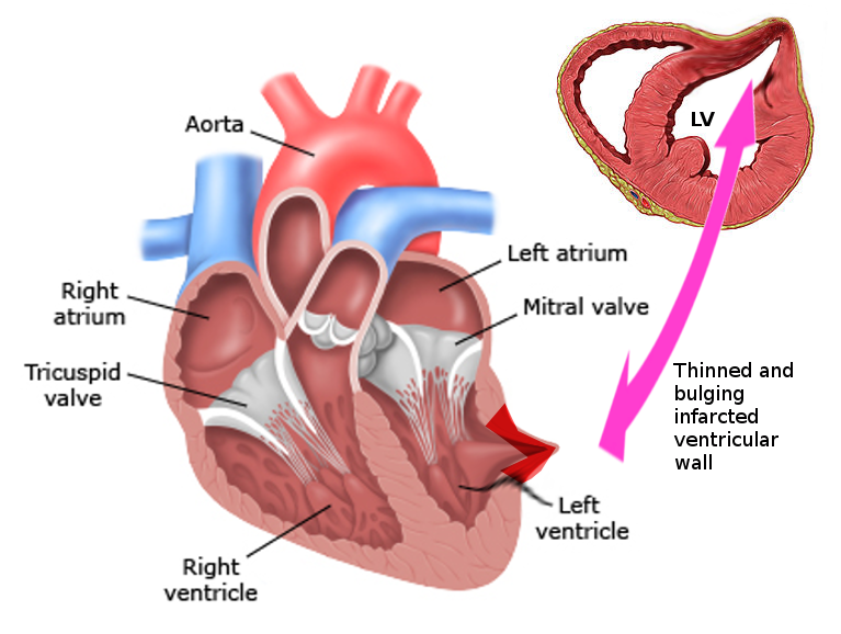
A left ventricular aneurysm (LVA) is a localised, thinned, scarred bulge in the wall of the heart’s main pumping chamber (the left ventricle). It most often develops after a large heart attack (myocardial infarction), when a section of heart muscle dies, loses its ability to contract, and is gradually replaced by fibrous scar tissue. Under the pressure generated inside the ventricle, this weakened patch stretches outward and forms a dyskinetic area — a region that balloons outward while the healthy muscle around it squeezes inward.
Most true LVAs involve the anterior wall and apex of the heart and follow a blockage of the left anterior descending (LAD) coronary artery. Because the aneurysmal wall does not contribute to pumping — and may even steal energy by expanding during each beat — a large aneurysm reduces the heart’s efficiency and can lead to heart failure, dangerous heart-rhythm disturbances, and blood clots that may travel to the brain or body.
An LVA is a chronic structural complication, distinct from the acute rupture that can occur in the first days after a heart attack. Many patients live for years with a small aneurysm, while larger ones may need medication, catheter procedures, or surgical ventricular restoration.
2. Key Facts at a Glance
| Key Fact | Detail |
|---|---|
| Also known as | LVA, ventricular aneurysm, cardiac aneurysm, post-infarction aneurysm |
| Body system affected | Cardiovascular system — left ventricle (main pumping chamber) |
| Common in | Adults after a large anterior heart attack, usually 50+ |
| Main cause | Prior myocardial infarction (LAD territory) |
| Severity range | From small and symptom-free to large aneurysms causing severe heart failure |
| Key treatments | Medications, ICD, catheter revascularisation, surgical ventricular restoration (Dor procedure) |
| Outlook | Good for small aneurysms; variable for large ones, improved by timely treatment |
3. Alternative Names and Medical Terminology
- Left ventricular aneurysm (LVA) — the standard clinical term.
- True aneurysm — a bulge whose wall still contains scarred heart muscle.
- False aneurysm (pseudoaneurysm) — a contained rupture walled off by pericardium, not muscle.
- Ventricular aneurysm / cardiac aneurysm — general synonyms.
- Dyskinetic or akinetic segment — descriptive terms for the non-contracting region.
- Post-infarction ventricular remodelling — the broader process that produces an aneurysm.
- Abbreviations you may see: LV (left ventricle), MI (myocardial infarction), EF (ejection fraction), SVR (surgical ventricular restoration).
4. Relevant Heart, Lung or Vascular Anatomy
The left ventricle is the thickest, most muscular chamber of the heart. It receives oxygen-rich blood from the left atrium and pumps it through the aortic valve into the aorta and the entire body. Its wall has three regions relevant to aneurysms: the anterior wall, the apex (the pointed tip), and the inferior/posterior wall.
Blood supply to this muscle comes from the coronary arteries — chiefly the left anterior descending (LAD) artery, which feeds the anterior wall, apex, and part of the septum. Because the apex and anterior wall are LAD territory, most aneurysms form here after an LAD blockage. The mitral valve and its supporting muscles sit within the ventricle and can be affected when the chamber enlarges. Surrounding the heart is the pericardium, a fibrous sac that can contain a rupture and form a false aneurysm.
5. How the Disease Affects the Body
When part of the ventricular wall becomes a scarred, thin aneurysm, the heart’s mechanics are disrupted in several ways. First, the aneurysmal segment is dyskinetic: during systole (contraction) it bulges outward instead of squeezing inward. This paradoxical motion wastes a portion of the ventricle’s pumping energy, so less blood is ejected with each beat and the ejection fraction falls.
Second, the enlarged, distorted chamber increases wall stress and oxygen demand on the remaining healthy muscle, which must work harder to maintain circulation. Over time this drives further remodelling — the ventricle dilates and becomes more spherical, worsening function in a self-reinforcing cycle that can lead to heart failure.
Third, the scarred border zone between dead and living muscle is an unstable electrical environment that can trigger ventricular tachycardia and other life-threatening arrhythmias. Fourth, blood tends to stagnate inside the aneurysm’s pouch, encouraging mural thrombus (clot) formation; pieces can break off and cause stroke or systemic embolism. Finally, ventricular dilation can stretch the mitral valve and cause mitral regurgitation. The combination of pump failure, arrhythmia and clot risk explains why a large aneurysm is far more than a cosmetic bulge.
6. Types and Classification
- True aneurysm — the wall is composed of scarred myocardium and fibrous tissue; it has a wide “neck,” bulges during systole, and rarely ruptures because it is still supported by residual muscle.
- False aneurysm (pseudoaneurysm) — a contained free-wall rupture; the wall is pericardium and clot only, with a narrow neck and a high risk of catastrophic rupture. This is a surgical emergency.
- Functional aneurysm — a forme fruste variant that protrudes during systole but not diastole.
- By location — most commonly anteroapical (LAD territory); less often inferior/posterior (right coronary or circumflex territory).
- By timing — acute/early (days after MI) versus chronic (weeks to months later, fully scarred).
7. Causes of the Disease
- Myocardial infarction — by far the leading cause, especially a large, transmural (full-thickness) anterior MI from LAD occlusion.
- Delayed or failed reperfusion — hearts that do not receive prompt angioplasty or clot-busting treatment are more likely to form an aneurysm.
- Chagas disease — a parasitic infection endemic in Latin America that classically causes apical aneurysms.
- Hypertrophic cardiomyopathy (apical variant) — can produce apical aneurysms.
- Chest trauma or cardiac surgery — can create false aneurysms.
- Infection (myocarditis) or sarcoidosis — rare inflammatory causes.
- Congenital aneurysm/diverticulum — an uncommon developmental form.
8. How the Disease Develops
An LVA is the end result of a process called adverse ventricular remodelling. When a coronary artery is blocked, the muscle it supplies is deprived of oxygen and begins to die within hours. If blood flow is not quickly restored, a full-thickness area of muscle is lost. Over the following days, the dead tissue is soft and vulnerable; over the following weeks it is cleared by the body and replaced with collagen scar.
Because scar tissue cannot contract, this patch of wall becomes passive. Each time the healthy ventricle generates pressure, the scarred region is stretched and gradually thins and bulges outward, a process governed by Laplace’s law (higher chamber size and pressure increase wall tension). The neighbouring healthy muscle enlarges to compensate, but this dilation increases wall stress further, drawing more muscle into dysfunction.
Within weeks to a few months, the aneurysm matures into a firm, fibrotic, well-defined bulge. The size of the aneurysm, the amount of remaining healthy muscle, and the promptness of the original heart-attack treatment all determine how severely function is affected.
9. Risk Factors
Modifiable:
- Untreated or poorly treated coronary artery disease.
- Delayed treatment of a heart attack — the single biggest risk factor.
- High blood pressure, which raises ventricular wall stress.
- Smoking, diabetes, high cholesterol and other drivers of coronary disease.
Non-modifiable / disease-related:
- A large, anterior, transmural MI.
- Single-vessel disease with poor collateral circulation (little back-up blood flow).
- Older age and prior heart attacks.
- Living in regions where Chagas disease is endemic.
10. Genetic and Family-History Factors
An LVA itself is not an inherited condition — it is a mechanical complication of muscle damage. However, the underlying coronary artery disease that causes most aneurysms has a strong hereditary component. A family history of early heart attacks, familial hypercholesterolaemia, diabetes, or hypertension raises a person’s lifetime risk of the MI that can later produce an aneurysm.
Certain aneurysm-associated conditions do have genetic links: hypertrophic cardiomyopathy (including the apical form) is often inherited in an autosomal-dominant pattern, and some arrhythmia syndromes cluster in families. Patients with a strong family history should discuss cardiac screening and risk-factor management with their doctor, but routine genetic testing is not part of standard LVA care.
11. Who Is Most at Risk?
- Survivors of a large anterior heart attack, particularly those who reached hospital late.
- Older adults, typically over 50, with established coronary disease.
- People with untreated or single-vessel LAD disease and poor collateral flow.
- Patients with persistent high blood pressure after their heart attack.
- Individuals from Chagas-endemic regions of Central and South America.
- Those with apical hypertrophic cardiomyopathy.
- People who did not receive timely angioplasty or thrombolysis during their infarction.
12. Prevalence and Epidemiology
Left ventricular aneurysm is now less common than it once was, largely because rapid angioplasty and clot-busting drugs limit the size of heart attacks and preserve muscle. In the era before routine reperfusion, aneurysms were reported in a substantial minority of patients after large anterior MIs; with modern treatment the frequency has fallen considerably, though exact figures vary between studies and regions.
Aneurysms are more common in men, reflecting the pattern of coronary disease, and typically appear in middle-aged and older adults. In parts of Latin America, Chagas disease remains an important non-ischaemic cause. Because many small aneurysms cause no symptoms and are found incidentally, the true prevalence is likely higher than clinical figures suggest. These numbers are approximate and should be interpreted as general trends rather than precise statistics.
13. Signs and Symptoms
The symptoms of a left ventricular aneurysm depend heavily on its size and location and on how much healthy heart muscle remains. Small aneurysms are frequently silent, discovered only when an echocardiogram or scan is performed for another reason. Larger aneurysms produce symptoms through three main pathways: heart failure, arrhythmia, and embolism.
Heart-failure symptoms are the most common and include breathlessness on exertion or when lying flat, fatigue, reduced exercise tolerance, and swelling of the ankles and legs. Arrhythmia symptoms include palpitations, a racing or irregular heartbeat, light-headedness, fainting (syncope), and in the worst cases sudden cardiac arrest from ventricular tachycardia or fibrillation. Embolic symptoms arise when a clot in the aneurysm breaks loose — most seriously a stroke (sudden weakness, speech difficulty, or vision loss), or a blocked artery in a limb or organ.
Some patients also experience angina (chest pain) from ongoing coronary disease, or a sense of a persistent abnormal heartbeat. Because these symptoms overlap with many other cardiac conditions, imaging is needed to confirm that an aneurysm is the cause.
14. Early-Stage Symptoms
- Often no symptoms at all, especially with a small aneurysm.
- Mild breathlessness or reduced stamina during exertion.
- Occasional palpitations or awareness of extra heartbeats.
- Lingering fatigue in the weeks and months after a heart attack.
- Persistent or slowly worsening angina if coronary disease is ongoing.
Because early symptoms are vague and easily attributed to normal post-heart-attack recovery, an aneurysm is often first suspected on a routine follow-up echocardiogram.
15. Advanced-Stage Symptoms
- Progressive heart failure: severe breathlessness, orthopnoea (breathless lying flat), waking at night short of breath, and leg swelling.
- Recurrent ventricular arrhythmias causing palpitations, dizziness, fainting, or cardiac arrest.
- Embolic events: stroke, transient ischaemic attack, or a suddenly cold, painful limb.
- Marked fatigue and exercise intolerance from a low ejection fraction.
- Symptoms of mitral regurgitation if the enlarged ventricle distorts the valve.
16. Symptoms in Women, Men and Older Adults
- Men are affected more often because coronary disease and large anterior MIs are more frequent in men; they tend to present with classic heart-failure or arrhythmia symptoms.
- Women may have more atypical heart-attack symptoms initially (fatigue, jaw or back discomfort, breathlessness rather than crushing chest pain), which can delay the original diagnosis and treatment.
- Older adults often have quieter, more generalised symptoms — confusion, weakness, poor appetite, or simply “slowing down” — and may have several coexisting conditions that mask the aneurysm. Falls or fainting from arrhythmia can be the first sign in this group.
17. Emergency Warning Signs
Call emergency services immediately if you or someone else has:
- Fainting or collapse, or a very fast, pounding heartbeat that does not settle.
- Signs of stroke — sudden facial droop, arm weakness, or slurred speech.
- Severe, sudden breathlessness or coughing up pink, frothy fluid.
- Crushing chest pain, which may signal a new heart attack.
- A suddenly cold, pale, painful limb (possible clot).
18. When to Seek Medical Help
Arrange a prompt (non-emergency) medical review if you notice new or worsening breathlessness, increasing ankle swelling, more frequent palpitations, unexplained dizziness, or reduced exercise tolerance — particularly if you have previously had a heart attack. Anyone recovering from a large anterior MI should attend all scheduled cardiology follow-ups, where surveillance imaging can detect an aneurysm before it causes serious problems. When in doubt, contact your doctor or use our contact page to arrange a specialist opinion.
19. Disease Stages, Grades and Severity
There is no single formal staging system for LVA; severity is judged by a combination of features:
- Size and extent — the proportion of the ventricle involved; larger aneurysms cause more dysfunction.
- Wall motion — akinetic (non-moving) segments are less harmful than dyskinetic (paradoxically bulging) ones.
- Ejection fraction — the lower the EF, the more severe the impact on pumping.
- True versus false — a false aneurysm (pseudoaneurysm) is high-risk regardless of size.
- Clinical burden — graded by NYHA heart-failure class, arrhythmia frequency, and any embolic events.
- Mural thrombus — its presence raises the severity and clot risk.
20. Disease Progression
The natural course of an LVA varies widely. After a heart attack, an aneurysm typically matures over several weeks to months as scar tissue forms. Once established, a small, stable aneurysm may cause little trouble for many years. Larger aneurysms, however, tend to promote ongoing adverse remodelling: the whole ventricle dilates, becomes more spherical, and loses efficiency, so heart failure gradually worsens.
Progression is driven by wall stress, uncontrolled blood pressure, continuing coronary disease, and the electrical instability of the scar border. Some patients remain stable on medication; others experience a stepwise decline punctuated by arrhythmias, embolic events, or episodes of decompensated heart failure. Timely revascularisation, good medical therapy, and — in selected cases — surgical restoration can slow or partially reverse this progression.
21. Possible Complications
- Heart failure — the most common serious complication.
- Ventricular arrhythmias — recurrent VT or VF, a leading cause of sudden death.
- Mural thrombus and embolism — stroke, limb or organ artery blockage.
- Mitral regurgitation — from ventricular enlargement and papillary muscle displacement.
- Rupture — rare for true aneurysms but a major risk for false aneurysms.
- Progressive ventricular dilation and low cardiac output.
- Angina from ongoing coronary disease.
22. Related and Associated Medical Conditions
- Coronary artery disease — the underlying cause in most patients (see isolated coronary artery disease).
- Ischaemic cardiomyopathy and other forms of heart failure.
- Post-infarction ventricular septal defect, another mechanical MI complication (more here).
- Atrial and ventricular arrhythmias (disorders of heart rhythm).
- Mitral valve disease from ventricular remodelling.
- Diabetes, hypertension, and hyperlipidaemia as shared risk factors.
23. Screening and Early Detection
There is no population-wide screening programme for LVA because it is a complication of a specific event — a heart attack. The key to early detection is structured cardiology follow-up after a large MI, during which an echocardiogram can identify abnormal wall motion and aneurysm formation well before symptoms appear. Patients with poor ventricular function may undergo further imaging (cardiac MRI) to characterise scar and viability. Anyone with Chagas disease or apical hypertrophic cardiomyopathy should also have periodic echocardiographic surveillance. Early detection allows anticoagulation, arrhythmia protection, and treatment planning to begin before complications occur.
24. How the Disease Is Diagnosed
Diagnosis of a left ventricular aneurysm brings together clinical suspicion and cardiac imaging. In someone with a history of anterior heart attack who develops heart failure, arrhythmia, or an embolic event, doctors specifically look for an aneurysm.
The pathway usually begins with an electrocardiogram (ECG), which may show persistent ST-segment elevation (“frozen” ST elevation) in the anterior leads long after the acute infarct — a classic clue. A chest X-ray may reveal an unusual bulge along the heart border. The cornerstone test is the transthoracic echocardiogram, which directly visualises the aneurysm, measures its size, assesses wall motion (dyskinetic vs akinetic), estimates ejection fraction, and detects any mural thrombus or mitral regurgitation.
When more detail is needed, cardiac MRI is the reference standard: it distinguishes true from false aneurysm and quantifies scar and viable muscle. Cardiac CT and left ventriculography (during coronary angiography) also delineate the aneurysm and map the coronary arteries so blockages can be treated at the same time.
25. Physical Examination and Medical History
The doctor will ask about previous heart attacks (especially anterior MI), the timeliness of any treatment received, and current symptoms of breathlessness, palpitations, fainting, or stroke-like episodes. Risk factors such as smoking, diabetes, hypertension, and family history are reviewed.
On examination, findings may include a displaced or abnormal apical impulse (sometimes a “double” or dyskinetic beat), a third or fourth heart sound, a mitral regurgitation murmur, and signs of heart failure such as raised jugular venous pressure, lung crackles, and leg swelling. The examination is often normal in small aneurysms, so imaging remains essential for confirmation.
26. Diagnostic Tests and Imaging
- ECG — persistent anterior ST elevation, Q waves, and signs of arrhythmia.
- Transthoracic echocardiography — first-line; shows the aneurysm, wall motion, EF, thrombus, and valve function.
- Cardiac MRI — gold standard for tissue characterisation, viability, scar, and true-vs-false distinction.
- Cardiac CT — high-resolution anatomy and coronary assessment.
- Coronary angiography with left ventriculography — defines coronary blockages and outlines the aneurysm before intervention.
- Chest X-ray — may show an abnormal cardiac contour or calcification.
- Holter/ambulatory monitoring — detects intermittent ventricular arrhythmias.
27. Blood Tests, Biomarkers and Genetic Testing
There is no blood test that diagnoses an aneurysm directly, but laboratory work supports overall assessment. BNP or NT-proBNP levels reflect the degree of heart strain and help gauge heart failure. Troponin is used to detect any new or ongoing muscle injury. Routine tests include kidney function and electrolytes (important before contrast imaging and surgery), a full blood count, a lipid profile, blood glucose/HbA1c, and coagulation studies for patients on or starting anticoagulation. Genetic testing is not routine, but may be considered if an inherited cardiomyopathy is suspected, and serology for Chagas disease is appropriate in patients from endemic areas.
28. Understanding Test Results
Interpreting results means combining several measurements. Ejection fraction (EF) describes the fraction of blood pumped out with each beat — a normal EF is roughly 55% or higher, and lower values indicate weaker pumping. The echo or MRI report will describe the aneurysm’s location and size, whether the wall motion is akinetic or dyskinetic, and whether a mural thrombus is present. Viability imaging shows how much muscle is scarred versus salvageable — important when deciding whether revascularisation will help. Elevated BNP/NT-proBNP points to significant heart strain. Your cardiologist will explain how these results fit together, but broadly: the larger the aneurysm, the lower the EF, and the more thrombus or arrhythmia present, the stronger the case for active treatment.
29. Differential Diagnosis
- False aneurysm (pseudoaneurysm) — must be distinguished from a true aneurysm because management differs sharply.
- Post-infarction ventricular septal defect — another mechanical MI complication.
- Left ventricular thrombus without aneurysm.
- Pericardial cyst or effusion producing an abnormal cardiac contour.
- Prominent LV diverticulum (congenital).
- Dilated or ischaemic cardiomyopathy causing global rather than localised dysfunction.
- Cardiac tumour or mass (tumours of the heart and lung).
30. Specialist and Multidisciplinary Evaluation
Managing an LVA is a team effort. A cardiologist coordinates diagnosis and medical therapy, while an interventional cardiologist addresses coronary blockages. A cardiac surgeon evaluates the need for and feasibility of ventricular restoration. An electrophysiologist manages arrhythmia risk and ICD decisions, a heart-failure specialist optimises medical therapy, and cardiac imaging experts provide the echo, MRI, and CT that guide planning. Anaesthetists, cardiac nurses, physiotherapists, and rehabilitation teams complete the group. This coordinated “heart-team” approach, standard at high-volume centres, produces the best outcomes. Browse experienced doctors and hospitals to find such a team.
31. Treatment Goals
- Relieve symptoms of heart failure, angina, and arrhythmia.
- Improve pumping efficiency by restoring a more normal ventricular shape and size.
- Prevent embolic events through anticoagulation or removal of thrombus.
- Reduce the risk of sudden cardiac death from ventricular arrhythmia.
- Treat underlying coronary disease to protect remaining muscle.
- Slow or reverse adverse remodelling and improve quality of life and survival.
32. When Is Treatment Required?
Treatment intensity is matched to the aneurysm and its effects. Small, stable, symptom-free true aneurysms may need only good medical therapy and monitoring. Active intervention is considered when there is significant heart failure, refractory (drug-resistant) ventricular arrhythmia, a mural thrombus or embolic event, severe mitral regurgitation, or a very large aneurysm compromising ventricular function. A false aneurysm (pseudoaneurysm) almost always requires prompt surgery because of its high rupture risk, regardless of symptoms. The decision balances the potential benefit against the patient’s overall fitness for surgery.
33. Active Monitoring and Watchful Waiting
For small, stable, asymptomatic true aneurysms, a strategy of watchful waiting is often appropriate. This means excellent control of blood pressure, cholesterol, and diabetes; guideline-based medications to protect the heart; and regular follow-up echocardiograms to track size, ejection fraction, and thrombus. Patients are taught to recognise warning symptoms and to report new breathlessness, palpitations, or swelling promptly. If surveillance shows the aneurysm enlarging or function declining, the plan shifts toward more active treatment. Watchful waiting is not “doing nothing” — it is structured, proactive observation.
34. Medications
Medications treat the aneurysm’s consequences and protect the rest of the heart:
- ACE inhibitors or ARBs (and sometimes ARNI) — reduce wall stress and limit remodelling.
- Beta-blockers — lower arrhythmia risk and improve heart-failure survival.
- Mineralocorticoid receptor antagonists (e.g., spironolactone/eplerenone) — for heart failure with reduced EF.
- SGLT2 inhibitors — increasingly used in heart failure.
- Diuretics — relieve fluid overload and swelling.
- Anticoagulants (e.g., warfarin) — prevent or treat mural thrombus and embolism.
- Antiplatelet agents and statins — manage the underlying coronary disease.
- Antiarrhythmic drugs (e.g., amiodarone) — for symptomatic ventricular arrhythmias.
35. Minimally Invasive Treatments
Compared with traditional open aneurysm repair, several less invasive options exist. Some surgical centres perform ventricular restoration through smaller incisions or off-pump techniques in selected patients, and hybrid programmes combine catheter and surgical steps to limit trauma. For coronary disease driving the aneurysm, minimally invasive coronary bypass may be feasible. Investigational percutaneous ventricular partitioning devices — a parachute-like implant placed by catheter to seal off the aneurysmal segment — aim to restore ventricular geometry without open surgery, though availability is limited and evidence is still evolving. Suitability depends on aneurysm anatomy and overall heart function. Explore minimally invasive cardiac procedures.
36. Catheter-Based and Endovascular Treatments
Catheter-based care addresses both the coronary cause and, in some cases, the aneurysm itself. Percutaneous coronary intervention (angioplasty and stenting) reopens blocked arteries to protect surviving muscle and relieve angina. Catheter ablation by an electrophysiologist can target the scar-border circuits that generate ventricular tachycardia. Investigational percutaneous LV partitioning/exclusion devices are deployed through a catheter to wall off the aneurysm and reduce chamber volume. These approaches avoid a full sternotomy and can be attractive for higher-risk patients, though not every aneurysm is suitable. See angioplasty and electrophysiological procedures.
37. Surgical Treatment Options
The definitive treatment for a large or complicated LVA is surgical ventricular restoration (SVR), which aims to remove the useless scarred bulge and rebuild the ventricle into a more efficient, cone-like shape. The best-known technique is the Dor procedure (endoventricular circular patch plasty), in which the aneurysm is opened, any clot removed, and a patch used to exclude the scar and restore near-normal chamber volume and geometry. Simpler linear aneurysmectomy (cutting out the aneurysm and closing the wall directly) is used in some cases, and modifications such as the Jatene and septal exclusion techniques exist.
Surgery is usually combined with coronary artery bypass grafting (CABG), and with mitral valve repair if significant regurgitation is present. In drug-resistant ventricular tachycardia, arrhythmia surgery or ablation and an ICD may be added.
For a false aneurysm, urgent surgical repair is required because of the rupture risk. When ventricular function is too poor for restoration, options extend to mechanical circulatory support (LVAD) or heart transplantation. The heart team individualises the operation to aneurysm size, muscle viability, coronary anatomy, and overall condition. Learn more about coronary artery bypass grafting and surgery options.
38. Advanced and Emerging Treatments
- Percutaneous ventricular partitioning devices — catheter-delivered implants to exclude the aneurysm and improve geometry.
- Regenerative and stem-cell therapies — experimental approaches aiming to repair or replace scarred muscle.
- Advanced 3D imaging and computer modelling — to plan the optimal ventricular reconstruction shape.
- Refined VT ablation and hybrid arrhythmia surgery — for scar-related arrhythmias.
- Next-generation LVADs and total artificial hearts — for end-stage disease.
These are offered selectively at specialist centres and within clinical trials; discuss eligibility with your cardiac team.
39. Treatment Options Compared
- Medical therapy alone — best for small, stable, low-symptom aneurysms; least invasive but does not remove the bulge.
- Catheter revascularisation / ablation — targets the coronary cause and arrhythmia with lower procedural risk; may not correct a very large aneurysm.
- Surgical ventricular restoration (Dor) ± CABG ± valve repair — most complete correction for large, symptomatic aneurysms; higher upfront risk but strong symptom and function benefit in the right patient.
- Advanced support (LVAD/transplant) — reserved for end-stage heart failure when restoration is not feasible.
The optimal choice balances aneurysm size, viable muscle, coronary anatomy, arrhythmia burden, and surgical fitness.
40. How Doctors Choose the Right Treatment
Doctors weigh the size, location, and type of the aneurysm (true vs false), the ejection fraction and amount of viable muscle, the severity of symptoms, the arrhythmia and clot burden, the coronary anatomy, and the presence of mitral regurgitation. Equally important are patient factors: age, kidney and lung function, frailty, other illnesses, and personal preferences. A false aneurysm generally mandates surgery; a small silent true aneurysm generally does not. The heart team integrates imaging, angiography, and clinical assessment to recommend the approach most likely to improve both quality and length of life.
41. Benefits and Risks of Treatment
Potential benefits:
- Relief of breathlessness, fatigue, and angina.
- Improved ejection fraction and exercise capacity after successful restoration.
- Fewer arrhythmias and lower stroke risk (with anticoagulation or thrombus removal).
- Better long-term survival in appropriately selected patients.
Potential risks:
- Surgical risks: bleeding, infection, arrhythmia, kidney injury, and, uncommonly, low-output heart failure or death.
- Anaesthetic and procedural complications.
- Bleeding risk from long-term anticoagulation.
- Incomplete symptom relief if muscle damage is extensive.
Careful patient selection and experienced surgical teams substantially reduce these risks.
42. What Happens If the Disease Is Left Untreated?
An untreated small, stable aneurysm may cause few problems for years. However, a large or complicated aneurysm left untreated tends to drive progressive heart failure as the ventricle dilates and weakens. The risk of life-threatening ventricular arrhythmias and sudden cardiac death remains, and stagnant blood in the pouch can throw off clots, causing stroke or organ infarction. A false aneurysm carries a serious risk of fatal rupture if not repaired. Left untreated, the combination of failing pump function, dangerous rhythms, and embolic events shortens life and reduces its quality — which is why timely assessment matters even when symptoms are mild.
43. Treatment Success and Expected Outcomes
Outcomes depend heavily on aneurysm size, remaining muscle, and the patient’s overall health. In well-selected patients, surgical ventricular restoration combined with bypass surgery can meaningfully improve heart-failure symptoms, raise ejection fraction, and enhance quality of life, with the majority experiencing durable benefit. Anticoagulation effectively lowers embolic risk, and an ICD protects against sudden arrhythmic death. Results are generally best when treatment is performed before the ventricle has dilated extensively and when surgery is done at high-volume centres by experienced teams. Outcomes are more guarded when ventricular function is already severely impaired. These are general expectations; your own prognosis should be discussed with your cardiac team.
44. Prognosis and Long-Term Outlook
The long-term outlook for a person with an LVA spans a wide range. Those with a small, stable aneurysm and preserved overall function often do well for many years with medication and monitoring. Those with a large aneurysm, low ejection fraction, and significant heart failure face a more serious prognosis, driven by pump failure, arrhythmia, and embolic risk.
The single most powerful factor is the amount of healthy, working heart muscle that remains. Modern care has improved outlooks considerably: prompt heart-attack treatment prevents many aneurysms, guideline-based medications limit remodelling, ICDs prevent sudden death, anticoagulation prevents strokes, and surgical restoration can restore function in suitable patients. With comprehensive care, many patients live active lives for years. Regular follow-up, strict risk-factor control, and treatment adherence are the keys to the best outcome; your cardiologist can give a personalised estimate.
45. Recovery and Rehabilitation
Recovery after ventricular restoration surgery typically involves a few days in intensive and step-down care followed by a hospital stay of about one to two weeks. Full recovery of energy and strength usually takes six to twelve weeks, during which activity is increased gradually and the breastbone is protected while it heals. Cardiac rehabilitation — a supervised programme of monitored exercise, education, and lifestyle support — is strongly recommended and improves both function and confidence. Medications are continued and adjusted, wounds are monitored, and follow-up imaging confirms the result. Patients who did not have surgery still benefit from a structured rehabilitation and risk-reduction programme.
46. Follow-Up Tests and Long-Term Monitoring
Long-term follow-up usually includes periodic clinical review and echocardiography to track ejection fraction, chamber size, and any residual or recurrent aneurysm or thrombus. ECGs and, when indicated, Holter monitoring assess for arrhythmia. Patients on anticoagulation need regular blood-test monitoring (e.g., INR for warfarin). Kidney function, electrolytes, and heart-failure biomarkers are checked as needed, and any implanted ICD is interrogated at routine intervals. The frequency of follow-up is tailored to aneurysm size and symptom stability.
47. Managing Recurrence or Disease Progression
Because an aneurysm reflects permanent muscle loss, the focus of long-term care is preventing further deterioration rather than “cure.” Progression is managed by optimising heart-failure medication, strictly controlling blood pressure, cholesterol, and diabetes, and treating any new coronary blockages promptly. Recurrent arrhythmias may need medication, ablation, or ICD adjustment; new or growing thrombus needs anticoagulation review. If heart failure advances despite maximal therapy, the team may reconsider surgical options, mechanical support, or transplantation. Consistent follow-up allows problems to be caught and addressed early.
48. Living with the Disease
Living with an LVA means combining medical treatment with heart-healthy habits. Most patients can return to a satisfying daily life with attention to a few essentials: taking medications reliably, attending follow-ups, monitoring for warning symptoms (breathlessness, swelling, palpitations), and maintaining a heart-protective lifestyle. Many people continue to work, travel, and enjoy hobbies, adjusting the intensity of activity to their heart’s capacity. Support from family, cardiac rehabilitation, and patient groups helps with the practical and emotional adjustments. Weighing yourself regularly and knowing your “action plan” for worsening symptoms are simple habits that prevent hospital admissions.
49. Diet and Nutrition Guidelines
- Follow a heart-healthy, Mediterranean-style diet rich in vegetables, fruit, whole grains, legumes, fish, and healthy oils.
- Limit salt to reduce fluid retention and control blood pressure — especially important with heart failure.
- Reduce saturated and trans fats and processed foods to manage cholesterol.
- Watch fluid intake if advised by your doctor for heart failure.
- Keep alcohol within recommended limits, or avoid it.
- If taking warfarin, keep vitamin K intake (leafy greens) consistent rather than fluctuating.
- Maintain a healthy weight and manage blood sugar if diabetic.
50. Exercise and Physical-Activity Guidelines
Regular, appropriately paced activity is beneficial, but intensity must match heart function. Cardiac rehabilitation provides a safe, supervised starting point and individualised exercise prescription. Most patients are encouraged to build up moderate aerobic activity such as walking or cycling, guided by symptoms and their care team. High-intensity or competitive exertion may be restricted in those with significant arrhythmia risk or low ejection fraction. Always warm up, avoid straining or heavy lifting soon after surgery, and stop and seek advice for chest pain, severe breathlessness, or fainting. Your cardiologist will tailor recommendations to your specific situation.
51. Medications, Activities and Habits to Avoid
- Do not stop prescribed cardiac medications without medical advice.
- Avoid smoking entirely and steer clear of second-hand smoke.
- Limit or avoid alcohol, which can worsen heart failure and arrhythmia.
- Be cautious with NSAIDs (e.g., ibuprofen), which cause fluid retention — check with your doctor.
- Avoid decongestants and stimulants that raise heart rate and blood pressure.
- Avoid heavy lifting and sudden intense exertion, especially during recovery.
- If on anticoagulants, avoid activities with high bleeding/injury risk and inform other clinicians before procedures.
52. Preventing the Disease or Reducing Its Risks
The best prevention is avoiding the large heart attack that causes most aneurysms. Key measures:
- Seek help immediately for heart-attack symptoms — fast reperfusion preserves muscle and prevents aneurysm.
- Control coronary risk factors: stop smoking, manage blood pressure, cholesterol, and diabetes.
- Take prescribed secondary-prevention medication (statins, antiplatelets, ACE inhibitors, beta-blockers) after a heart attack.
- Maintain a heart-healthy diet, regular exercise, and healthy weight.
- Attend cardiac follow-up so that remodelling is detected and treated early.
- In endemic regions, prevent and treat Chagas disease.
53. Pregnancy and the Disease
LVA is uncommon in women of childbearing age because it usually follows heart attacks in older adults, but when it does occur, pregnancy poses significant added strain on an already weakened heart. Any woman with a ventricular aneurysm or reduced ejection fraction should have pre-pregnancy counselling with a cardiologist and, if she becomes pregnant, be managed by a specialist maternal-cardiac team. Some heart medications must be changed before conception because they are unsafe in pregnancy. Decisions are highly individual and should always involve expert advice.
54. Disease in Children and Young Adults
True post-infarction aneurysms are rare in children and young adults, since coronary heart attacks are uncommon in these age groups. When aneurysms do appear in the young, causes are more likely to be congenital ventricular aneurysms or diverticula, Kawasaki disease with coronary involvement, myocarditis, Chagas disease, or hypertrophic cardiomyopathy. Evaluation and management in these cases are led by paediatric or adult congenital cardiac specialists, and treatment is tailored to the underlying cause. Related congenital conditions are discussed in our congenital heart disease section.
55. Disease in Older Adults
Older adults make up the largest group with LVA because coronary disease and prior heart attacks accumulate with age. In this group, aneurysms often coexist with other conditions — kidney disease, diabetes, lung disease, and frailty — that influence treatment choices. Symptoms may be subtle or atypical, and surgical risk is generally higher, so the balance between benefit and risk is weighed carefully. Many older patients are managed very effectively with optimised medication, anticoagulation, ICDs, and catheter procedures, reserving major surgery for carefully selected individuals. Comprehensive geriatric-aware assessment improves outcomes.
56. Emotional Health and Patient Support
A diagnosis involving the heart, and the memory of a heart attack, can bring anxiety, low mood, and fear of sudden events — this is common and understandable. Emotional wellbeing directly affects recovery and adherence to treatment. Helpful steps include cardiac rehabilitation (which addresses psychological as well as physical recovery), talking openly with the care team, connecting with patient support groups, and seeking counselling or psychological support when needed. Family involvement, stress-reduction techniques, and clear information about the condition all reduce fear. If you feel persistently anxious or depressed, tell your doctor — effective support is available.
57. Preparing for Your Specialist Appointment
- Bring a list of all your medications and doses.
- Gather previous test results and imaging (echo, MRI, angiogram reports) and details of any past heart attack.
- Write down your symptoms, when they occur, and what triggers them.
- Note your medical history and family history of heart disease.
- Prepare your questions in advance (see next section).
- Bring a family member or friend to help remember information.
- Know your insurance or medical-travel arrangements if seeking treatment abroad.
58. Questions to Ask Your Doctor
- Is my aneurysm a true or false aneurysm, and how large is it?
- What is my ejection fraction, and how much healthy muscle remains?
- Is there a blood clot inside the aneurysm, and do I need anticoagulation?
- Am I at risk of dangerous heart rhythms, and do I need an ICD?
- Do I need surgery, a catheter procedure, or medication alone?
- What are the benefits and risks of each treatment option for me?
- What will recovery and rehabilitation involve?
- How will this affect my daily activities, work, and life expectancy?
- How often will I need follow-up tests?
- Would treatment abroad be a safe and cost-effective option for me?
59. Cost of Diagnosis and Treatment
Costs vary widely by country, hospital, and the exact procedures needed. The figures below are approximate ranges for surgical ventricular restoration (often with bypass) and should be confirmed with each centre.
| Region | Approx. cost of surgery (USD) | Notes |
|---|---|---|
| United States | $70,000 – $200,000+ | Highest; varies by insurance |
| United Kingdom / Western Europe | $40,000 – $90,000 | Private; NHS covered for residents |
| Singapore | $30,000 – $60,000 | High-quality regional hub |
| Thailand | $18,000 – $40,000 | Popular medical-tourism destination |
| Turkey | $15,000 – $35,000 | Growing cardiac-tourism sector |
| India | $7,000 – $18,000 | Often 50–90% less than US/UK |
Diagnostic tests (echo, MRI, angiography) add further cost. Medical tourism to India, Turkey, or Thailand can substantially reduce total expense while maintaining high standards at accredited hospitals. Explore destinations and request a personalised quote via our contact page.
60. Factors Affecting Treatment Cost
- Type and complexity of surgery (restoration alone vs combined with CABG and valve repair).
- Country and hospital chosen, and whether it is a private or public facility.
- Surgeon and centre reputation and volume.
- Length of hospital and ICU stay and any complications.
- Implants and devices (ICD, patches, valve prostheses).
- Diagnostic imaging and pre-operative tests.
- Cardiac rehabilitation and follow-up care.
- For international patients: travel, accommodation, translation, and aftercare arrangements.
61. Choosing the Right Specialist
Look for a cardiac surgeon and cardiology team with specific experience in ventricular restoration and complex ischaemic heart disease. Helpful criteria include board certification, a high annual volume of similar operations, published or audited outcomes, and access to a full heart team (interventional cardiology, electrophysiology, heart failure, imaging). Good communication, willingness to explain options, and support for second opinions are all positive signs. Browse experienced doctors and surgeons on our directory to compare credentials and locations.
62. Choosing the Right Hospital or Treatment Centre
- Choose a centre with internationally recognised accreditation (e.g., JCI) and strong cardiac-surgery credentials.
- Prefer high-volume heart centres with dedicated cardiac ICUs and a full heart team.
- Review outcome and complication data where available.
- Check availability of advanced imaging, ICD/electrophysiology, and mechanical support.
- For international patients, confirm international patient services, language support, and structured aftercare.
- Compare accredited hospitals and destinations on our site.
63. Getting a Second Medical Opinion
Because treatment of an LVA involves major decisions — whether to operate, which technique to use, and whether treatment abroad is appropriate — a second opinion is valuable and encouraged. Another experienced heart team may confirm the plan, suggest alternatives, or offer newer options, giving you confidence and clarity. Bring your imaging and reports so the reviewing team has full information. We can help arrange an independent expert opinion — visit our contact page or request a second opinion.
64. Treatment Abroad and Medical-Travel Considerations
Many patients travel for high-quality, more affordable cardiac surgery at accredited hospitals in India, Turkey, Thailand, and Singapore. When planning treatment abroad, consider: the hospital’s accreditation and cardiac-surgery track record; the surgeon’s experience; clear, itemised cost estimates; fitness to fly before and after surgery; length of stay and aftercare arrangements; visa, translation, and travel logistics; and coordination of follow-up back home with your local doctor. A well-planned medical journey can combine excellent care with significant savings. Explore destinations and treatments, or contact us for tailored guidance.
65. Frequently Asked Questions
Is a left ventricular aneurysm the same as an aortic aneurysm? No. An LVA is a bulge in the heart’s pumping chamber, usually after a heart attack, while an aortic aneurysm is a widening of the aorta. They are different conditions with different treatments.
Will a true aneurysm burst? True aneurysms rarely rupture because their wall contains supporting scar tissue. False aneurysms, however, have a high rupture risk and usually need urgent surgery.
Do I need surgery? Not always. Small, symptom-free aneurysms are often managed with medication and monitoring. Surgery is considered for large aneurysms causing heart failure, dangerous arrhythmias, clots, or a false aneurysm.
Why am I on blood thinners? Blood can pool and clot inside the aneurysm; anticoagulation reduces the risk of that clot causing a stroke or blocking another artery.
Can my heart function improve after treatment? In suitable patients, surgical ventricular restoration can improve ejection fraction and symptoms, especially when done before the ventricle has dilated extensively.
Can I exercise? Yes, usually within limits set by your cardiologist and ideally starting with cardiac rehabilitation. Activity level depends on your heart function and arrhythmia risk.
Is treatment abroad safe? At accredited, high-volume centres it can be very safe and cost-effective. Choose carefully and plan follow-up with your home doctor.
66. Patient Stories and Treatment Experiences
The following are representative, anonymised examples for illustration only.
-
Rahul, India: After a large anterior heart attack, Rahul developed breathlessness and was found to have an anteroapical aneurysm with a low ejection fraction. He underwent a Dor procedure combined with bypass surgery at an accredited centre and, after cardiac rehabilitation, returned to work with markedly improved stamina.
-
Margaret, United Kingdom: Margaret’s small, symptom-free aneurysm was discovered on a routine follow-up echo. Her team chose medication and anticoagulation with regular monitoring. Years later she remains stable and active.
-
Ahmed, travelled to Turkey: Seeking affordable expert care, Ahmed travelled abroad for ventricular restoration surgery after recurrent ventricular tachycardia. He received an ICD, had a smooth recovery, and coordinated follow-up with his doctor at home.
67. Latest Research and Clinical Trials
Research continues to refine the management of ventricular aneurysm and post-infarction heart failure. Active areas include percutaneous ventricular partitioning devices that reshape the ventricle without open surgery; regenerative and cell-based therapies aiming to repair scarred muscle; improved imaging and 3D modelling to plan the ideal ventricular shape; and advances in VT ablation and hybrid arrhythmia surgery. Ongoing work also compares surgical restoration plus bypass against medical therapy in selected patients, and explores newer heart-failure medications. Patients interested in trials should ask their cardiac team about eligibility and reputable studies registered with recognised trial registries. (No specific unpublished results are cited here.)
68. Related Diseases and Conditions
- Isolated Coronary Artery Disease
- Post-Infarction Ventricular Septal Defect
- Complex Coronary Artery Disease
- Ischemic Cardiomyopathy
- Heart Failure
- Disorders of Heart Rhythm
69. Related Treatments and Procedures
- Coronary Artery Bypass Grafting
- Angioplasty
- Minimally Invasive Cardiac Surgery
- Electrophysiological Procedures
- Mitral Valve Procedures
70. Medical Glossary
- Left ventricle (LV): the heart’s main pumping chamber.
- Aneurysm: a localised bulge in a weakened wall.
- True aneurysm: a bulge whose wall still contains scarred muscle.
- False aneurysm (pseudoaneurysm): a contained rupture walled off by pericardium; high rupture risk.
- Myocardial infarction (MI): a heart attack; death of heart muscle from blocked blood flow.
- Ejection fraction (EF): the percentage of blood pumped out with each beat.
- Dyskinetic: a wall segment that bulges outward during contraction.
- Akinetic: a wall segment that does not move.
- Mural thrombus: a blood clot on the inner wall of the heart.
- Remodelling: change in the heart’s size and shape after injury.
- Surgical ventricular restoration (SVR): surgery to rebuild a more normal ventricular shape.
- Dor procedure: endoventricular circular patch plasty to exclude the aneurysm.
- CABG: coronary artery bypass grafting.
- ICD: implantable cardioverter-defibrillator, which treats dangerous rhythms.
- Ventricular tachycardia (VT): a fast, dangerous rhythm arising from the ventricle.
71. Medical Review, Editorial Policy and Last Updated Date
Last updated: 11 July 2026.
This article is written for patient education and is reviewed by qualified medical professionals for accuracy and clarity. Our editorial policy is to align content with established cardiology guidance from bodies such as the ACC/AHA, ESC, NHS, and STS, to use cautious, qualified language, and to avoid fabricated statistics or citations. Content is updated periodically as practice evolves.
Disclaimer: This information is educational and is not a substitute for professional medical advice, diagnosis, or treatment. Always consult a qualified healthcare provider about your individual condition.
72. Clinical Guidelines and Medical References
This content draws on general, widely accepted knowledge from recognised sources, including:
- American College of Cardiology / American Heart Association (ACC/AHA) guidelines on coronary disease and heart failure.
- European Society of Cardiology (ESC) guidelines on heart failure and myocardial revascularisation.
- Society of Thoracic Surgeons (STS) resources on cardiac surgery.
- National Health Service (NHS) patient information.
- Standard cardiology and cardiac-surgery textbooks.
Specific study titles, DOIs, and figures are not cited here; consult your cardiologist and current guidelines for the latest evidence.
73. Book an Appointment or Request a Second Opinion
If you or a loved one has a left ventricular aneurysm, expert help is available. Our network connects you with experienced cardiologists, cardiac surgeons, and accredited hospitals worldwide for diagnosis, treatment, and second opinions — including affordable, high-quality options abroad.
- Book an appointment or request a second opinion: /contact/
- Contact our team for personalised guidance and quotes: /contact/
- Explore top hospitals, doctors, and destinations for cardiac care.
Take the next step toward expert heart care today.