1. Disease Overview
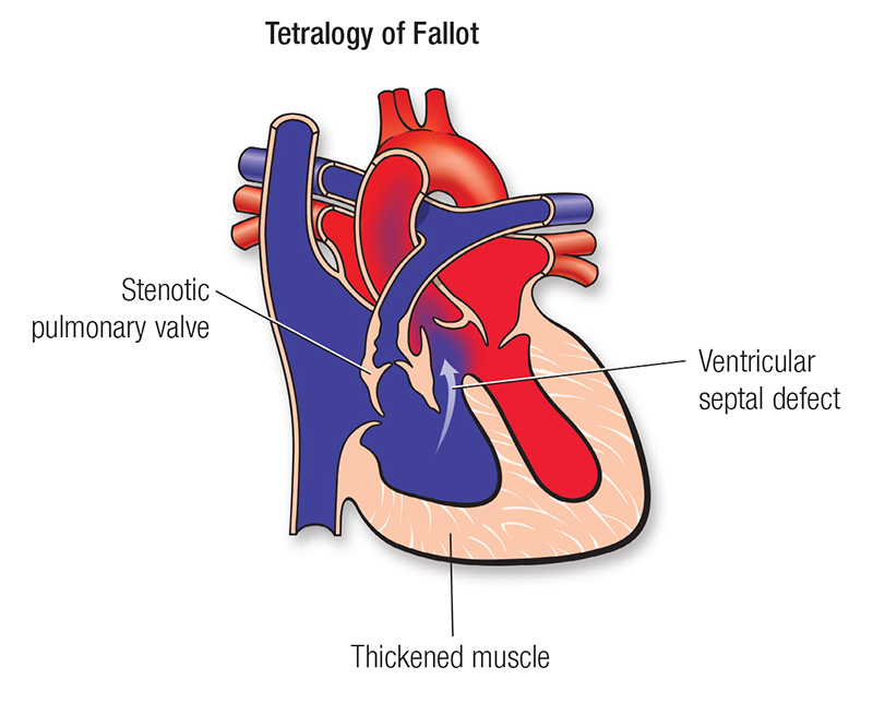
Tetralogy of Fallot (ToF) is the most common cyanotic (blue-baby) congenital heart defect, present at birth and formed by a combination of four structural abnormalities of the heart. Its name reflects those four features, first described together by the French physician Étienne-Louis Arthur Fallot in 1888. The four components are a large ventricular septal defect (VSD), an overriding aorta that sits above the hole in the wall between the ventricles, pulmonary stenosis (narrowing of the outflow to the lungs), and right ventricular hypertrophy (thickening of the right pumping chamber).
The core problem is that narrowing below and at the pulmonary valve limits blood flow to the lungs, while the large VSD and overriding aorta allow oxygen-poor (blue) blood to bypass the lungs and enter the body’s circulation. This produces low blood-oxygen levels and the bluish skin tint (cyanosis) that many, though not all, affected infants show.
Today, ToF is a highly treatable condition. With complete surgical repair, usually performed in infancy, the great majority of children survive and grow into active adults. Modern care has effectively turned a once-fatal defect into a lifelong-managed condition, making adult congenital heart disease follow-up—particularly for late pulmonary valve problems—a central part of care. Learn more about related conditions on our congenital heart disease hub.
2. Key Facts at a Glance
| Fact | Detail |
|---|---|
| Also known as | ToF, Fallot’s tetralogy, “blue baby” syndrome (historical) |
| Body system affected | Heart (right ventricular outflow, ventricular septum, aortic root, pulmonary valve) |
| Common in | Newborns and infants; the most common cyanotic congenital heart defect |
| Severity range | Mild (“pink” ToF) to severe (ductal-dependent, ToF with pulmonary atresia) |
| Key treatments | Complete open-heart surgical repair in infancy; later pulmonary valve replacement |
| Outlook | Excellent after repair; most patients reach adulthood with good quality of life and need lifelong cardiology follow-up |
3. Alternative Names and Medical Terminology
- Tetralogy of Fallot (ToF or TOF) — the standard medical name.
- Fallot’s tetralogy — the possessive form.
- “Blue baby” disease — an older lay term (also applied to other cyanotic defects).
- ToF with pulmonary atresia (ToF/PA) — a severe end of the spectrum with no forward flow through the pulmonary valve.
- ToF with absent pulmonary valve — a rare variant with a rudimentary or missing valve.
- Related abbreviations: VSD (ventricular septal defect), RVOT (right ventricular outflow tract), RVH (right ventricular hypertrophy), PR/PI (pulmonary regurgitation/insufficiency), PVR (pulmonary valve replacement).
4. Relevant Heart, Lung or Vascular Anatomy
The heart has four chambers: two atria (upper) and two ventricles (lower). Normally, the right ventricle pumps oxygen-poor blood through the pulmonary valve and pulmonary arteries to the lungs, while the left ventricle pumps oxygen-rich blood through the aorta to the body. A muscular and membranous interventricular septum separates the two ventricles.
In Tetralogy of Fallot, four related features disturb this arrangement:
- Ventricular septal defect (VSD): a large opening in the septum, usually just below the aortic valve.
- Overriding aorta: the aorta is shifted rightward and sits astride the VSD, receiving blood from both ventricles.
- Right ventricular outflow tract (RVOT) obstruction: narrowing from thickened muscle below the valve (infundibular stenosis), a small or thickened pulmonary valve, and sometimes small pulmonary arteries.
- Right ventricular hypertrophy: the right ventricle thickens because it must pump against the obstruction.
The underlying developmental cause is thought to be anterior malalignment of the conal (outflow) septum during fetal heart formation.
5. How the Disease Affects the Body
The four defects act together. Because the RVOT is narrowed, the right ventricle faces high resistance pumping blood to the lungs. The large VSD connects the two ventricles so that pressures equalize, and the overriding aorta is positioned to receive blood from both sides. When the resistance to lung flow is higher than the resistance to the body, deoxygenated blood takes the path of least resistance and shunts right-to-left—from the right ventricle, across the VSD, into the aorta and out to the body without first being oxygenated.
The result is hypoxemia (low blood oxygen) and, when severe, cyanosis—a bluish tint to the lips, nail beds and skin. The degree of blueness depends chiefly on the severity of the outflow obstruction: a child with mild obstruction may be “pink,” while one with severe obstruction is markedly cyanotic.
A hallmark is the hypercyanotic (“tet”) spell: a sudden episode—often with crying, feeding or waking—where infundibular muscle spasm and falling systemic resistance abruptly increase right-to-left shunting, causing deepening blueness, rapid deep breathing, irritability and, if severe, limpness or loss of consciousness. Over time, chronic low oxygen drives compensations such as increased red-cell production. Untreated, sustained hypoxemia strains the heart and other organs, which is why timely repair is so important.
6. Types and Classification
Tetralogy of Fallot spans a spectrum, usually classified by the degree and nature of RVOT obstruction:
- Classic ToF with pulmonary stenosis — the most common form, with variable narrowing.
- “Pink” (acyanotic) ToF — mild obstruction; oxygen levels are near-normal early, with mostly left-to-right shunting.
- ToF with pulmonary atresia (ToF/PA) — the valve is completely closed; lung blood flow depends on a patent ductus arteriosus or collateral vessels (MAPCAs).
- ToF with absent pulmonary valve syndrome — a rudimentary valve causing severe pulmonary regurgitation and dilated pulmonary arteries that may compress the airways.
- ToF with complete atrioventricular septal defect — associated with Down syndrome.
Anatomic details—pulmonary artery size, coronary artery course, and additional defects—further guide surgical planning.
7. Causes of the Disease
Tetralogy of Fallot is a congenital defect that develops during the first eight weeks of fetal heart formation, when the heart’s outflow tract and septum are dividing. In most cases the exact cause is unknown (multifactorial), arising from a mix of genetic and environmental influences. Identifiable contributors include:
- Genetic and chromosomal conditions, such as 22q11.2 deletion (DiGeorge) syndrome, Down syndrome, and Alagille syndrome.
- Maternal factors during pregnancy, including poorly controlled diabetes, phenylketonuria, rubella and some other viral illnesses.
- Prenatal exposures, such as heavy alcohol use or certain medications.
- Advanced maternal age in some studies.
Importantly, ToF is not caused by anything the parents did or failed to do, and in the majority of families there is no identifiable trigger.
8. How the Disease Develops
Tetralogy of Fallot originates in early fetal life. During normal development the conotruncus (the common outflow channel) divides into the aorta and pulmonary artery, and the outflow (conal) septum aligns with the muscular septum to close the space between the ventricles. In ToF, the conal septum is displaced anteriorly and superiorly (malaligned). This single developmental error explains all four features: the malalignment leaves a gap (the VSD), crowds and narrows the right ventricular outflow (pulmonary stenosis), lets the aorta shift over the septum (overriding aorta), and—because the right ventricle must work harder against the obstruction—causes it to thicken (right ventricular hypertrophy).
In the womb, the defect usually does not harm the fetus, because oxygenation occurs through the placenta and fetal circulation naturally bypasses the lungs. Problems emerge after birth, when the lungs must take over gas exchange. If the RVOT obstruction is fixed and severe, it may also be progressive: infundibular muscle can thicken over the first months of life, so a baby who is pink at birth can become increasingly cyanotic. This natural progression is one reason repair is generally planned in infancy rather than delayed.
9. Risk Factors
Risk factors are largely non-modifiable and relate to the pregnancy and the developing fetus:
- Family history of congenital heart disease.
- Chromosomal/genetic syndromes (22q11.2 deletion, Down, Alagille).
- Maternal diabetes that is poorly controlled around conception.
- Maternal phenylketonuria without dietary control.
- Maternal rubella or certain other infections in early pregnancy.
- Maternal alcohol use or exposure to some medications/teratogens.
- Advanced maternal age (a modest association in some data).
Because ToF forms so early, risk reduction focuses on preconception and early-pregnancy health, not on anything done after birth.
10. Genetic and Family-History Factors
Roughly a quarter of ToF cases are linked to an identifiable genetic or chromosomal condition. The best known is 22q11.2 deletion syndrome (DiGeorge/velocardiofacial syndrome), which can also involve immune, calcium, palate and learning differences. Down syndrome (trisomy 21) and Alagille syndrome are also associated. Most cases, however, are sporadic, with no clear inherited pattern.
The recurrence risk for a future sibling is modestly increased above the general-population baseline but remains low in most families; risk is higher when a syndrome is present. Families are often offered genetic counselling and testing (including microarray for 22q11.2) after diagnosis, and fetal echocardiography in subsequent pregnancies.
11. Who Is Most at Risk?
- Newborns and infants, since ToF is present from birth and typically declares itself early.
- Children with 22q11.2 deletion, Down or Alagille syndrome.
- Babies of mothers with poorly controlled diabetes or phenylketonuria, or with rubella in early pregnancy.
- Infants with a family history of congenital heart disease.
- Both boys and girls are affected, with only a slight male predominance in most series.
12. Prevalence and Epidemiology
Tetralogy of Fallot occurs in roughly 3 to 5 of every 10,000 live births and accounts for a notable share of all cyanotic congenital heart defects—making it the most common cyanotic lesion beyond the newborn period. Congenital heart disease overall affects close to 1% of newborns, and ToF is one of its more familiar forms.
Thanks to successful surgery, there is now a large and growing adult population living with repaired ToF; adults with the condition may outnumber children in some regions. These figures are approximate and vary with population, screening practices and reporting.
13. Signs and Symptoms
Symptoms depend heavily on the severity of the outflow obstruction. Common features include:
- Cyanosis — bluish lips, tongue, nail beds or skin, sometimes only during exertion or crying.
- Heart murmur — often noted at birth or in the first days of life, caused by the narrowed outflow.
- Rapid or labored breathing, especially with feeding or activity.
- Poor feeding and slow weight gain in infancy.
- Tiring easily, fussiness or fatigue.
- Hypercyanotic “tet” spells — sudden episodes of intense blueness, deep rapid breathing and irritability.
- Squatting — older, untreated children may instinctively squat during play, which reduces shunting and eases symptoms.
Some infants with mild disease (“pink” ToF) have few symptoms at first, and the defect may be found because of a murmur.
14. Early-Stage Symptoms
In the newborn and early-infancy period, the earliest clues are often a murmur detected on routine examination and mild or intermittent cyanosis—for example, blueness that appears only when the baby cries or feeds. Low readings on pulse-oximetry screening may be the first objective sign. Feeding difficulties, sweating during feeds, breathlessness and slow weight gain can also appear early. In “pink” ToF, the child may look well initially and become more cyanotic over the first weeks or months as infundibular muscle thickens.
15. Advanced-Stage Symptoms
If ToF is untreated or severe, features become more pronounced:
- Persistent, deep cyanosis at rest.
- Frequent or severe tet spells, which can cause fainting, seizures or, rarely, become life-threatening.
- Clubbing of the fingers and toes (rounded, bulbous nail beds) from chronic low oxygen.
- Exercise intolerance and marked fatigue.
- Polycythemia (thickened blood from excess red cells), raising clot and stroke risk.
- Delayed growth and development.
In repaired adults, “advanced” concerns shift to the long term—chiefly the effects of severe pulmonary regurgitation, right-heart enlargement, arrhythmias and reduced exercise capacity.
16. Symptoms in Women, Men and Older Adults
ToF presents in infancy and childhood, so it is not a sex-specific “adult-onset” disease, and boys and girls show similar features. In repaired adults, however, sex can matter for later management: women with repaired ToF need specialized care around pregnancy, when the extra demands on the heart can unmask right-ventricular strain or arrhythmia. Older adults with repaired ToF—an increasingly common group—more often develop pulmonary regurgitation, right-heart dilation, atrial or ventricular arrhythmias, and may need late reinterventions such as pulmonary valve replacement. Unrepaired ToF surviving to older adulthood is now rare.
17. Emergency Warning Signs
Seek emergency care immediately for a child with ToF who has:
- A tet spell — sudden deepening blueness, rapid deep breathing, extreme irritability or inconsolable crying, going limp, or losing consciousness.
- Seizures or unresponsiveness.
- Severe breathing difficulty or gasping.
For repaired adults: fainting, sustained palpitations, chest pain, or severe breathlessness warrant urgent assessment.
18. When to Seek Medical Help
Contact a doctor promptly if an infant shows bluish skin or lips, poor feeding, breathlessness, unusual fussiness, sweating with feeds, or poor weight gain. Any newly detected murmur or low pulse-oximetry reading should be evaluated. Repaired patients should keep regular cardiology follow-up and report new symptoms—reduced stamina, palpitations, swelling or fainting—between visits. When in doubt about a spell or breathing problem, treat it as urgent.
19. Disease Stages, Grades and Severity
Tetralogy of Fallot is not “staged” like cancer; severity is judged mainly by the degree of RVOT obstruction and resulting oxygen levels:
- Mild (“pink” ToF): minimal obstruction, near-normal oxygen, mainly left-to-right shunt.
- Moderate: intermittent cyanosis, especially with exertion or crying.
- Severe: marked resting cyanosis, frequent tet spells.
- Extreme (ductal-dependent): ToF with pulmonary atresia or critical obstruction, where lung blood flow depends on the ductus arteriosus or collaterals.
In repaired adults, severity is graded by pulmonary regurgitation, right-ventricular size and function, arrhythmia burden and exercise capacity, often measured by cardiac MRI.
20. Disease Progression
Before repair, ToF tends to worsen over time as infundibular muscle thickens, increasing obstruction and cyanosis; tet spells may become more frequent. This natural progression is a key reason for early surgical repair.
After successful repair, the heart usually functions well for years or decades. However, most repairs leave some pulmonary regurgitation (leakiness), which the right ventricle tolerates for a long time but which can gradually cause the chamber to dilate and weaken. Late progression may bring arrhythmias and declining exercise capacity, driving the need for pulmonary valve replacement. Lifelong monitoring catches these changes early.
21. Possible Complications
Before or without repair:
- Severe or fatal tet spells.
- Polycythemia with clot, stroke or brain abscess risk.
- Infective endocarditis (heart-lining infection).
- Growth delay and, over years, heart failure.
After repair (long-term):
- Chronic pulmonary regurgitation and right-ventricular dilation.
- Arrhythmias (atrial flutter/fibrillation; ventricular arrhythmias), with a small risk of sudden cardiac death.
- Residual VSD or residual outflow obstruction.
- Aortic root dilation and aortic regurgitation.
- Need for reoperation or catheter reintervention.
22. Related and Associated Medical Conditions
- 22q11.2 deletion (DiGeorge) syndrome — immune, calcium, palate and learning issues.
- Down syndrome and Alagille syndrome.
- Right-sided aortic arch (present in a minority) and coronary artery anomalies that affect surgical technique.
- Additional VSDs, atrial septal defects or a patent ductus arteriosus.
- Absent pulmonary valve syndrome with airway compression.
- Later in life, atrial and ventricular arrhythmias, right-heart failure and infective endocarditis risk.
Explore related conditions such as ventricular septal defects and pulmonary valve disease.
23. Screening and Early Detection
- Prenatal screening: ToF can often be detected on a routine mid-pregnancy anomaly ultrasound, prompting a detailed fetal echocardiogram by a specialist.
- Newborn pulse-oximetry screening: measuring blood oxygen shortly after birth helps catch cyanotic defects, including ToF, before discharge.
- Newborn examination: a murmur or visible cyanosis leads to referral and echocardiography.
- Genetic testing (e.g., for 22q11.2 deletion) is offered when a syndrome is suspected.
Early detection allows planned delivery at or near a cardiac center and timely treatment.
24. How the Disease Is Diagnosed
Diagnosis usually begins soon after birth—or even before birth—and combines clinical assessment with imaging. The pathway typically includes:
- Prenatal detection on anomaly ultrasound, confirmed by fetal echocardiography.
- Physical examination, noting cyanosis and a characteristic systolic ejection murmur at the upper-left chest from the narrowed outflow.
- Pulse oximetry, showing low oxygen saturation.
- Echocardiography — the cornerstone test, which visualizes all four defects: the VSD, overriding aorta, RVOT obstruction and right-ventricular thickening, and estimates the severity of narrowing.
- Electrocardiogram (ECG), typically showing right-axis deviation and right-ventricular hypertrophy.
- Chest X-ray, which may show the classic “boot-shaped” heart (coeur en sabot) and reduced lung markings.
- Cardiac MRI or CT, used especially in older children and adults to assess pulmonary arteries, right-ventricular volumes and regurgitation.
- Cardiac catheterization, reserved for complex anatomy—for instance, to map coronary arteries or collateral vessels (MAPCAs) before surgery.
Echocardiography is usually sufficient to confirm the diagnosis and plan repair; advanced imaging refines the surgical roadmap. See more about our diagnostic services and hospitals.
25. Physical Examination and Medical History
The history focuses on cyanosis (when it appears, triggers, spells), feeding and growth, breathing, family history of heart disease, and pregnancy details. On examination, clinicians look for bluish discoloration of lips and nail beds, clubbing in older untreated children, and signs of respiratory distress. A key finding is a harsh systolic ejection murmur heard best at the upper-left sternal border, arising from the narrowed pulmonary outflow (not the VSD). During a tet spell, the murmur may paradoxically soften as outflow flow falls. A single second heart sound is common.
26. Diagnostic Tests and Imaging
- Echocardiography (transthoracic): first-line; defines all four features and severity.
- ECG: right-axis deviation, right-ventricular hypertrophy; used long-term to watch for arrhythmia and QRS widening.
- Chest X-ray: boot-shaped heart, decreased pulmonary vascularity, possible right aortic arch.
- Cardiac MRI: the gold standard for quantifying pulmonary regurgitation and right-ventricular volumes in repaired patients—key to timing valve replacement.
- Cardiac CT: detailed anatomy of pulmonary arteries, aorta and coronaries.
- Cardiac catheterization/angiography: for complex anatomy, pressure measurement and pre-intervention mapping.
- Exercise testing and Holter monitoring: assess functional capacity and rhythm in follow-up.
27. Blood Tests, Biomarkers and Genetic Testing
There is no single blood test that diagnoses ToF. Useful labs include a complete blood count (which may show polycythemia from chronic low oxygen), iron studies, and coagulation and kidney/liver function before surgery. Pulse-oximetry provides the key bedside oxygen measurement. In follow-up, BNP/NT-proBNP may support assessment of right-heart strain. Genetic testing—chromosomal microarray for 22q11.2 deletion and evaluation for Down or Alagille syndromes—is offered because it affects family counselling and broader medical care.
28. Understanding Test Results
- Low oxygen saturation on pulse oximetry signals right-to-left shunting; the lower the value, the more severe the obstruction tends to be.
- Echocardiography describing a large VSD, overriding aorta, RVOT narrowing and right-ventricular hypertrophy confirms ToF; the gradient across the outflow gauges severity.
- ECG showing right-ventricular hypertrophy is expected; in repaired adults, a widening QRS can flag arrhythmia risk.
- Cardiac MRI reports right-ventricular end-diastolic volume and pulmonary regurgitant fraction; rising values guide the timing of pulmonary valve replacement.
Your cardiologist interprets these together with symptoms—no single number decides care.
29. Differential Diagnosis
Other causes of a blue baby or a murmur must be distinguished from ToF:
- Transposition of the great arteries — a different cyanotic defect.
- Pulmonary atresia with intact septum, tricuspid atresia, truncus arteriosus.
- Double-outlet right ventricle and Ebstein anomaly.
- Severe isolated pulmonary stenosis or a large VSD without the full tetralogy.
- Total anomalous pulmonary venous return.
Echocardiography reliably separates these conditions by showing the specific combination that defines ToF.
30. Specialist and Multidisciplinary Evaluation
ToF care is delivered by a multidisciplinary team: a pediatric (and later adult) congenital cardiologist, a congenital cardiac surgeon, cardiac anesthesiologists and intensivists, specialist nurses, cardiac imaging experts, and—when a syndrome is present—geneticists, immunologists and developmental specialists. For adults, an Adult Congenital Heart Disease (ACHD) program coordinates lifelong care, including electrophysiology for arrhythmia and maternal-fetal medicine for pregnancy. You can find experienced congenital heart specialists and accredited hospitals through our directory.
31. Treatment Goals
- Restore normal blood flow to the lungs and correct the mixing of blue and red blood.
- Eliminate cyanosis and abolish tet spells.
- Close the VSD and relieve the RVOT obstruction.
- Preserve right-ventricular function and, wherever possible, the pulmonary valve.
- Support normal growth and development.
- Prevent long-term complications (arrhythmia, right-heart failure) through lifelong follow-up and timely reintervention.
32. When Is Treatment Required?
Essentially all children with ToF need surgical repair—the condition does not resolve on its own. The main question is timing. Most centers perform complete repair electively in infancy, commonly between about 3 and 12 months of age, when the child is stable and growing. Repair is done sooner if there is severe cyanosis, frequent tet spells, or ductal-dependent lung flow (ToF with pulmonary atresia). In some infants, a temporary shunt (see below) is placed first to secure lung blood flow before a later full repair.
33. Active Monitoring and Watchful Waiting
Pure “watchful waiting” is not a long-term strategy for unrepaired ToF, but short-term monitoring has a role—for example, watching a stable “pink” infant to reach an optimal weight and age for surgery, while managing symptoms medically. In the meantime, families are taught to recognize and respond to tet spells (knee-to-chest positioning, calming the child, seeking help). After repair, care shifts to lifelong active surveillance rather than watchful waiting, with periodic imaging to detect late pulmonary regurgitation before it causes harm.
34. Medications
Medications do not cure ToF but support the child before and around surgery:
- Prostaglandin E1 (alprostadil): keeps the ductus arteriosus open in ductal-dependent newborns to maintain lung blood flow until surgery.
- Beta-blockers (e.g., propranolol): may reduce infundibular spasm and the frequency of tet spells.
- During a tet spell: oxygen, calming, knee-to-chest positioning, intravenous fluids, morphine, and sometimes phenylephrine or beta-blockade.
- After repair (as needed): medications for arrhythmia or heart failure, and anticoagulation/antiplatelet therapy in selected cases (e.g., after certain valve procedures).
- Endocarditis prophylaxis with antibiotics before some dental/surgical procedures in defined situations.
35. Minimally Invasive Treatments
Traditional ToF repair is open-heart surgery, but less-invasive options exist in specific situations:
- Transcatheter procedures (below) can secure or improve lung blood flow without open surgery in selected infants.
- For repaired adults, transcatheter pulmonary valve replacement (TPVR) delivers a new valve via a catheter, avoiding repeat open-heart surgery in suitable anatomy.
- Balloon dilation or stenting of narrowed pulmonary arteries or the RVOT can be done percutaneously.
Explore minimally invasive cardiac surgery and hybrid cardiac procedures.
36. Catheter-Based and Endovascular Treatments
Catheter-based (percutaneous) techniques are increasingly important:
- Ductal stenting or RVOT stenting to maintain lung blood flow as a temporizing measure in small or high-risk infants.
- Balloon pulmonary valvuloplasty to enlarge a narrowed valve in selected cases.
- Balloon angioplasty and stenting of stenosed branch pulmonary arteries.
- Transcatheter pulmonary valve replacement (TPVR/TPVI) in older children and adults to treat late pulmonary regurgitation without open surgery.
- Device closure of a residual defect in selected patients.
See our overview of catheter and endovascular stenting procedures.
37. Surgical Treatment Options
Complete surgical repair is the definitive treatment and is performed on cardiopulmonary bypass (open-heart surgery), usually in infancy. The operation has two core steps: closing the VSD with a patch so the aorta draws only from the left ventricle, and relieving the RVOT obstruction by removing thickened muscle and enlarging the outflow. Depending on anatomy, the surgeon may:
- Preserve and repair the pulmonary valve (valve-sparing repair) when possible, to limit later regurgitation.
- Use a transannular patch across the valve ring when the valve/annulus is too small—this relieves obstruction but leaves significant pulmonary regurgitation to manage later.
- Place an RV-to-pulmonary-artery conduit in complex cases such as ToF with pulmonary atresia or anomalous coronaries.
In some infants, a palliative shunt (classically a modified Blalock-Taussig-Thomas shunt) is placed first to boost lung blood flow, with full repair later. Later in life, many patients need pulmonary valve replacement to treat chronic regurgitation and protect the right ventricle. Learn more about congenital heart procedures and browse leading cardiac surgery hospitals. Outcomes are excellent at experienced, high-volume centers.
38. Advanced and Emerging Treatments
- Transcatheter pulmonary valves with new designs and delivery systems to fit a wider range of native and patched outflow tracts.
- Self-expanding and larger-diameter valves extending percutaneous options to more patients.
- Advanced cardiac MRI and 3D/4D flow imaging, plus 3D-printed and computer-simulated models, to individualize surgical and interventional planning.
- Tissue-engineered and growth-accommodating valves/conduits under investigation, aiming to reduce reoperations in growing children.
- Refined arrhythmia mapping and catheter ablation for late ventricular arrhythmias.
Many advances focus on fewer, less-invasive reinterventions across a patient’s lifetime.
39. Treatment Options Compared
- Complete primary repair (infancy): the standard; corrects the defect in one operation with excellent long-term results.
- Staged approach (shunt then repair): used for small/unstable infants or complex anatomy; secures lung flow first, adds a second procedure.
- Transannular patch vs. valve-sparing repair: patch reliably relieves obstruction but causes more pulmonary regurgitation; valve-sparing preserves the valve when anatomy allows.
- Surgical vs. transcatheter pulmonary valve replacement (later): surgery suits any anatomy and can address multiple issues; TPVR avoids repeat open surgery in selected patients with faster recovery.
The best choice depends on anatomy, age, size and symptoms, decided by the heart team.
40. How Doctors Choose the Right Treatment
Key decision factors include the severity of RVOT obstruction, pulmonary artery and valve size, coronary artery anatomy, the presence of pulmonary atresia or collaterals, the child’s age, weight and stability, and any associated defects or syndromes. For late reinterventions, doctors weigh right-ventricular size and function, pulmonary regurgitant fraction, symptoms and arrhythmia. Decisions are made by a multidisciplinary heart team together with the family, balancing durability, risk and the goal of minimizing lifetime reoperations.
41. Benefits and Risks of Treatment
Benefits: repair relieves cyanosis, abolishes tet spells, restores near-normal circulation, supports normal growth, and allows most children an active life with excellent long-term survival.
Risks (as with any open-heart surgery): bleeding, infection, arrhythmia, residual VSD or outflow obstruction, complete heart block occasionally needing a pacemaker, and—inherent to many repairs—long-term pulmonary regurgitation requiring later valve replacement. Serious operative complications are uncommon at experienced centers, and the benefits of repair far outweigh the risks of leaving ToF untreated.
42. What Happens If the Disease Is Left Untreated?
Untreated ToF carries a poor prognosis. Cyanosis and tet spells tend to worsen, and complications accumulate—polycythemia with clot and stroke risk, brain abscess, infective endocarditis, growth failure and progressive heart strain. Historically, a substantial proportion of unrepaired children did not survive childhood, and few reached adulthood without repair. With modern surgery this natural history is almost never seen in countries with access to cardiac care, which is why timely treatment is essential.
43. Treatment Success and Expected Outcomes
Complete repair of ToF is one of the success stories of pediatric cardiac surgery. At experienced centers, operative survival is very high and most children leave hospital with a well-functioning heart, normal oxygen levels and no cyanosis. The great majority grow, play and attend school like their peers. Outcomes are best when repair is done at the right time by a high-volume team. Success is not “one and done,” however: because many repairs leave some pulmonary regurgitation, a proportion of patients will need a planned pulmonary valve replacement years or decades later. Outcome figures are general and vary by anatomy and center.
44. Prognosis and Long-Term Outlook
The long-term outlook after ToF repair is generally very good. Most patients survive well into adulthood and enjoy a good quality of life, working, exercising and raising families. Lifelong follow-up is essential because late issues are common but usually manageable: chronic pulmonary regurgitation and right-ventricular dilation are the leading concerns, and arrhythmias (atrial and ventricular) become more frequent with age, carrying a small risk of sudden cardiac death.
Modern care mitigates these risks through regular imaging (especially cardiac MRI), timely pulmonary valve replacement, and arrhythmia management including ablation or, in selected high-risk patients, an implantable defibrillator. Prognosis is more guarded in complex forms such as ToF with pulmonary atresia and MAPCAs, or when the right ventricle has been damaged by long-standing severe regurgitation. Overall, the aim of lifelong ACHD care is to keep the right ventricle healthy, catch problems early, and intervene at the optimal moment. With this approach, most people with repaired ToF can expect a long and active life. See our congenital heart disease resources for more.
45. Recovery and Rehabilitation
After complete repair, infants typically spend a few days in intensive care and about a week in hospital. Early recovery focuses on breathing support, pain control, feeding and monitoring for arrhythmia or fluid changes. At home the incision heals over several weeks, and most infants feed and grow better than before surgery. Older children and adults undergoing valve replacement recover over several weeks and may benefit from cardiac rehabilitation, with developmental support offered when needed.
46. Follow-Up Tests and Long-Term Monitoring
ToF requires lifelong cardiology follow-up, even after a successful repair. Typical surveillance includes periodic clinical review, ECG, echocardiography, and—especially in adolescents and adults—cardiac MRI to measure right-ventricular size and pulmonary regurgitation. Holter monitoring and exercise testing assess rhythm and fitness. The frequency is individualized, often yearly or every few years, and increases when problems emerge. This monitoring guides the timing of pulmonary valve replacement and detection of arrhythmia before complications arise.
47. Managing Recurrence or Disease Progression
ToF does not “recur,” but residual and progressive problems are managed over a lifetime. The most common is worsening pulmonary regurgitation with right-ventricular enlargement, addressed by surgical or transcatheter pulmonary valve replacement at the optimal time. Residual VSDs or outflow obstruction may need catheter or surgical treatment. Arrhythmias are treated with medication, catheter ablation, pacemakers or defibrillators as appropriate. The guiding principle is proactive, well-timed reintervention informed by regular imaging, rather than waiting for severe symptoms.
48. Living with the Disease
Most people with repaired ToF live full, active lives—attending school, working and participating in sports within individualized limits. Key elements of daily life include keeping regular ACHD follow-up, taking any prescribed medications, practicing good dental hygiene (to reduce endocarditis risk), and carrying medical information about the heart condition. Children generally reach normal milestones, though those with syndromes may need extra developmental support. Adults should discuss exercise, career, contraception and pregnancy with their cardiologist. With modern care, ToF is a manageable lifelong condition, not a barrier to a fulfilling life.
49. Diet and Nutrition Guidelines
Before repair, feeding support is important, as cyanotic infants tire easily and gain weight slowly; small, frequent feeds and occasional high-calorie supplementation help them reach a good surgical weight. After repair, most children eat and grow normally on a standard balanced, age-appropriate diet. Adults with repaired ToF benefit from general heart-healthy nutrition—vegetables and fruit, whole grains, lean protein, limited salt—and adequate iron intake, especially after polycythemia. There is no special ToF diet; individualized advice from the care team is best.
50. Exercise and Physical-Activity Guidelines
After successful repair, most children and adults can be physically active, and regular exercise is encouraged. The appropriate level is individualized with the cardiologist, based on right-ventricular function, residual lesions, exercise-test results and arrhythmia risk. Many patients play recreational and even competitive sport; some with significant residual disease, arrhythmia or marked right-ventricular dilation are advised to avoid intense or high-static exertion. Formal exercise testing helps set safe limits, and cardiac rehabilitation can guide return to activity after any procedure.
51. Medications, Activities and Habits to Avoid
- Avoid dehydration, which can worsen shunting or arrhythmia; maintain good fluid intake.
- Avoid smoking and excess alcohol, which harm the heart and vessels.
- Discuss high-intensity or competitive sport with your cardiologist before participating.
- Do not skip follow-up or stop prescribed medications without advice.
- Maintain excellent dental hygiene and follow guidance on antibiotic prophylaxis before certain procedures to reduce endocarditis risk.
- Certain stimulants or over-the-counter drugs may aggravate arrhythmia—check first.
- In infants prone to tet spells, caregivers learn to avoid and manage triggers (dehydration, agitation).
52. Preventing the Disease or Reducing Its Risks
ToF itself usually cannot be prevented, because it forms early in fetal development from causes that are often unknown. However, good preconception and pregnancy care may reduce risk: controlling maternal diabetes and phenylketonuria, ensuring rubella immunity, avoiding alcohol and unsafe medications, and taking folic acid as advised. Genetic counselling helps families with a syndrome or prior affected child, and fetal echocardiography enables early detection and planning. After repair, “prevention” focuses on avoiding complications—through follow-up, dental care, endocarditis precautions and healthy living.
53. Pregnancy and the Disease
Many women with repaired ToF can have successful pregnancies, but care should be coordinated in advance with a cardiologist and maternal-fetal medicine specialist. Pregnancy increases blood volume and cardiac work, which can unmask right-ventricular strain, pulmonary regurgitation or arrhythmia. Preconception counselling assesses risk, reviews medications, and offers genetic counselling (recurrence risk, 22q11.2 testing) and fetal echocardiography for the baby. Women with significant residual disease may be advised to treat it (for example, valve replacement) before pregnancy. With planning and specialist care, outcomes are usually good.
54. Disease in Children and Young Adults
ToF is fundamentally a childhood diagnosis, and most affected people are children or young adults living with a repaired heart. In this group, the priorities are normal growth and development, school and sports participation, and transition to adult (ACHD) care in the late teens. Young adults should understand their specific anatomy and repair, keep follow-up, and plan for contraception and pregnancy. Those with associated syndromes may need developmental, immune or learning support. Establishing lifelong self-care habits early leads to the best long-term outcomes.
55. Disease in Older Adults
Thanks to decades of successful surgery, a growing number of adults—including older adults—are living with repaired ToF. In this population, the leading issues are chronic pulmonary regurgitation, right-heart dilation and dysfunction, atrial and ventricular arrhythmias, and aortic root enlargement. Many older patients ultimately need pulmonary valve replacement (surgical or transcatheter) and arrhythmia management. Coexisting acquired heart disease (such as coronary artery disease or hypertension) adds complexity. Care is best delivered by a dedicated ACHD team experienced in late ToF management.
56. Emotional Health and Patient Support
A congenital heart diagnosis can be stressful for families and patients alike. Parents may feel anxious about surgery, while those living with ToF can worry about activity limits, scars, fertility or prognosis. Emotional support matters: counselling, peer and family support groups, and connection with congenital heart networks all help. School and workplace understanding, and open discussion with the care team, ease the psychological burden and are an important part of comprehensive care.
57. Preparing for Your Specialist Appointment
- Bring all prior records: operative notes, echo/MRI reports, ECGs and a list of past procedures.
- Note current medications and doses, allergies and other conditions.
- Write down symptoms—reduced stamina, palpitations, breathlessness, swelling, fainting—and when they occur.
- For children, record feeding, growth and any spells.
- Prepare your questions in advance (see next section).
- Bring a support person and, for adults, be ready to discuss exercise, work and pregnancy plans.
Our team can help you prepare and connect with specialists.
58. Questions to Ask Your Doctor
- What type and severity of ToF do I (or my child) have?
- What surgery or procedure is recommended, and when?
- Will the repair be valve-sparing or use a transannular patch, and what does that mean for the future?
- What are the risks and expected outcomes at your center?
- Will a pulmonary valve replacement likely be needed later, and when?
- What follow-up schedule and tests (echo, MRI, Holter) will be needed?
- What activity, sport and lifestyle limits apply?
- Are there genetic or syndrome concerns, and should we have genetic testing?
- What are the signs of complications I should watch for?
- For adults: what should I know about pregnancy, contraception and endocarditis prevention?
59. Cost of Diagnosis and Treatment
Costs vary widely by country, hospital and complexity. The figures below are broad approximations for complete ToF repair and related care; medical-tourism destinations are often 50–90% less than US/UK prices.
| Region | Approx. cost of complete ToF repair (USD) |
|---|---|
| United States | ~$60,000 – $250,000+ |
| United Kingdom / Western Europe (private) | ~$40,000 – $120,000 |
| Singapore | ~$25,000 – $60,000 |
| Thailand | ~$15,000 – $40,000 |
| Turkey | ~$12,000 – $35,000 |
| India | ~$5,000 – $15,000 |
Diagnostic tests (echo, MRI, catheterization) and later pulmonary valve replacement add cost. Always confirm current, itemized quotes. Explore affordable medical tourism destinations.
60. Factors Affecting Treatment Cost
- Complexity of anatomy (e.g., ToF with pulmonary atresia/MAPCAs, coronary anomalies).
- Type of procedure (primary repair, staged shunt, valve-sparing vs. patch, later valve replacement).
- Hospital and surgeon experience, accreditation and location.
- Length of ICU and hospital stay and any complications.
- Imaging and diagnostics (MRI, catheterization) and follow-up.
- Implants/conduits/valves used.
- For international patients: travel, accommodation, interpreter and post-op stay costs.
61. Choosing the Right Specialist
Look for a congenital cardiac surgeon and congenital cardiologist (pediatric or ACHD) with:
- Specific, high-volume experience in ToF repair and reoperations.
- Strong published outcomes and low complication rates.
- Access to a full multidisciplinary team and modern imaging.
- Clear communication and willingness to discuss options and a second opinion.
- For adults, affiliation with a recognized ACHD program.
Browse experienced heart specialists in our directory.
62. Choosing the Right Hospital or Treatment Centre
Prioritize centers with:
- Accreditation (e.g., JCI for international patients) and a dedicated congenital/pediatric cardiac program.
- High surgical volume and documented outcomes for ToF.
- Pediatric cardiac ICU, advanced imaging (echo, MRI, cath lab), and ACHD services for lifelong care.
- Multidisciplinary teams and experience with complex variants.
- Good international-patient support if traveling.
Compare leading cardiac surgery hospitals and top destinations.
63. Getting a Second Medical Opinion
A second opinion is worthwhile for ToF—especially for complex anatomy, decisions about valve-sparing vs. patch repair, or the timing of pulmonary valve replacement. Another expert may confirm the plan or offer alternatives, and reviewing imaging at a high-volume center can add valuable perspective. Seeking a second opinion is routine and does not offend good clinicians. We can help you arrange an expert second opinion.
64. Treatment Abroad and Medical-Travel Considerations
Many families travel abroad for ToF care to access experienced surgeons at lower cost—India, Turkey, Thailand and Singapore are popular for pediatric and congenital cardiac surgery. When planning treatment abroad, consider:
- Center accreditation and ToF-specific outcomes.
- Clear, itemized cost estimates and what they include.
- Travel readiness of a cyanotic infant and airline/medical clearance.
- Length of stay for surgery, recovery and post-op checks.
- Continuity of care—sharing records with your home cardiologist for lifelong follow-up.
- Language, logistics and insurance.
Explore trusted medical tourism destinations and hospitals, and contact us for guidance.
65. Frequently Asked Questions
Is Tetralogy of Fallot curable? Complete surgical repair corrects the defects and lets most children live active lives, but it is best described as a lifelong-managed condition, since some patients need later pulmonary valve replacement and ongoing follow-up.
What are the four defects in ToF? A ventricular septal defect, an overriding aorta, pulmonary (right-outflow) stenosis, and right-ventricular hypertrophy.
What is a “tet spell”? A sudden episode of deepening blueness, rapid breathing and irritability from increased right-to-left shunting. Knee-to-chest positioning and prompt medical care help; frequent spells prompt earlier surgery.
When is surgery done? Usually in infancy, often between about 3 and 12 months, and sooner if cyanosis or spells are severe.
Will my child need another operation? Many patients need a pulmonary valve replacement years or decades later because of leaky valve flow (pulmonary regurgitation). Regular imaging determines the timing.
Can adults with repaired ToF exercise and have children? Yes—most can be active and many women have successful pregnancies, with activity levels and pregnancy planned individually with a cardiologist.
Is ToF genetic? About a quarter of cases are linked to a genetic condition such as 22q11.2 deletion; most are sporadic. Genetic counselling is offered.
66. Patient Stories and Treatment Experiences
The following stories are representative illustrations, not real individuals, provided for education.
-
Aarav, India (infant): Aarav was blue from birth and had a murmur; echocardiography confirmed ToF. He had complete repair at six months. His parents describe him now as a thriving, energetic toddler who eats and grows well.
-
Sofia, Spain (young adult): Repaired as a baby, Sofia developed significant pulmonary regurgitation in her twenties. A transcatheter pulmonary valve replacement relieved her symptoms with a short recovery, and she returned to running.
-
Daniel, UK (adult): Living with repaired ToF, Daniel noticed palpitations in his thirties. His ACHD team treated an arrhythmia with catheter ablation and continues to monitor his right ventricle, allowing him to stay active at work and with his family.
67. Latest Research and Clinical Trials
Research in ToF focuses on lifelong outcomes and less-invasive reinterventions. Active areas include the optimal timing of pulmonary valve replacement to protect the right ventricle, valve-sparing repair techniques, and next-generation transcatheter pulmonary valves that fit more anatomies. Advanced cardiac MRI, 3D/4D flow imaging and computer-simulated and 3D-printed planning are improving individualized care, while tissue-engineered valves and conduits aim to reduce reoperations in growing children. Arrhythmia research explores better risk stratification and ablation. Patients interested in trials should ask their ACHD center; reputable information is available from recognized cardiology and congenital heart organizations.
68. Related Diseases and Conditions
- Congenital Heart Disease (overview)
- Ventricular Septal Defects
- Transposition of the Great Arteries
- Single Ventricle Physiological Conditions
- Pulmonary Valve Disease
- Atrioventricular Septal Defect
69. Related Treatments and Procedures
- Congenital Heart Procedures
- Aortic Valve Procedures
- Minimally Invasive Cardiac Surgery
- Hybrid Cardiac Procedures
- Endovascular Stenting
70. Medical Glossary
- Tetralogy of Fallot (ToF): a congenital defect combining four heart abnormalities.
- Ventricular septal defect (VSD): a hole in the wall between the two ventricles.
- Overriding aorta: an aorta positioned over the VSD, receiving blood from both ventricles.
- Pulmonary stenosis: narrowing of the outflow from the right ventricle to the lungs.
- Right ventricular hypertrophy (RVH): thickening of the right pumping chamber.
- RVOT (right ventricular outflow tract): the channel from the right ventricle to the pulmonary artery.
- Infundibulum: the muscular region below the pulmonary valve that is often narrowed in ToF.
- Cyanosis: bluish skin/lips from low blood oxygen.
- Tet (hypercyanotic) spell: a sudden episode of worsening cyanosis.
- Transannular patch: a patch enlarging the outflow across the valve ring, often causing pulmonary regurgitation.
- Pulmonary regurgitation (PR): backward leakage through the pulmonary valve.
- Pulmonary valve replacement (PVR): surgical or catheter placement of a new pulmonary valve.
- Blalock-Taussig-Thomas shunt: a palliative connection to increase lung blood flow.
- MAPCAs: major aortopulmonary collateral arteries supplying the lungs in severe forms.
- ACHD: adult congenital heart disease care.
71. Medical Review, Editorial Policy and Last Updated Date
Last updated: 11 July 2026.
This article was written for patient education and reviewed for general accuracy against mainstream cardiology sources (including ACC/AHA and ESC guidance on congenital and adult congenital heart disease). BestHeartSurgery.com content is created to be clear, balanced and current, and is periodically updated. Disclaimer: this information is educational and is not a substitute for professional medical advice, diagnosis or treatment. Always consult a qualified cardiologist or cardiac surgeon about your specific situation.
72. Clinical Guidelines and Medical References
Guidance on ToF and its lifelong management draws on reputable bodies, including:
- American College of Cardiology (ACC) and American Heart Association (AHA) — congenital and adult congenital heart disease guidelines.
- European Society of Cardiology (ESC) — guidelines for adult congenital heart disease.
- The Society of Thoracic Surgeons (STS) — congenital cardiac surgery data and standards.
- NHS and WHO — patient information and global health resources.
- Standard pediatric and congenital cardiology textbooks.
These general references inform the content; consult your specialist and current guidelines for individual decisions.
73. Book an Appointment or Request a Second Opinion
Ready to take the next step for yourself or your child? Our team can connect you with experienced congenital heart specialists and accredited hospitals worldwide, and help arrange an expert second opinion.
- Book now: Get started
- Contact our team: Reach us here
- Explore doctors, hospitals and destinations for ToF care.