1. Disease Overview
Atrioventricular septal defect (AVSD) is a congenital heart condition in which the central part of the heart — the tissue that normally separates the upper chambers (atria) from the lower chambers (ventricles) and forms the two atrioventricular (AV) valves — fails to develop completely. Instead of a solid dividing wall and two distinct valves (mitral and tricuspid), the heart has a hole in its centre and, in the most complete form, a single common AV valve shared between both sides.
This defect arises from incomplete fusion of the endocardial cushions, embryonic tissue that ordinarily builds the lower atrial septum, the upper ventricular septum, and the inflow valves. Because of that shared origin, AVSD is also called an endocardial cushion defect or AV canal defect.
The result is abnormal mixing of blood, typically a left-to-right shunt in which oxygen-rich blood is recycled uselessly back to the lungs, plus leakage across the malformed valve. Over time this floods the lungs, enlarges the heart, and — if untreated — can cause heart failure in infancy and irreversible pulmonary hypertension. AVSD has a strong association with Down syndrome (trisomy 21). The great majority of children today are treated successfully with surgical repair, usually within the first year of life, and go on to lead full, active lives.
2. Key Facts at a Glance
| Feature | Detail |
|---|---|
| Also known as | AV canal defect, endocardial cushion defect, atrioventricular canal defect |
| Body system affected | Heart — central septum and AV valves; secondarily the lungs |
| Common in | Newborns and infants; strongly associated with Down syndrome |
| Severity range | Partial (milder) to complete (serious, needs early surgery) |
| Key treatments | Surgical patch repair with AV valve reconstruction; supportive medication before surgery |
| Outlook | Very good after successful repair; lifelong cardiology follow-up needed |
3. Alternative Names and Medical Terminology
- Atrioventricular septal defect (AVSD) — the current preferred term.
- Atrioventricular canal defect / AV canal defect — widely used clinical synonym.
- Endocardial cushion defect — refers to the embryological origin.
- Complete AVSD (CAVSD), partial AVSD (PAVSD), and transitional or intermediate AVSD — describe the anatomical subtypes.
- A partial AVSD is sometimes described as a primum atrial septal defect with a cleft mitral valve.
4. Relevant Heart, Lung or Vascular Anatomy
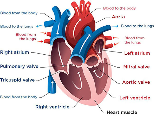
The heart has four chambers: two atria on top and two ventricles below. Normally the atrial septum separates the atria, the ventricular septum separates the ventricles, and two separate valves — the mitral valve on the left and the tricuspid valve on the right — control blood flow from atria to ventricles.
The centre of the heart, where all four chambers meet, is the atrioventricular junction or “crux”. In AVSD this region is deficient. Key features include a primum atrial septal defect (a hole low in the atrial septum), often an inlet ventricular septal defect, and a malformed valve apparatus. In complete AVSD a single common atrioventricular valve with five leaflets bridges both ventricles rather than two separate valves. The left ventricular outflow tract is also elongated (“goose-neck” appearance) and prone to narrowing. The lungs receive blood through the pulmonary arteries, which bear the brunt of the excess flow.
5. How the Disease Affects the Body
In a normal heart, oxygen-poor blood returns to the right side and is pumped to the lungs, while oxygen-rich blood returns to the left side and is pumped to the body — the two circuits are kept separate. In AVSD, the central hole and shared valve break down that separation.
Because pressure is higher on the left side of the heart, blood flows abnormally from the left chambers to the right chambers through the defect — a left-to-right shunt. This extra volume is pumped again and again to the lungs, causing pulmonary overcirculation. The lungs become congested, breathing becomes fast and laboured, and the heart works harder, leading to congestive heart failure in infants with complete AVSD.
The malformed common valve often fails to close properly, so blood leaks backward (AV valve regurgitation), adding to the volume overload. Over months, the small pulmonary blood vessels react to the high flow and pressure by thickening and narrowing. If this continues, pulmonary vascular disease develops and can become irreversible, eventually reversing the shunt (Eisenmenger syndrome) and causing low oxygen levels. This is why early surgical repair — usually before six months of age in complete AVSD — is so important.
6. Types and Classification
AVSD is classified mainly by how much of the septum is missing and how the AV valve is arranged:
- Complete AVSD (CAVSD): a common AV valve, a primum atrial septal defect, and an inlet ventricular septal defect. This is the most serious form and is strongly linked to Down syndrome. It is further sub-typed (Rastelli A, B, C) by how the bridging leaflets attach.
- Partial AVSD (PAVSD): a primum atrial septal defect with a cleft in the left AV (mitral) valve but two separate valve orifices and little or no ventricular-level shunt.
- Transitional / intermediate AVSD: features between the two — two valve orifices but a small, restrictive ventricular defect.
Balance is also described as balanced (both ventricles well developed) or unbalanced (one ventricle dominant, sometimes needing single-ventricle management).
7. Causes of the Disease
AVSD is a congenital defect — present from birth — caused by abnormal development of the endocardial cushions during the fourth to eighth week of pregnancy. In most cases the exact trigger is unknown and results from a combination of genetic and environmental factors.
- Chromosomal abnormalities, especially trisomy 21 (Down syndrome), are the single most important associated cause.
- Genetic syndromes such as heterotaxy (abnormal organ arrangement) and some single-gene mutations.
- Maternal factors during pregnancy, including poorly controlled diabetes, certain infections, and some medications or teratogen exposures.
It is important for families to understand that nothing a parent did or did not do usually “causes” AVSD; it reflects an early developmental event.
8. How the Disease Develops
During normal fetal development, two mounds of tissue called the endocardial cushions grow toward each other in the centre of the developing heart and fuse. This fusion closes the lower atrial septum, completes the top of the ventricular septum, and splits the single AV canal into two separate valves.
In AVSD, this fusion is incomplete. The septa remain deficient in the middle, and the valve tissue does not divide into distinct mitral and tricuspid valves, leaving a common valve or a cleft valve.
Before birth, the defect causes few problems because the lungs are not yet used for breathing and pressures on both sides of the heart are similar. After birth, pulmonary pressures fall over the first weeks of life, and the left-to-right shunt grows. Symptoms of heart failure therefore typically appear at six to eight weeks of age in complete AVSD, as blood floods the lungs. Partial forms may cause no symptoms for years. Untreated high pulmonary flow gradually damages the pulmonary arteries, setting up irreversible pulmonary vascular disease.
9. Risk Factors
- Down syndrome (trisomy 21) — the strongest risk factor; roughly two in five children with complete AVSD have Down syndrome.
- Other chromosomal or genetic syndromes, including heterotaxy syndromes.
- Maternal pre-gestational diabetes, especially if poorly controlled.
- Family history of congenital heart disease.
- Maternal exposure to certain teratogens, alcohol, or specific medications during early pregnancy.
- Maternal rubella or other infections in the first trimester.
Many affected children have no identifiable risk factor beyond the developmental event itself.
10. Genetic and Family-History Factors
Genetics play a central role in AVSD. The clearest link is trisomy 21, but AVSD also occurs in heterotaxy syndrome and with mutations in genes controlling heart-tube looping and septation (for example genes such as CRELD1 have been implicated in research).
There is a modestly increased risk when a parent or sibling has had a congenital heart defect. Families with a child who has AVSD, or a parent who was themselves born with it, are usually offered genetic counselling and fetal echocardiography in future pregnancies. When AVSD is diagnosed, testing for chromosomal conditions (such as a karyotype for Down syndrome) is routinely offered.
11. Who Is Most at Risk?
- Infants with Down syndrome, in whom AVSD is one of the most common heart defects.
- Babies of mothers with pre-existing diabetes.
- Children with heterotaxy or other complex congenital syndromes.
- Those with a family history of congenital heart disease.
Complete AVSD is diagnosed slightly more often in this high-risk population, whereas partial AVSD can appear in otherwise healthy children and may go undetected until later childhood or adulthood.
12. Prevalence and Epidemiology
Congenital heart defects affect roughly 1 in 100 (about 1%) of newborns worldwide, and AVSD accounts for a small but significant share — on the order of a few percent of all congenital heart lesions. It occurs in all populations and affects boys and girls at broadly similar rates.
Its epidemiology is dominated by its association with Down syndrome: a substantial minority of children with trisomy 21 have some form of AVSD, most often the complete type. Because prenatal screening now detects many cases before birth, diagnosis increasingly happens antenatally. These figures are approximate and vary between regions and reporting systems.
13. Signs and Symptoms
Symptoms depend heavily on the type and size of the defect. Complete AVSD usually causes noticeable problems in the first weeks to months of life, while partial AVSD may cause few or no symptoms until later.
Common signs and symptoms include:
- Fast or laboured breathing (tachypnoea), especially during feeding.
- Poor feeding and slow weight gain (“failure to thrive”).
- Sweating, particularly on the forehead during feeds.
- Easy tiring and reduced energy.
- Frequent chest infections.
- A heart murmur heard by the doctor.
- Pallor or, in advanced cases with reversed shunting, a bluish tinge (cyanosis).
In older children and adults with partial AVSD, symptoms may be limited to exertional breathlessness, palpitations, or fatigue. A doctor often first suspects the condition because of a murmur or abnormal heart findings during a routine examination.
14. Early-Stage Symptoms
In complete AVSD, the earliest clues appear at a few weeks of age as pulmonary pressures fall:
- Rapid, effortful breathing.
- Tiring or sweating during feeds, taking a long time to finish a bottle.
- Poor weight gain.
- A heart murmur detected at a newborn or infant check.
Partial AVSD may be silent early on, sometimes discovered only when a murmur is heard incidentally.
15. Advanced-Stage Symptoms
If AVSD is untreated, advanced signs reflect worsening heart failure and pulmonary vascular disease:
- Marked breathlessness, difficulty feeding, and failure to thrive.
- Recurrent respiratory infections.
- Enlarged liver and swelling from fluid retention.
- In late, irreversible pulmonary hypertension: cyanosis (blue lips and fingertips), reduced exercise tolerance, and features of Eisenmenger syndrome.
- In adults with long-standing partial AVSD: atrial arrhythmias and progressive breathlessness.
16. Symptoms in Women, Men and Older Adults
AVSD is chiefly diagnosed in infancy, so classic sex-based differences seen in adult heart disease do not apply in the same way. In babies, boys and girls present similarly.
Adults living with an unrepaired or repaired partial AVSD — including women considering pregnancy — may experience exertional breathlessness, palpitations from atrial arrhythmias, or fatigue. Older adults with previously undiagnosed partial AVSD can present for the first time with atrial fibrillation, a heart murmur, or unexplained breathlessness, sometimes mistaken for other heart or lung conditions.
17. Emergency Warning Signs
Seek emergency care for an infant who has:
- Severe difficulty breathing, grunting, or blue/grey lips, tongue or skin (cyanosis).
- Refusal to feed with lethargy or unresponsiveness.
- A racing heart with poor colour and cold, mottled skin.
These suggest heart failure or a cardiac emergency and require immediate assessment.
18. When to Seek Medical Help
Contact a doctor promptly if an infant tires or sweats during feeds, is not gaining weight, breathes rapidly, or has recurrent chest infections. Any heart murmur noted at a baby check should be evaluated. Adults with a known AVSD who develop new breathlessness, palpitations, or reduced exercise tolerance should see their cardiologist. When in doubt about a struggling or blue baby, seek urgent care.
19. Disease Stages, Grades and Severity
AVSD is not “staged” like a cancer; severity is judged by anatomy and its physiological effects:
- Anatomical type: partial, transitional, or complete (most severe).
- Balance: balanced versus unbalanced ventricles.
- Degree of AV valve regurgitation: mild, moderate, or severe leakage.
- Pulmonary artery pressure/resistance: normal early, then rising; the key marker of urgency and, if very high and fixed, of inoperability.
Severity guides both the timing and type of surgery.
20. Disease Progression
The natural course depends on the subtype. Complete AVSD progresses quickly: pulmonary overcirculation and heart failure develop within weeks to months, and — if untreated — pulmonary vascular disease can become irreversible within the first one to two years of life.
Partial AVSD progresses slowly; some patients remain well into adulthood, but chronic left AV valve leak and atrial-level shunting can gradually enlarge the heart, cause arrhythmias, and lead to breathlessness over years. Successful surgical repair halts this progression, though some patients need later reoperation for valve issues.
21. Possible Complications
- Congestive heart failure in infancy (complete AVSD).
- Pulmonary hypertension and, if untreated, irreversible pulmonary vascular disease / Eisenmenger syndrome.
- AV valve regurgitation (leaky valve), which may require repair or replacement.
- Residual or recurrent shunts after surgery.
- Left ventricular outflow tract obstruction (“goose-neck” narrowing).
- Heart rhythm problems, including heart block after surgery and later atrial arrhythmias.
- Infective endocarditis risk.
22. Related and Associated Medical Conditions
AVSD frequently coexists with:
- Down syndrome (trisomy 21) — the leading associated condition.
- Heterotaxy syndromes and other complex congenital heart defects, such as tetralogy of Fallot or double-outlet right ventricle.
- Patent ductus arteriosus and additional septal defects.
- Pulmonary hypertension as a secondary consequence.
Children with Down syndrome also commonly have other health issues (thyroid, gastrointestinal, and airway conditions) that influence overall care.
23. Screening and Early Detection
Screening occurs at several points:
- Prenatal ultrasound and fetal echocardiography, which can detect AVSD before birth — particularly when Down syndrome is suspected or there is a family history.
- Newborn pulse-oximetry screening, which can flag some heart defects.
- Routine newborn and infant examinations, where a murmur or signs of heart failure prompt referral.
Because of the strong Down-syndrome link, all babies with trisomy 21 are routinely evaluated with echocardiography.
24. How the Disease Is Diagnosed
Diagnosis follows a stepwise pathway that combines clinical assessment with imaging. It often begins when a heart murmur or signs of heart failure are noticed in an infant, or when a routine check raises concern in an older child.
The doctor takes a history (feeding, breathing, weight gain, family history, Down syndrome) and performs a physical examination. Key tests include:
- Echocardiography (echo): the cornerstone of diagnosis, showing the septal defect, the common or cleft valve, valve leakage, ventricular balance, and pulmonary pressures.
- Electrocardiogram (ECG): typically shows a characteristic left-axis deviation and conduction changes.
- Chest X-ray: may show an enlarged heart and increased lung blood flow.
- Cardiac MRI or CT: provides detailed anatomy when echo is inconclusive or in complex cases.
- Cardiac catheterization: reserved for measuring pulmonary artery pressure and resistance when operability is uncertain, especially in older infants who present late.
Prenatally, fetal echocardiography can establish the diagnosis before birth, allowing families and teams to plan. A definitive diagnosis integrates all these findings to define the type, severity, and best timing for treatment.
25. Physical Examination and Medical History
On examination, a doctor may find a heart murmur (from the shunt and valve leak), a prominent or heaving chest, rapid breathing, an enlarged liver, and poor weight gain. Signs of Down syndrome may be evident. The history focuses on feeding difficulty, sweating, breathlessness, growth, frequency of chest infections, family history of congenital heart disease, and pregnancy factors such as maternal diabetes. In older patients, the history covers exercise tolerance and palpitations.
26. Diagnostic Tests and Imaging
- Echocardiography — primary tool; defines anatomy, valve function, shunt direction, and pressures.
- ECG — left-axis deviation, first-degree heart block, and chamber enlargement patterns.
- Chest X-ray — cardiomegaly and pulmonary plethora.
- Cardiac MRI — precise anatomy, ventricular volumes, and shunt quantification without radiation.
- Cardiac CT — rapid detailed imaging of complex anatomy.
- Cardiac catheterization — measures pulmonary vascular resistance to confirm operability in selected cases.
27. Blood Tests, Biomarkers and Genetic Testing
Blood tests do not diagnose AVSD itself but support management. BNP or NT-proBNP may be raised in heart failure. Routine bloods (full blood count, kidney and liver function, electrolytes) guide medication and surgery. Oxygen saturation monitoring helps assess shunting. Genetic testing — a karyotype or microarray — is offered to confirm Down syndrome or other chromosomal conditions and to guide counselling. In selected families, targeted gene testing and genetic counselling are arranged.
28. Understanding Test Results
- Echo describes the type (partial/complete), size of the defect, severity of valve leak, ventricular balance, and estimated pulmonary pressure — the factors that decide surgical timing.
- A large left-to-right shunt with high pulmonary flow signals the need for early repair.
- High, fixed pulmonary vascular resistance on catheterization may indicate that surgery is unsafe.
- ECG left-axis deviation is a classic supportive finding.
Your cardiac team will explain what each result means for your child’s specific situation.
29. Differential Diagnosis
Conditions that can mimic or accompany AVSD include:
- Isolated ventricular septal defect or atrial septal defect.
- Patent ductus arteriosus and other left-to-right shunts.
- Cleft mitral valve without a septal defect.
- Complex lesions such as tetralogy of Fallot or single-ventricle physiology, which may coexist.
- Non-cardiac causes of an infant’s breathlessness and poor feeding (lung disease, sepsis).
Echocardiography reliably distinguishes AVSD from these.
30. Specialist and Multidisciplinary Evaluation
AVSD is managed by a paediatric cardiology and congenital cardiac surgery team. The wider team may include cardiac anaesthetists, intensivists, specialist nurses, geneticists, feeding and speech therapists, and — where Down syndrome is present — paediatricians coordinating associated conditions. For adults, a specialist in adult congenital heart disease (ACHD) leads care. You can explore experienced doctors and specialist hospitals through this site.
31. Treatment Goals
- Close the abnormal openings between the heart chambers.
- Reconstruct the AV valve(s) so they open and close normally, minimising leakage.
- Protect the lungs from high pressure and flow by repairing early enough.
- Relieve heart failure and restore normal growth.
- Preserve long-term heart function and quality of life, with lifelong follow-up.
32. When Is Treatment Required?
Complete AVSD almost always requires surgical repair in infancy, typically between three and six months of age, before pulmonary vascular disease develops. Symptoms of heart failure, poor growth, or rising pulmonary pressures may prompt earlier surgery.
Partial or transitional AVSD is repaired electively, often between one and three years of age or when significant valve leak or shunting is confirmed, even if symptoms are mild. Some adults with previously undiagnosed partial AVSD need surgery when the atrial shunt or valve leak causes heart enlargement or arrhythmia.
33. Active Monitoring and Watchful Waiting
Watchful waiting has a limited role in AVSD because the defect does not close on its own. Small, well-tolerated partial defects may be observed with regular echocardiograms while awaiting the optimal time for surgery. Before surgery, infants with complete AVSD are monitored closely for heart failure, growth, and feeding, and medical therapy is used to stabilise them. Continued observation without repair is not appropriate for significant complete AVSD.
34. Medications
Medication does not cure AVSD but controls symptoms and stabilises a baby before and after surgery:
- Diuretics (such as furosemide) to reduce fluid overload and ease breathing.
- ACE inhibitors to reduce the heart’s workload and the shunt.
- Digoxin in some cases to support heart function.
- Nutritional support, including higher-calorie feeds, to promote weight gain.
- Treatment of chest infections as needed.
These measures buy time and improve the child’s condition ahead of definitive surgical repair.
35. Minimally Invasive Treatments
The definitive treatment of AVSD is open-heart surgical repair, and there is currently no reliable device that can close a complete AVSD through a catheter alone. Some centres offer less-invasive surgical approaches, such as smaller incisions in selected older or partial cases. Minimally invasive and robotic congenital techniques continue to develop; you can read more under minimally invasive cardiac surgery. Most infants, however, are best served by conventional repair through a standard incision.
36. Catheter-Based and Endovascular Treatments
Cardiac catheterization in AVSD is used mainly for diagnosis — measuring pulmonary pressures and resistance — rather than for closing the defect, because the malformed valve and complex anatomy are not suited to a simple closure device. Catheter techniques may treat associated lesions (for example a patent ductus arteriosus) or address residual problems after surgery, and balloon procedures occasionally play a role in staged or unbalanced cases. Overall, catheter therapy complements rather than replaces surgery.
37. Surgical Treatment Options
Surgical repair is the mainstay of treatment for AVSD and offers excellent results in experienced hands. It is performed through open-heart surgery using the heart-lung (cardiopulmonary bypass) machine, usually in infancy.
The goals are to close the atrial and ventricular openings and to reconstruct the valves. Common techniques include:
- Complete repair (one- or two-patch technique): the surgeon uses one or two patches (of pericardium or synthetic material) to close the atrial and ventricular components of the defect and to separate the common valve into left and right valves. The cleft in the left AV valve is sutured to reduce leakage, and the valves are reconstructed to function as normally as possible. Care is taken to avoid the heart’s conduction tissue and prevent heart block.
- Partial AVSD repair: closure of the primum atrial defect with a patch and repair of the mitral (left AV) valve cleft.
- Staged approaches: in unbalanced AVSD or when a baby is too unwell, an initial pulmonary artery band may be placed to protect the lungs, with full repair or single-ventricle-type surgery later.
Repair is typically timed before six months of age in complete AVSD to protect the lungs. Explore congenital heart procedures and connect with specialist surgery centres for more detail. Most children need only one operation, though some later require valve reoperation.
38. Advanced and Emerging Treatments
Advances focus on improving valve repair and reducing reoperation. These include refined valve reconstruction techniques, better imaging with 3D echocardiography and cardiac MRI for surgical planning, and growing experience with less-invasive and robotic approaches in suitable patients. Research into bioengineered patches and valve materials that can grow with the child, and into optimising the timing of surgery, continues. For adults, transcatheter valve therapies are evolving for selected residual valve problems.
39. Treatment Options Compared
- Medication alone: controls symptoms temporarily but never corrects the defect; used only as a bridge to surgery.
- Pulmonary artery banding (palliation): protects the lungs when full repair must be delayed, but requires a later definitive operation.
- Complete surgical repair: the definitive, curative-intent option with excellent long-term outcomes; the standard of care for most patients.
- Catheter procedures: helpful for associated lesions or residual defects, not for primary AVSD closure.
For the great majority, timely surgical repair is clearly the best option.
40. How Doctors Choose the Right Treatment
Decisions depend on:
- Type of AVSD (partial, transitional, complete) and ventricular balance.
- Severity of valve leak and pulmonary pressures.
- Age, weight, and overall health, including Down syndrome and other conditions.
- Symptoms and growth.
- Presence of associated defects.
The team weighs the urgency of protecting the lungs against the technical benefits of operating on a slightly larger, stronger baby, then plans the timing and technique accordingly.
41. Benefits and Risks of Treatment
Benefits: correction of the shunt, relief of heart failure, protection of the lungs from irreversible damage, normal growth, and a good quality of life.
Risks of surgery include bleeding, infection, residual shunt or valve leak, heart block requiring a pacemaker, arrhythmias, left ventricular outflow obstruction, and the general risks of anaesthesia and bypass. Some patients need reoperation later, most often for the left AV valve. In experienced congenital centres, surgical risk is low and the benefits far outweigh the risks, particularly when repair is done at the right time.
42. What Happens If the Disease Is Left Untreated?
Untreated complete AVSD usually leads to progressive heart failure in infancy, poor growth, and repeated infections. Persistent high pulmonary blood flow causes irreversible pulmonary vascular disease within the first one to two years, eventually producing Eisenmenger syndrome — reversed shunting, chronic low oxygen levels, and a markedly shortened life expectancy. Untreated partial AVSD tends to cause gradual heart enlargement, valve leak, arrhythmias, and breathlessness over years. Timely surgery prevents these outcomes.
43. Treatment Success and Expected Outcomes
Outcomes after modern surgical repair are very good. Most children recover well, resume normal growth, and have few restrictions. Operative survival in experienced centres is high, and the majority enjoy a good long-term result.
The main issue affecting long-term outcome is the left AV valve: some patients develop leakage over time and a proportion need a further valve operation. Regular follow-up detects and manages these issues early. Overall, most patients live full, active lives after successful repair.
44. Prognosis and Long-Term Outlook
The long-term outlook for children who have their AVSD repaired at the right time is excellent, and the great majority reach adulthood with good heart function. Prognosis is best when surgery is performed before pulmonary vascular disease develops and when the AV valve can be repaired effectively.
Key factors influencing the outlook include the completeness of repair, the degree of any residual valve leak or narrowing, the presence of pulmonary hypertension, and any associated conditions such as Down syndrome. A minority of patients require reoperation in childhood or adulthood, most often for the left AV valve or for left ventricular outflow obstruction, and a small number develop arrhythmias or need a pacemaker.
Lifelong follow-up with a congenital or adult congenital cardiologist is essential, even for those who feel completely well, to monitor valve function and rhythm. With good care, most people who had AVSD as children can expect to work, exercise, and — with appropriate planning — have families of their own.
45. Recovery and Rehabilitation
After surgery, infants spend time in a paediatric cardiac intensive care unit followed by a ward stay, together often around one to two weeks depending on progress. Early recovery focuses on breathing support, pain control, feeding, and monitoring for rhythm problems. At home, families support feeding and gradual return of energy; most infants recover quickly and resume normal development within weeks. Older children and adults follow a graded return to activity. Wound care and follow-up echocardiograms are part of the recovery plan; you can learn more about cardiac treatments and aftercare through this site.
46. Follow-Up Tests and Long-Term Monitoring
Lifelong cardiology follow-up is recommended. Typical monitoring includes periodic echocardiograms to assess valve function and any residual shunt, ECGs to check rhythm and detect heart block, and clinical review of growth, exercise tolerance, and symptoms. Frequency is greater in the first year after surgery and then spaced out for stable patients. Adults transition to an adult congenital heart disease service.
47. Managing Recurrence or Disease Progression
AVSD does not “come back”, but residual or new problems can develop, chiefly left AV valve leak, outflow tract narrowing, or arrhythmias. Management includes ongoing surveillance, medication for symptoms or rhythm, and reoperation when valve leak or obstruction becomes significant. Early detection through regular follow-up allows timely, planned intervention rather than emergency treatment.
48. Living with the Disease
Most children and adults after successful repair live normal, active lives. Practical considerations include keeping up with follow-up appointments, maintaining good dental hygiene and endocarditis awareness, staying physically active within any advised limits, and informing new doctors of the cardiac history. Families of children with Down syndrome coordinate cardiac care with their other health needs. With good self-management and monitoring, the condition rarely limits daily life.
49. Diet and Nutrition Guidelines
Before surgery, infants with heart failure often need high-calorie, nutrient-dense feeding — sometimes via nasogastric tube — to support growth despite tiring during feeds. Dietitian input is valuable. After repair, most children eat and grow normally; a balanced, heart-healthy diet is encouraged. In older children and adults, standard cardiovascular nutrition applies: plenty of fruit, vegetables, and whole grains, moderate salt (especially if any heart failure), and healthy weight maintenance.
50. Exercise and Physical-Activity Guidelines
After successful repair with good heart function, most children and adults can be physically active and take part in sports, which is encouraged for overall health. Recommendations are individualised by a cardiologist based on valve function, pulmonary pressures, rhythm, and exercise-test results. Those with significant residual valve leak, pulmonary hypertension, or arrhythmias may need to limit high-intensity or competitive sport. Regular, moderate activity is beneficial for nearly everyone.
51. Medications, Activities and Habits to Avoid
- Avoid stopping prescribed medications (such as diuretics) without medical advice.
- Maintain good dental hygiene and discuss endocarditis prophylaxis with your cardiologist for certain procedures.
- Those with pulmonary hypertension should avoid activities and medications that worsen it and seek specialist advice before pregnancy or high-altitude travel.
- Adults should avoid smoking and excess alcohol.
- Always tell healthcare providers about the congenital heart history before any surgery or new medication.
52. Preventing the Disease or Reducing Its Risks
AVSD usually cannot be prevented because it forms early in pregnancy. Risk reduction focuses on healthy pregnancy practices: good control of maternal diabetes before and during pregnancy, avoiding alcohol and unnecessary medications, ensuring rubella immunity, taking recommended folic acid, and attending antenatal care. Genetic counselling helps families with a history of congenital heart disease or Down syndrome understand and plan for recurrence risk.
53. Pregnancy and the Disease
Women who had AVSD repaired in childhood usually tolerate pregnancy well if heart and valve function are good, but should have pre-pregnancy counselling and specialist care. Pregnancy raises the workload on the heart, so significant residual valve leak, arrhythmias, or — especially — pulmonary hypertension increase risk; pregnancy may be strongly advised against in Eisenmenger syndrome. There is also a modestly increased chance of congenital heart disease in the baby, so fetal echocardiography is offered. Care should be shared between a cardiologist and an obstetric team.
54. Disease in Children and Young Adults
AVSD is fundamentally a childhood diagnosis, most often repaired in infancy. Children with Down syndrome need coordinated care for associated conditions alongside their heart. After repair, most children grow and develop normally, attend school, and play sport. Young adults transition to adult congenital heart disease services and should understand their history, follow-up needs, and any activity or contraception/pregnancy considerations. Explore congenital heart disease resources for more.
55. Disease in Older Adults
Older adults may present with a previously undiagnosed partial AVSD, sometimes discovered when atrial fibrillation, a murmur, or breathlessness prompts investigation. Adults who had childhood repair can develop late complications — valve leak, arrhythmias, or heart failure — decades later. Surgery in older adults carries somewhat higher risk and is planned carefully, weighing pulmonary pressures and overall health. Ongoing specialist follow-up is important throughout adult life.
56. Emotional Health and Patient Support
A diagnosis of a heart defect in a baby is stressful for families, and a coexisting diagnosis of Down syndrome can add complexity. Emotional support, clear information, and connection with parent support groups and congenital heart charities help greatly. Older children and adults living with a repaired heart may have concerns about activity, appearance of scars, and the future; counselling and peer support are valuable. Care teams and specialist nurses can signpost resources.
57. Preparing for Your Specialist Appointment
- Bring your child’s medical records, previous echo/ECG reports, and a list of medications.
- Note symptoms: feeding, breathing, sweating, weight gain, energy.
- Write down family history of heart disease and any pregnancy factors.
- Prepare your questions in advance (see next section).
- Bring a support person and, for older patients, a summary of their surgical history.
Good preparation helps you get the most from the consultation. You can arrange an appointment via the contact page.
58. Questions to Ask Your Doctor
- What type of AVSD does my child have, and how severe is it?
- Is there associated Down syndrome or other conditions we should test for?
- How is the valve affected, and how much is it leaking?
- What are the pulmonary artery pressures, and are the lungs at risk?
- When should surgery be done, and why at that time?
- What surgical technique do you recommend, and what are its risks?
- What are the chances my child will need another operation later?
- What is the expected recovery and long-term outlook?
- What follow-up and monitoring will be needed for life?
- Are there activity, dental, or (later) pregnancy considerations we should know about?
59. Cost of Diagnosis and Treatment
Costs vary widely by country, hospital, and case complexity. The figures below are broad approximate ranges for complete AVSD repair including hospital stay; they should be confirmed with individual centres.
| Region | Approximate cost of AVSD repair (USD) |
|---|---|
| United States | $60,000 – $200,000+ |
| United Kingdom / Western Europe | $40,000 – $120,000 |
| Singapore | $30,000 – $70,000 |
| Thailand | $15,000 – $35,000 |
| Turkey | $12,000 – $30,000 |
| India | $6,000 – $15,000 |
Medical-tourism destinations such as India, Turkey, and Thailand often cost roughly 50–90% less than the US or UK for comparable, accredited care. Explore destinations and hospitals for current quotes.
60. Factors Affecting Treatment Cost
- Type and complexity of AVSD (complete versus partial; balanced versus unbalanced).
- Need for staged surgery or additional procedures.
- Length of ICU and hospital stay and any complications.
- Surgeon and hospital reputation and accreditation.
- Country and city, and local cost of living.
- Diagnostic tests, medications, and follow-up care.
- For international patients: travel, accommodation, and interpreter services.
61. Choosing the Right Specialist
Look for a paediatric/congenital cardiac surgeon and cardiologist with:
- Specific, high-volume experience in AVSD and AV valve repair.
- Good published or audited outcomes.
- Work within a dedicated congenital heart team and ICU.
- Clear communication and willingness to answer questions.
Browse experienced doctors and read about surgeon credentials before deciding.
62. Choosing the Right Hospital or Treatment Centre
Prioritise centres with:
- International accreditation (such as JCI) and strong safety records.
- A dedicated paediatric cardiac programme with congenital surgery, cardiac ICU, and specialist anaesthesia.
- High case volumes and good reported outcomes for congenital repair.
- Support services for international patients and long-term follow-up.
Compare accredited hospitals and surgery centres through this site.
63. Getting a Second Medical Opinion
A second opinion is reasonable and often reassuring before major heart surgery, especially for complex or unbalanced AVSD or when the timing or type of repair is uncertain. Share your echo and reports with another congenital specialist to confirm the diagnosis, plan, and timing. Reputable teams welcome second opinions. You can request one through our contact page.
64. Treatment Abroad and Medical-Travel Considerations
Many families travel abroad for high-quality, lower-cost congenital heart surgery. When planning:
- Choose a JCI-accredited centre with a proven congenital programme.
- Confirm the surgeon’s AVSD experience and outcomes.
- Plan for a stay long enough to cover surgery, ICU recovery, and early follow-up.
- Arrange records transfer, interpreter services, and a plan for follow-up back home.
- Clarify total costs and what is included.
Explore recommended destinations and connect with accredited hospitals to plan treatment abroad.
65. Frequently Asked Questions
Is AVSD serious? Complete AVSD is serious and needs surgery in infancy, but outcomes after timely repair are very good.
Can AVSD close on its own? No. Unlike some small holes in the heart, AVSD requires surgical repair.
Is AVSD always linked to Down syndrome? No, but it is strongly associated; many children with AVSD have Down syndrome, and many with Down syndrome have AVSD.
When is surgery usually done? Complete AVSD is typically repaired between three and six months of age; partial AVSD is repaired electively, often in early childhood.
Will my child need more than one operation? Most need only one, but some later require a further operation, usually on the left AV valve.
Can my child live a normal life afterwards? Yes — most children grow, play, and live full lives after successful repair, with lifelong cardiology follow-up.
Is the repair a cure? It corrects the defect and relieves symptoms; lifelong monitoring is still needed to watch the valve and rhythm.
66. Patient Stories and Treatment Experiences
The following are representative, anonymised illustrations, not specific individuals.
- Aisha, from Kenya: Her daughter was born with Down syndrome and complete AVSD and struggled to feed. After medication to stabilise her, she had patch repair at four months in an accredited centre abroad. She now feeds well and is growing steadily.
- Ravi, from India: Diagnosed with partial AVSD as a young adult after breathlessness and a murmur, he had elective repair of his atrial defect and mitral cleft. He returned to work and light sport within weeks.
- Marta, from Poland: Her son’s AVSD was detected before birth on fetal echo, allowing the family to plan surgery in the first months of life. She valued knowing the team in advance and having a clear follow-up plan.
67. Latest Research and Clinical Trials
Research continues to improve AVSD care without changing its surgical foundation. Active areas include refining AV valve repair to reduce late leakage and reoperation, optimising the timing of surgery, and using 3D echocardiography and cardiac MRI for better planning. Investigators are studying patch and valve materials that may grow with the child, strategies to prevent heart block, and outcomes in children with Down syndrome and pulmonary hypertension. Families interested in trials should ask their specialist centre; guidance evolves as evidence accumulates.
68. Related Diseases and Conditions
- Atrial septal defects
- Ventricular septal defects
- Patent ductus arteriosus
- Tetralogy of Fallot
- Single ventricle physiological conditions
- Congenital heart disease overview
69. Related Treatments and Procedures
- Congenital heart procedures
- Mitral valve procedures
- Minimally invasive cardiac surgery
- Hybrid cardiac procedures
- Other procedures
70. Medical Glossary
- Atrioventricular (AV) valve: valve between an atrium and a ventricle (mitral on the left, tricuspid on the right).
- Endocardial cushions: embryonic tissue that forms the central heart septum and AV valves.
- Primum atrial septal defect: a hole low in the atrial septum, seen in AVSD.
- Inlet ventricular septal defect: a hole in the inflow part of the ventricular septum.
- Common AV valve: a single shared valve in complete AVSD instead of two separate valves.
- Left-to-right shunt: abnormal flow of blood from the left to the right side of the heart.
- Pulmonary hypertension: raised blood pressure in the lung arteries.
- Eisenmenger syndrome: irreversible pulmonary vascular disease with reversed shunting and low oxygen.
- Cleft mitral valve: a split in the left AV valve leaflet causing leakage.
- Cardiopulmonary bypass: the heart-lung machine used during open-heart surgery.
- Heart block: a conduction problem that can slow the heartbeat, sometimes needing a pacemaker.
- Pulmonary artery banding: a palliative operation to reduce lung blood flow.
71. Medical Review, Editorial Policy and Last Updated Date
Last updated: 11 July 2026.
This article was prepared by the BestHeartSurgery.com editorial team and reviewed for general accuracy against established cardiology sources and guidelines. Our editorial policy emphasises clear, patient-friendly, evidence-aligned information, with periodic review and updates. This content is provided for education only and is not a substitute for professional medical advice, diagnosis, or treatment. Always consult a qualified cardiologist or congenital heart specialist about your individual situation.
72. Clinical Guidelines and Medical References
This information aligns with the general guidance of leading bodies, including the American College of Cardiology (ACC) and American Heart Association (AHA), the European Society of Cardiology (ESC) guidelines on adult congenital heart disease, the Society of Thoracic Surgeons (STS), the UK NHS, and the World Health Organization (WHO). It reflects widely accepted congenital cardiology and cardiac surgery knowledge. It does not cite specific studies; please consult these organisations and your care team for detailed, current guidance.
73. Book an Appointment or Request a Second Opinion
If your child or you have been diagnosed with an atrioventricular septal defect, our network can connect you with experienced congenital heart specialists and accredited hospitals worldwide.
- Book an appointment: /contact/
- Request a second opinion or ask a question: visit our contact page.
You can also explore doctors, hospitals, and destinations to plan your care with confidence.