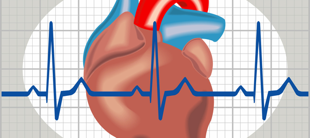
1. Disease Overview
Disorders of heart rhythm, known medically as cardiac arrhythmias, are conditions in which the heart beats too fast, too slowly, or irregularly. A healthy adult heart usually beats 60–100 times per minute at rest, following an orderly electrical sequence that keeps the upper chambers (atria) and lower chambers (ventricles) working in coordination. In an arrhythmia, that electrical signalling is disrupted — the impulse may start in the wrong place, travel along abnormal pathways, or be blocked entirely.
Many arrhythmias are harmless and occur in people with completely healthy hearts. An occasional skipped or extra beat is common and rarely dangerous. However, some rhythm disorders are serious. Atrial fibrillation can raise the risk of stroke; slow rhythms (bradyarrhythmias) can cause fainting; and fast, chaotic ventricular rhythms such as ventricular fibrillation can cause sudden cardiac arrest within minutes.
Arrhythmias range from a fluttering sensation in the chest to life-threatening emergencies. Because symptoms overlap and some dangerous rhythms cause few warning signs, accurate diagnosis with tools such as the ECG, Holter monitor, and electrophysiology (EP) study is essential. Modern treatment — from medication and catheter ablation to pacemakers and implantable defibrillators (ICDs) — allows most patients to live full, active lives.
2. Key Facts at a Glance
| Fact | Detail |
|---|---|
| Also known as | Cardiac arrhythmia, dysrhythmia, irregular heartbeat, heart rhythm disorder |
| Body system affected | Heart’s electrical conduction system |
| Common in | Older adults; those with heart disease, high blood pressure or thyroid disorders |
| Main types | Atrial fibrillation/flutter, SVT, bradyarrhythmia/heart block, ventricular tachycardia/fibrillation |
| Severity range | Harmless extra beats to life-threatening sudden cardiac arrest |
| Key treatments | Antiarrhythmic drugs, catheter ablation, pacemakers, ICDs, cardioversion |
| Outlook | Excellent for most; depends on type and underlying heart health |
3. Alternative Names and Medical Terminology
- Arrhythmia / dysrhythmia — the general term for abnormal rhythm.
- AF or AFib — atrial fibrillation.
- Aflutter — atrial flutter.
- SVT — supraventricular tachycardia.
- VT — ventricular tachycardia; VF — ventricular fibrillation.
- Bradycardia — slow heart rate; tachycardia — fast heart rate.
- Heart block / AV block — impaired conduction between atria and ventricles.
- Ectopic beats / PVCs / PACs — premature (extra) beats.
- Palpitations — the subjective awareness of the heartbeat.
4. Relevant Heart, Lung or Vascular Anatomy
The heart’s rhythm is controlled by its electrical conduction system. Each beat normally begins in the sinoatrial (SA) node, the natural pacemaker in the wall of the right atrium. The impulse spreads across both atria, causing them to contract, then reaches the atrioventricular (AV) node, which briefly delays the signal.
From the AV node the impulse travels down the bundle of His, splits into the right and left bundle branches, and finally reaches the Purkinje fibres that trigger coordinated ventricular contraction. Disruption at any point — the SA node, atrial tissue, AV node, or ventricular muscle — produces a specific type of arrhythmia. Structures such as the pulmonary veins (where they enter the left atrium) are frequent sources of the abnormal electrical activity that drives atrial fibrillation.
5. How the Disease Affects the Body
Arrhythmias interfere with the heart’s ability to pump blood efficiently. When the heart beats too fast, the ventricles have too little time to fill, so each beat ejects less blood; sustained fast rhythms can weaken the heart muscle over time (tachycardia-induced cardiomyopathy). When the heart beats too slowly, the body may not receive enough oxygen-rich blood, causing fatigue, dizziness, or fainting.
In atrial fibrillation, the atria quiver rather than contract, so blood can pool and form clots — particularly in the left atrial appendage. A clot that breaks free may travel to the brain and cause a stroke, one of the most serious consequences of arrhythmia. Irregular rhythms also reduce the coordinated “atrial kick” that normally boosts ventricular filling, lowering cardiac output by up to 20–30% in some patients.
The most dangerous arrhythmias, ventricular tachycardia and ventricular fibrillation, cause the ventricles to contract chaotically or too rapidly to pump effectively. Blood flow to the brain and vital organs stops, producing cardiac arrest and loss of consciousness within seconds. Without immediate defibrillation and CPR, this is rapidly fatal — which is why some rhythm disorders are medical emergencies.
6. Types and Classification
Arrhythmias are grouped by their origin and their effect on rate:
- Supraventricular arrhythmias (arising above the ventricles): atrial fibrillation, atrial flutter, atrial tachycardia, AV nodal re-entrant tachycardia (AVNRT), and Wolff-Parkinson-White syndrome.
- Ventricular arrhythmias (arising in the ventricles): premature ventricular contractions (PVCs), ventricular tachycardia, and ventricular fibrillation.
- Bradyarrhythmias (slow rhythms): sinus node dysfunction (sick sinus syndrome) and AV blocks (first-, second-, and third-degree/complete heart block).
- Premature (ectopic) beats: PACs and PVCs — usually benign.
By rate, arrhythmias are broadly classed as tachycardias (over 100 bpm) or bradycardias (under 60 bpm).
7. Causes of the Disease
- Coronary artery disease and prior heart attack (scar tissue disrupts conduction).
- High blood pressure causing atrial enlargement.
- Heart valve disease and heart failure.
- Thyroid disorders (both overactive and underactive).
- Electrolyte imbalances — low or high potassium, magnesium, or calcium.
- Stimulants — caffeine, alcohol, nicotine, recreational drugs, and some medications.
- Congenital heart conditions and inherited electrical disorders.
- Obstructive sleep apnoea, infection, fever, and severe stress.
Many arrhythmias, especially benign extra beats, occur with no identifiable cause.
8. How the Disease Develops
Arrhythmias arise through three main electrical mechanisms. In enhanced automaticity, heart cells fire abnormally on their own — for example, irritable tissue near the pulmonary veins triggering atrial fibrillation. In re-entry, an electrical impulse loops repeatedly around a circuit of tissue, sustaining a fast rhythm; this underlies atrial flutter, AVNRT, and many ventricular tachycardias. In triggered activity, abnormal after-depolarisations set off extra beats.
These mechanisms often develop against a background of structural change. Long-standing high blood pressure or valve disease stretches and scars the atria, creating the diseased tissue in which re-entry circuits form. A heart attack leaves ventricular scar that becomes a substrate for dangerous VT. Over years, atrial fibrillation itself remodels the atria — “AF begets AF” — so brief episodes become more frequent and eventually persistent.
Inherited channelopathies such as long QT or Brugada syndrome alter the ion channels that govern each cell’s electrical recovery, predisposing to sudden ventricular arrhythmias even in structurally normal hearts. Understanding the mechanism guides whether a patient benefits most from drugs, ablation, or a device.
9. Risk Factors
Modifiable:
- High blood pressure and untreated coronary disease
- Obesity and obstructive sleep apnoea
- Excess alcohol (“holiday heart”) and caffeine
- Smoking and stimulant drug use
- Uncontrolled diabetes and thyroid disease
- Electrolyte imbalance and physical or emotional stress
Non-modifiable:
- Increasing age
- Existing structural heart disease or prior heart surgery
- Congenital and inherited electrical disorders
- Family history of arrhythmia or sudden cardiac death
10. Genetic and Family-History Factors
Some arrhythmias run in families. Inherited channelopathies — including long QT syndrome, short QT syndrome, Brugada syndrome, and catecholaminergic polymorphic VT — arise from mutations in genes controlling cardiac ion channels and can cause sudden death in young, otherwise healthy people. Arrhythmogenic right ventricular cardiomyopathy (ARVC) and familial forms of atrial fibrillation also have a hereditary basis.
A family history of unexplained sudden death, fainting, or early pacemaker/ICD use should prompt evaluation. Genetic testing and family (cascade) screening are recommended when an inherited arrhythmia syndrome is suspected, so at-risk relatives can be identified and protected before a first dangerous event.
11. Who Is Most at Risk?
- Older adults — atrial fibrillation and conduction disease rise sharply with age.
- People with coronary artery disease, heart failure, or valve disease.
- Those with high blood pressure, diabetes, or obesity.
- Patients with sleep apnoea or thyroid disorders.
- Athletes (both benign slow rhythms and rare inherited risks).
- People with a family history of arrhythmia or sudden cardiac death.
- Heavy alcohol users and those on certain medications.
12. Prevalence and Epidemiology
Arrhythmias are extremely common. Occasional premature beats occur in most people at some point and are usually harmless. Atrial fibrillation is the most common sustained arrhythmia, affecting a growing number of people worldwide — its prevalence rises steeply after age 65 and is expected to increase as populations age and survive other heart conditions.
Bradyarrhythmias requiring pacemakers also become more frequent with age. Life-threatening ventricular arrhythmias are less common but account for a substantial share of sudden cardiac deaths globally. Exact figures vary by region and by how actively populations are screened, so published rates should be read as approximate.
13. Signs and Symptoms
Many arrhythmias cause no symptoms and are found incidentally. When symptoms do occur, they reflect either the abnormal rhythm itself or reduced blood flow. Common features include:
- Palpitations — awareness of skipped, extra, fluttering, or pounding beats.
- Dizziness or light-headedness.
- Shortness of breath, especially on exertion.
- Chest discomfort or tightness.
- Fatigue and reduced exercise tolerance.
- Fainting (syncope) or near-fainting.
- Anxiety accompanying the sensation of an irregular heartbeat.
The nature of symptoms offers clues: a sudden, regular racing pulse suggests SVT; an irregular “flip-flopping” beat suggests atrial fibrillation or ectopics; and unexplained fainting raises concern for a dangerous brady- or ventricular arrhythmia. Because symptoms come and go, patients may feel completely well between episodes, which can make diagnosis challenging.
14. Early-Stage Symptoms
Early or mild arrhythmias often announce themselves subtly:
- Occasional skipped beats or a brief “flutter” in the chest.
- Short, self-limiting episodes of a racing heart.
- Mild light-headedness that quickly passes.
- A sense that the heart “flip-flops” or pounds after caffeine, alcohol, or stress.
At this stage many people ignore the symptoms or attribute them to anxiety. Early atrial fibrillation may be paroxysmal — coming and going on its own — so it is easy to miss without monitoring.
15. Advanced-Stage Symptoms
As arrhythmias become more frequent, persistent, or occur against weakened heart muscle, symptoms intensify:
- Persistent breathlessness and marked fatigue.
- Reduced exercise capacity and swelling of the legs (if heart failure develops).
- Frequent or prolonged palpitations.
- Recurrent fainting or blackouts.
- Symptoms of stroke (from AF-related clots): sudden weakness, slurred speech, facial droop.
Sustained fast rhythms may cause tachycardia-induced cardiomyopathy, while advanced conduction disease can produce profound slowing and collapse.
16. Symptoms in Women, Men and Older Adults
- Women more often report palpitations, anxiety, and fatigue; certain SVTs (such as AVNRT) are more common in women, and some are influenced by hormonal cycles.
- Men have a higher incidence of atrial fibrillation and of ventricular arrhythmias linked to coronary disease.
- Older adults frequently have silent or atypical presentations — vague tiredness, confusion, falls, or breathlessness rather than classic palpitations. Silent atrial fibrillation is common in this group and may first present as a stroke, underscoring the value of pulse checks and screening.
17. Emergency Warning Signs
Call emergency services immediately for:
- Collapse or loss of consciousness with no or abnormal breathing (possible cardiac arrest).
- Sustained, very fast heartbeat with chest pain, severe breathlessness, or fainting.
- Signs of stroke — sudden face droop, arm weakness, or slurred speech.
These may indicate ventricular tachycardia/fibrillation or an AF-related stroke and require urgent defibrillation, CPR, or clot treatment.
18. When to Seek Medical Help
See a doctor promptly if you have recurrent palpitations, episodes of a racing or very slow pulse, unexplained dizziness or fainting, or breathlessness that is new or worsening. Anyone with a family history of sudden cardiac death or who faints during exercise should be evaluated. Do not wait for symptoms to become severe — early assessment can prevent stroke and other complications.
19. Disease Stages, Grades and Severity
Arrhythmias are not “staged” like cancer, but several are classified by pattern and severity. Atrial fibrillation is described as paroxysmal (self-terminating within 7 days), persistent (lasting longer or needing intervention), long-standing persistent (over a year), or permanent (accepted, no further rhythm-control attempts).
Heart block is graded first-degree (delayed conduction), second-degree (Mobitz I and II — intermittent blocked beats), and third-degree/complete (no conduction between atria and ventricles). Ventricular arrhythmias are classed by whether they are non-sustained or sustained and whether they cause haemodynamic collapse. Severity ultimately depends on symptoms, stroke risk, and the health of the underlying heart.
20. Disease Progression
Progression varies by type. Atrial fibrillation tends to advance over time: occasional paroxysmal episodes become more frequent, longer, and eventually persistent or permanent as the atria remodel. Conduction disease (heart block) is often gradual, progressing from mild delay to complete block that requires a pacemaker.
Some arrhythmias remain stable or even resolve when a trigger (thyroid disease, alcohol, medication) is corrected. Ventricular arrhythmias linked to progressive heart failure or expanding scar can worsen as the underlying condition advances. Early rhythm control and treatment of the underlying cause can slow or halt progression.
21. Possible Complications
- Stroke and systemic embolism — chiefly from atrial fibrillation and flutter.
- Heart failure — from sustained fast rhythms (tachycardia-induced cardiomyopathy) or loss of atrial kick.
- Sudden cardiac arrest — from ventricular tachycardia/fibrillation.
- Fainting and injury — from bradyarrhythmias or complete heart block.
- Reduced quality of life — recurrent symptoms, anxiety, and hospital visits.
- Cognitive decline — linked to AF and small strokes over time.
22. Related and Associated Medical Conditions
Arrhythmias often coexist with, and are driven by, other conditions:
- Coronary artery disease and prior heart attack.
- Heart failure and cardiomyopathies.
- Valvular heart disease, especially mitral valve disease.
- High blood pressure and diabetes.
- Thyroid dysfunction.
- Obstructive sleep apnoea.
- Chronic kidney disease and electrolyte disturbance.
Treating these associated conditions is central to controlling the rhythm disorder itself.
23. Screening and Early Detection
Because atrial fibrillation is frequently silent, opportunistic screening is encouraged — simple pulse checks during routine visits, blood-pressure monitors that detect irregular rhythm, and increasingly wearable devices and smartwatches with ECG functions. Targeted screening is recommended for people over 65 and those with risk factors.
For inherited arrhythmia syndromes, family (cascade) screening with ECG and genetic testing identifies at-risk relatives. Athletes in some countries undergo pre-participation ECG screening to detect conditions predisposing to sudden death. Any device-detected irregular rhythm should be confirmed with a formal ECG before treatment.
24. How the Disease Is Diagnosed
Diagnosing an arrhythmia centres on capturing the abnormal rhythm on a recording and identifying its cause. The process usually begins with a detailed history — the pattern, triggers, and duration of symptoms — and a physical examination that includes checking the pulse and heart sounds.
The cornerstone test is the 12-lead ECG, which records the heart’s electrical activity and can identify many arrhythmias, conduction blocks, and clues to underlying disease. Because episodes are often intermittent, ambulatory monitoring is frequently needed: a Holter monitor worn for 24–48 hours, event or patch monitors worn for weeks, or an implantable loop recorder placed under the skin for long-term surveillance of infrequent symptoms.
Further tests define the cause and consequences: an echocardiogram assesses heart structure and function; blood tests check thyroid function and electrolytes; and exercise testing can provoke exertion-related rhythms. When precise localisation is needed — for example before ablation — an invasive electrophysiology (EP) study maps the heart’s electrical pathways using catheters, deliberately triggering and pinpointing the abnormal circuit. Together these tools confirm the diagnosis and guide treatment.
25. Physical Examination and Medical History
The doctor asks about the character of the palpitations (fast or slow, regular or irregular, sudden or gradual), triggers, duration, associated fainting or chest pain, medications, caffeine and alcohol intake, and family history of arrhythmia or sudden death. Examination includes checking the pulse rate and regularity, blood pressure, heart sounds and murmurs, and signs of heart failure such as raised neck veins or leg swelling. Sometimes the doctor can feel an irregular pulse or find clues to an underlying valve or thyroid problem that points to the cause.
26. Diagnostic Tests and Imaging
- 12-lead ECG — the primary tool for identifying rhythm and conduction abnormalities.
- Holter / ambulatory monitors — 24-hour to multi-week recordings to catch intermittent arrhythmias.
- Event recorders and implantable loop recorders — for infrequent symptoms.
- Echocardiogram — evaluates heart structure, valves, and pumping function.
- Exercise stress test — provokes exertion-related arrhythmias and assesses coronary disease.
- Cardiac CT or MRI — assesses structure, scar, and cardiomyopathy.
- Electrophysiology (EP) study — invasive mapping to localise and characterise the arrhythmia before ablation.
27. Blood Tests, Biomarkers and Genetic Testing
- Thyroid function tests — overactive thyroid is a common, treatable cause of AF.
- Electrolytes (potassium, magnesium, calcium) — imbalances trigger arrhythmias.
- Full blood count — to check for anaemia or infection.
- Kidney and liver function — relevant to drug and anticoagulant dosing.
- Cardiac biomarkers (troponin) — if a heart attack is suspected.
- BNP/NT-proBNP — supports assessment of associated heart failure.
- Genetic testing — for suspected inherited channelopathies or cardiomyopathies.
28. Understanding Test Results
An ECG shows the rate, rhythm, and pattern of electrical activity; a cardiologist interprets features such as the absence of P-waves (atrial fibrillation), a “sawtooth” pattern (atrial flutter), a prolonged PR interval (first-degree block), or wide, fast complexes (ventricular tachycardia). Holter and loop-recorder reports quantify how often abnormal beats occur and whether they correlate with symptoms.
Echocardiography reports describe chamber size, valve function, and ejection fraction, which help judge stroke risk and treatment options. Normal blood tests do not exclude an arrhythmia — many patients have entirely normal labs. Results are always interpreted together with symptoms; your specialist will explain what each finding means for you.
29. Differential Diagnosis
Symptoms of arrhythmia overlap with other conditions that must be excluded:
- Anxiety and panic attacks (a very common cause of palpitations).
- Overactive thyroid and low blood sugar.
- Anaemia and dehydration.
- Panic-related hyperventilation.
- Coronary artery disease and heart failure.
- Structural heart disease and valve problems.
- Effects of caffeine, alcohol, stimulants, or medications.
Distinguishing a benign cause from a dangerous rhythm relies on capturing the event on an ECG or monitor.
30. Specialist and Multidisciplinary Evaluation
Complex arrhythmias are managed by a cardiologist and, for interventions, an electrophysiologist — a cardiologist specialising in heart rhythm. Care often involves a wider team: cardiac imaging specialists, cardiac surgeons (for surgical ablation or device complications), anticoagulation and stroke-prevention services, heart failure specialists, and endocrinologists for thyroid-driven arrhythmia. Genetic counsellors join the team when an inherited syndrome is suspected. This multidisciplinary approach ensures both the rhythm and its underlying cause are addressed. You can find experienced doctors and specialist hospitals through our directory.
31. Treatment Goals
- Relieve symptoms — palpitations, breathlessness, fatigue, and fainting.
- Control the heart rate and, where appropriate, restore normal rhythm.
- Prevent stroke in atrial fibrillation and flutter.
- Prevent sudden cardiac death in high-risk ventricular arrhythmias.
- Protect heart function and prevent tachycardia-induced heart failure.
- Treat the underlying cause (thyroid disease, valve disease, sleep apnoea).
- Improve quality of life and reduce hospital admissions.
32. When Is Treatment Required?
Not every arrhythmia needs treatment. Occasional benign ectopic beats in a healthy heart usually require only reassurance. Treatment is indicated when an arrhythmia causes troublesome symptoms, poses a stroke risk (atrial fibrillation/flutter), threatens life (ventricular tachycardia/fibrillation), causes fainting or very slow rates (heart block), or impairs heart function. The decision weighs symptom burden, arrhythmia type, and the health of the underlying heart, and is made jointly by patient and specialist.
33. Active Monitoring and Watchful Waiting
For low-risk arrhythmias — such as infrequent PVCs or well-tolerated paroxysmal SVT — a strategy of monitoring rather than immediate intervention is often appropriate. This includes lifestyle advice (reducing caffeine, alcohol, and stress), correcting triggers, and periodic ECG or Holter checks. Patients are taught to recognise warning signs and when to seek help. If symptoms worsen, episodes become more frequent, or heart function declines, treatment is stepped up. Watchful waiting avoids unnecessary drug side effects and procedures in people at low risk.
34. Medications
Several drug classes are used, chosen by arrhythmia type:
- Rate-control drugs — beta-blockers and certain calcium-channel blockers (diltiazem, verapamil) slow the heart rate in AF.
- Antiarrhythmic drugs — flecainide, propafenone, sotalol, amiodarone, and dronedarone help restore or maintain normal rhythm.
- Digoxin — used in selected patients for rate control.
- Anticoagulants — warfarin or direct oral anticoagulants (apixaban, rivaroxaban, dabigatran, edoxaban) prevent AF-related stroke.
- Adenosine — given intravenously to terminate certain SVTs.
- Atropine / isoprenaline — used acutely for severe slow rhythms.
Drug choice balances effectiveness against side effects, kidney and liver function, and other heart conditions. Take medications exactly as prescribed and never stop them abruptly.
35. Minimally Invasive Treatments
Beyond drugs, several less-invasive options exist. Electrical cardioversion delivers a synchronised shock under brief sedation to reset AF or flutter to normal rhythm. Vagal manoeuvres (bearing down, carotid massage) and adenosine can stop some SVT episodes. Catheter ablation, performed through veins in the groin without open surgery, is a mainstay minimally invasive cure for many arrhythmias. Device implantation — pacemakers and ICDs — is done through a small incision below the collarbone. These approaches offer effective treatment with short recovery times compared with open surgery.
36. Catheter-Based and Endovascular Treatments
Catheter ablation is a cornerstone of arrhythmia care. Thin catheters are guided through blood vessels to the heart, where the electrophysiologist maps the abnormal circuit and destroys the culprit tissue using radiofrequency energy (heat) or cryoablation (freezing). For atrial fibrillation, pulmonary vein isolation electrically isolates the veins that trigger the arrhythmia; newer pulsed-field ablation offers a tissue-selective alternative. Ablation is highly effective and often curative for SVT, atrial flutter, and many cases of AF and VT. For patients with AF who cannot take anticoagulants, left atrial appendage occlusion — a catheter-delivered device that seals off the clot-forming pouch — reduces stroke risk. Learn more about electrophysiological procedures.
37. Surgical Treatment Options
Most arrhythmias are treated without open surgery, but surgical options are important in selected cases. The Maze procedure creates a pattern of scar lines in the atria that channel electrical impulses along a controlled path, curing atrial fibrillation; it is often performed as a concomitant procedure during open-heart surgery for valve or bypass operations. A less-invasive mini-Maze uses small chest incisions and thoracoscopic access.
Device implantation is a surgical mainstay for rhythm disorders. Pacemakers treat slow rhythms and heart block by delivering timed electrical impulses; implantable cardioverter-defibrillators (ICDs) monitor for dangerous ventricular arrhythmias and deliver a shock to restore normal rhythm, preventing sudden death. Cardiac resynchronisation therapy (CRT) devices coordinate the ventricles in selected heart-failure patients. Surgical ablation of ventricular tachycardia and surgical left atrial appendage removal are options in complex cases.
Surgery is chosen when catheter approaches are unsuitable, when the patient is already undergoing open-heart surgery for another reason, or when device therapy is required. Our surgery and procedures sections describe these interventions in detail.
38. Advanced and Emerging Treatments
- Pulsed-field ablation (PFA) — a newer, tissue-selective energy source that may improve safety and speed of AF ablation.
- Leadless pacemakers — self-contained capsules placed directly in the heart, avoiding leads and pockets.
- Subcutaneous and extravascular ICDs — defibrillators without leads inside the heart.
- High-density 3D electroanatomical mapping and robotic/magnetic navigation for precise ablation.
- Stereotactic radioablation — investigational non-invasive radiotherapy for refractory VT.
- Conduction system pacing (His-bundle and left bundle branch pacing) for more physiological pacing.
- AI-based rhythm detection in wearables and implants.
39. Treatment Options Compared
- Medications — non-invasive and widely available, but may cause side effects and are often not curative; suit many patients as first-line therapy.
- Cardioversion — quickly restores rhythm but is not a lasting cure on its own.
- Catheter ablation — often curative for SVT, flutter, and effective for AF and VT; higher upfront cost and small procedural risk, but lasting benefit.
- Pacemakers — definitive for slow rhythms and heart block.
- ICDs — life-saving for high-risk ventricular arrhythmias.
- Surgical Maze — durable AF cure, usually combined with other heart surgery.
The best option depends on arrhythmia type, symptoms, and patient preference.
40. How Doctors Choose the Right Treatment
Treatment selection considers the type and mechanism of the arrhythmia, its symptom burden and stroke or sudden-death risk, the health of the underlying heart (structure and ejection fraction), the patient’s age, other conditions, and preferences, and the likely durability of each option. For example, a young patient with SVT may be offered curative ablation; an older patient with AF may start with rate control and anticoagulation; and a heart-attack survivor with poor ejection fraction may need an ICD to prevent sudden death. Shared decision-making ensures the plan fits the individual.
41. Benefits and Risks of Treatment
Benefits: relief of symptoms, restored normal rhythm, prevention of stroke and sudden cardiac death, protection of heart function, and improved quality of life. Many procedures are curative.
Risks: antiarrhythmic drugs can cause side effects and, rarely, provoke new arrhythmias; anticoagulants increase bleeding risk. Catheter ablation carries small risks of bleeding, vascular injury, and, rarely, damage to nearby structures. Device implantation can be complicated by infection, lead problems, or bleeding. Overall, serious complications are uncommon in experienced centres, and the benefits usually outweigh the risks for appropriately selected patients.
42. What Happens If the Disease Is Left Untreated?
Untreated arrhythmias carry variable but potentially serious consequences. Untreated atrial fibrillation substantially raises the risk of stroke and can weaken the heart over time. Persistent fast rhythms may cause tachycardia-induced cardiomyopathy and heart failure. Untreated complete heart block can cause fainting, injury, and death. Most dangerously, untreated ventricular tachycardia or fibrillation leads to sudden cardiac arrest. Some benign arrhythmias, by contrast, cause no harm if left alone — which is why accurate diagnosis matters so much.
43. Treatment Success and Expected Outcomes
Outcomes are generally very good and depend on the arrhythmia type. Catheter ablation cures the large majority of patients with SVT and typical atrial flutter, and significantly reduces or eliminates episodes in most AF patients, though some need a repeat procedure. Pacemakers reliably abolish symptoms of slow rhythms. ICDs effectively terminate dangerous ventricular arrhythmias and prevent sudden death in high-risk patients. Drug therapy controls symptoms in many people. Success is measured not only by rhythm control but by fewer symptoms, prevented strokes, and restored quality of life. Reported success rates are approximate and vary with patient and centre.
44. Prognosis and Long-Term Outlook
For most people, the long-term outlook is excellent. Many arrhythmias are benign and require only reassurance or simple treatment. With appropriate stroke prevention, people with atrial fibrillation can live full lives, and the risk of AF-related stroke falls markedly with anticoagulation. Curative ablation gives many SVT and flutter patients a normal life expectancy free of the arrhythmia.
Prognosis is more guarded when arrhythmias arise from serious underlying heart disease — advanced heart failure, extensive coronary disease, or inherited cardiomyopathies — because the outlook is then linked to the underlying condition as much as the rhythm itself. Even here, modern devices such as ICDs and CRT, combined with optimal medical therapy, substantially improve survival and quality of life.
Key factors shaping prognosis include the type of arrhythmia, the health of the heart muscle, how promptly stroke and sudden-death risks are addressed, and adherence to treatment. Regular follow-up and control of underlying conditions such as blood pressure, thyroid disease, and sleep apnoea further improve long-term outcomes. With today’s therapies, most patients can expect good symptom control and a favourable outlook.
45. Recovery and Rehabilitation
Recovery depends on the treatment. After catheter ablation, most patients go home the same or next day and resume normal activities within about a week, avoiding heavy lifting briefly to protect the groin puncture sites. After pacemaker or ICD implantation, patients typically stay one night and avoid raising the arm above shoulder height and heavy lifting on the device side for a few weeks while the leads settle. Wound care, gradual return to activity, and attending follow-up device checks are important. Cardiac rehabilitation and lifestyle support help those with associated heart disease recover confidence and fitness.
46. Follow-Up Tests and Long-Term Monitoring
Long-term follow-up includes periodic ECGs and Holter monitoring to confirm rhythm control, and echocardiography to check heart function. Patients on anticoagulants have bleeding and (for warfarin) INR monitoring. Pacemakers and ICDs are checked regularly in clinic or by remote monitoring, which transmits device data and alerts the team to problems or arrhythmia recurrences. Follow-up also reviews medication tolerance and control of underlying conditions such as blood pressure and thyroid function.
47. Managing Recurrence or Disease Progression
Arrhythmias can recur — particularly atrial fibrillation, which may return after ablation or drug therapy. Management includes repeat ablation, adjusting or changing antiarrhythmic drugs, reinforcing lifestyle measures (weight loss, reduced alcohol, treating sleep apnoea, blood-pressure control), and ensuring ongoing stroke prevention. For progressive conduction disease or worsening heart failure, therapy may be escalated to a pacemaker, ICD, or CRT device. Regular review allows treatment to be tailored as the condition evolves.
48. Living with the Disease
Most people with a rhythm disorder live active, normal lives. Key habits help: taking medications reliably, monitoring your pulse, recognising and recording symptoms, and attending follow-up. Learn your triggers — caffeine, alcohol, poor sleep, and stress — and manage them. If you have a device, carry an identification card, understand which environments to avoid (strong magnetic fields), and keep device checks up to date. Many patients use wearables to track their rhythm. With good self-management, arrhythmias rarely need to dominate daily life.
49. Diet and Nutrition Guidelines
- Follow a heart-healthy diet rich in vegetables, fruit, whole grains, and lean protein.
- Limit alcohol — even modest amounts can trigger AF (“holiday heart”).
- Moderate caffeine; note whether it provokes your palpitations.
- Maintain adequate potassium and magnesium through a balanced diet; correct deficiencies as advised.
- Reduce salt to help control blood pressure.
- If taking warfarin, keep vitamin-K (green leafy vegetable) intake consistent.
- Achieve and maintain a healthy weight — obesity worsens AF.
50. Exercise and Physical-Activity Guidelines
Regular moderate exercise benefits most people with arrhythmias and improves overall heart health. Aim for the generally recommended 150 minutes per week of moderate activity, tailored to your condition. However, some arrhythmias — particularly certain inherited channelopathies and those causing exertional fainting — require activity restrictions and specialist clearance before competitive or intense sport. If you have a device or exertion-triggered symptoms, agree an exercise prescription with your cardiologist. Stop and seek advice if exercise brings on palpitations, dizziness, chest pain, or fainting.
51. Medications, Activities and Habits to Avoid
- Avoid excess alcohol and stimulant drugs, which trigger arrhythmias.
- Limit high caffeine and energy drinks if they provoke symptoms.
- Be cautious with decongestants and some over-the-counter cold remedies — always check with your pharmacist.
- Certain drugs prolong the QT interval and can be dangerous in susceptible people; tell every prescriber about your arrhythmia.
- Do not stop antiarrhythmics or anticoagulants abruptly.
- Treat sleep apnoea and avoid smoking.
- ICD/pacemaker patients should avoid strong magnetic and electromagnetic fields as advised.
52. Preventing the Disease or Reducing Its Risks
Many arrhythmias, especially atrial fibrillation, can be prevented or delayed by controlling risk factors:
- Keep blood pressure well controlled.
- Maintain a healthy weight and stay physically active.
- Limit alcohol and avoid smoking and stimulant drugs.
- Diagnose and treat sleep apnoea.
- Manage diabetes and thyroid disease.
- Attend regular checks if you have heart disease or a family history.
These measures also reduce recurrence after treatment.
53. Pregnancy and the Disease
Pregnancy increases heart rate and blood volume and can unmask or worsen arrhythmias; benign palpitations and some SVTs are relatively common. Most are manageable, but care requires a cardiologist-obstetric team. Some antiarrhythmic and anticoagulant drugs are not safe in pregnancy, so medications may need adjusting before conception and during pregnancy. Women with inherited arrhythmia syndromes or existing devices should have pre-pregnancy counselling. Cardioversion and, if essential, ablation can be performed safely in selected cases. Most women with arrhythmias have successful pregnancies with appropriate monitoring.
54. Disease in Children and Young Adults
In children and young adults, arrhythmias are often SVT (including Wolff-Parkinson-White syndrome) or arise from congenital heart disease or inherited channelopathies. Sudden fainting during exercise, or a family history of sudden death, needs urgent evaluation. Many childhood SVTs are curable by catheter ablation once the child is old enough. Young athletes may undergo pre-participation screening. Early diagnosis of inherited syndromes protects both the child and at-risk relatives.
55. Disease in Older Adults
Arrhythmias are most common in older adults. Atrial fibrillation and conduction disease (needing pacemakers) rise sharply with age, and AF is often silent, first presenting as a stroke. Treatment must balance stroke prevention against bleeding risk, since older patients are more prone to both. Multiple medications, kidney impairment, and frailty complicate drug choice. Nonetheless, ablation, pacemakers, and ICDs are safely used in carefully selected older patients, and stroke prevention is especially valuable in this group.
56. Emotional Health and Patient Support
Living with an unpredictable heartbeat can cause anxiety, fear, and low mood, and palpitations and panic can reinforce each other. Fear of shocks is common in ICD patients. Support helps: education about the condition, relaxation and stress-management techniques, cardiac rehabilitation, and peer or patient support groups. Some people benefit from counselling or psychological therapy. Reassurance that most arrhythmias are manageable, and knowing when symptoms are and are not dangerous, greatly reduces distress.
57. Preparing for Your Specialist Appointment
- Keep a symptom diary — when episodes occur, how long they last, and any triggers.
- Note your pulse rate during episodes if you can, or use a wearable/ECG app.
- Bring a list of all medications, doses, and supplements.
- List your medical history and any family history of arrhythmia or sudden death.
- Bring previous ECGs, monitor reports, or device cards.
- Write down your questions in advance.
- Consider bringing a family member to help remember information.
58. Questions to Ask Your Doctor
- What type of arrhythmia do I have, and is it dangerous?
- What is causing it, and can the cause be treated?
- Am I at risk of stroke, and do I need blood-thinning medication?
- What are my treatment options — medication, ablation, or a device?
- What are the benefits and risks of each option?
- Is catheter ablation likely to cure my arrhythmia?
- Will I need a pacemaker or defibrillator?
- What activities, foods, or medications should I avoid?
- What warning signs mean I should seek emergency help?
- How often will I need follow-up and monitoring?
59. Cost of Diagnosis and Treatment
Costs vary widely by country, hospital, and complexity. The figures below are approximate US-dollar ranges for orientation only.
| Treatment | US / UK / Western Europe | India / Turkey / Thailand |
|---|---|---|
| ECG / Holter / basic workup | $200 – $2,000 | $50 – $500 |
| Electrophysiology study | $8,000 – $20,000 | $1,500 – $4,000 |
| Catheter ablation (SVT/AF) | $20,000 – $60,000 | $4,000 – $12,000 |
| Pacemaker implantation | $20,000 – $50,000 | $3,500 – $10,000 |
| ICD implantation | $30,000 – $100,000 | $8,000 – $20,000 |
Medical-tourism destinations such as India, Turkey, Thailand, and Singapore often cost 50–90% less than the US or UK for comparable quality care. Explore destinations for details.
60. Factors Affecting Treatment Cost
- Type of procedure — device implants and complex AF/VT ablation cost more than simple SVT ablation.
- Device type — ICDs and CRT devices are far costlier than basic pacemakers.
- Hospital and country, and level of accreditation.
- Surgeon/electrophysiologist experience and hospital volume.
- Length of hospital stay and use of intensive care.
- Pre-operative tests and imaging.
- Follow-up, device monitoring, and medications.
- Insurance coverage and, for international patients, travel and accommodation.
61. Choosing the Right Specialist
Look for a board-certified cardiologist, and for procedures a specialist electrophysiologist with specific training in ablation and device implantation. Consider the specialist’s procedure volume and outcomes, experience with your particular arrhythmia, and access to modern mapping and ablation technology. Good communication, willingness to explain options, and support for shared decision-making matter. Our doctors directory lists experienced cardiac specialists.
62. Choosing the Right Hospital or Treatment Centre
Choose a centre with a dedicated cardiac electrophysiology (EP) lab, a high volume of ablation and device procedures, and strong published outcomes. International patients should look for JCI or equivalent accreditation, 24/7 cardiac emergency cover, and multidisciplinary teams including cardiac surgery back-up. Availability of advanced technology — 3D mapping, cryo- and pulsed-field ablation, and remote device monitoring — is a marker of quality. Browse accredited hospitals on our site.
63. Getting a Second Medical Opinion
A second opinion is worthwhile before major decisions — such as choosing between long-term drugs, ablation, or an implanted device, or when a diagnosis is uncertain. Another specialist may confirm the diagnosis, suggest alternative options, or offer newer techniques. Bring your ECGs, monitor reports, echocardiogram, and device information. Reputable specialists welcome second opinions. You can request a second opinion through our service.
64. Treatment Abroad and Medical-Travel Considerations
Many patients travel abroad for arrhythmia care to access high-quality treatment at lower cost and shorter waiting times. Destinations such as India, Turkey, Thailand, and Singapore offer JCI-accredited hospitals and experienced electrophysiologists. Plan carefully: verify credentials and outcomes, arrange pre-travel records and consultations, confirm device follow-up and remote monitoring in your home country, and allow enough recovery time before flying, especially after device implantation. Consider language support, aftercare, and how anticoagulation will be managed during travel. Our destinations section can help you plan.
65. Frequently Asked Questions
Are palpitations always dangerous? No. Most palpitations are harmless, especially brief skipped beats. They warrant assessment if frequent, prolonged, or accompanied by fainting, chest pain, or breathlessness.
Can atrial fibrillation be cured? AF can often be controlled and sometimes effectively eliminated with catheter ablation, though it may recur. Stroke prevention remains important regardless.
Will I need a pacemaker? Only if you have a slow heart rhythm or heart block causing symptoms. Most arrhythmia patients do not need one.
Is catheter ablation safe? Yes — in experienced centres it is a well-established, low-risk procedure, and it is often curative for SVT and atrial flutter.
Why do I need blood thinners for AF? AF allows clots to form in the heart that can cause stroke. Anticoagulants greatly reduce this risk.
Can I exercise with an arrhythmia? Usually yes, and exercise is beneficial — but some rhythm disorders need specialist clearance first.
Can stress or caffeine trigger arrhythmias? They can trigger palpitations and some arrhythmias in susceptible people; identifying and moderating triggers helps.
Are smartwatches reliable for detecting arrhythmias? They can flag irregular rhythms and prompt evaluation, but any alert should be confirmed with a formal ECG.
66. Patient Stories and Treatment Experiences
The following are representative, anonymised examples for illustration.
Ravi, India — In his 30s, Ravi had sudden episodes of a racing heart for years. An EP study diagnosed SVT, and a single catheter ablation cured it. He has been episode-free since and returned to playing cricket.
Margaret, United Kingdom — Diagnosed with atrial fibrillation at 68 after a routine pulse check, Margaret started an anticoagulant and rate-control medication. Her stroke risk fell substantially, and a later ablation reduced her episodes.
Ahmed, UAE — After surviving a heart attack, Ahmed had a weakened heart and dangerous ventricular arrhythmias. An ICD was implanted; it later delivered a life-saving shock, and he continues to live actively with regular remote monitoring.
67. Latest Research and Clinical Trials
Arrhythmia care is advancing rapidly. Pulsed-field ablation is a major development, offering tissue-selective energy that may make AF ablation safer and faster. Leadless pacemakers and extravascular/subcutaneous ICDs reduce lead-related complications. Conduction-system pacing aims to pace the heart more naturally. Research also focuses on earlier AF detection through wearables and AI, better stroke-prevention strategies, and stereotactic radioablation for otherwise untreatable VT. Trials continue to refine when to prefer rhythm control over rate control. Ask your specialist whether any trials are suitable for you.
68. Related Diseases and Conditions
- Coronary Artery Disease — a major cause of ventricular arrhythmias.
- Heart Failure — closely linked with AF and dangerous rhythms.
- Valvular Heart Disease — especially mitral disease driving AF.
- Infective Endocarditis — can involve the conduction system.
- Dilated Cardiomyopathy — associated with arrhythmias and sudden death.
- Ischemic Cardiomyopathy — scar-related ventricular arrhythmias.
69. Related Treatments and Procedures
- Electrophysiological Procedures — EP studies, ablation, and device implantation.
- Minimally Invasive Cardiac Surgery — including mini-Maze for AF.
- Hybrid Cardiac Procedures — combined catheter and surgical AF treatment.
- Coronary Artery Bypass Grafting — often combined with surgical ablation.
- Other Procedures — additional device and rhythm interventions.
70. Medical Glossary
- Arrhythmia — any abnormal heart rhythm.
- Atrial fibrillation (AF) — rapid, irregular quivering of the atria.
- Atrial flutter — a fast, organised atrial rhythm with a sawtooth ECG pattern.
- Bradycardia — a slow heart rate (under 60 bpm).
- Tachycardia — a fast heart rate (over 100 bpm).
- SVT — supraventricular tachycardia, a fast rhythm from above the ventricles.
- Ventricular fibrillation (VF) — chaotic ventricular activity causing cardiac arrest.
- Heart block — impaired conduction between atria and ventricles.
- Ablation — targeted destruction of tissue causing an arrhythmia.
- Cardioversion — restoring normal rhythm with a synchronised shock.
- Pacemaker — a device that treats slow heart rhythms.
- ICD — implantable cardioverter-defibrillator that treats dangerous fast rhythms.
- Electrophysiology (EP) study — invasive mapping of the heart’s electrical system.
- Anticoagulant — a blood-thinning drug that prevents clots and stroke.
- Ejection fraction — the percentage of blood pumped out of the ventricle with each beat.
71. Medical Review, Editorial Policy and Last Updated Date
Last updated: 11 July 2026.
This article is reviewed for accuracy against guidance from bodies including the American College of Cardiology (ACC), American Heart Association (AHA), European Society of Cardiology (ESC), Heart Rhythm Society (HRS), and NHS. Our editorial policy emphasises evidence-based, patient-friendly information, using ranges and qualifiers rather than invented statistics.
Disclaimer: This content is for educational purposes only and is not a substitute for professional medical advice, diagnosis, or treatment. Always consult a qualified cardiologist or healthcare provider about your individual condition.
72. Clinical Guidelines and Medical References
General guidance for this topic is drawn from reputable sources, including:
- American College of Cardiology (ACC) / American Heart Association (AHA) / Heart Rhythm Society (HRS) guidelines on atrial fibrillation and management of ventricular arrhythmias.
- European Society of Cardiology (ESC) guidelines on atrial fibrillation, supraventricular tachycardia, and cardiac pacing.
- NHS and World Health Organization patient information on arrhythmias and stroke prevention.
- Standard cardiology and electrophysiology textbooks.
These are cited as general references; always rely on current guidelines and your treating specialist for clinical decisions.
73. Book an Appointment or Request a Second Opinion
If you or a loved one has a heart rhythm disorder, expert help is available. Our network connects you with experienced electrophysiologists and accredited hospitals worldwide for diagnosis, ablation, and device therapy.
- Book an appointment: Get started here
- Request a second opinion or ask a question: Contact us
Take the first step toward controlling your heart rhythm and protecting your long-term health.