1. Disease Overview
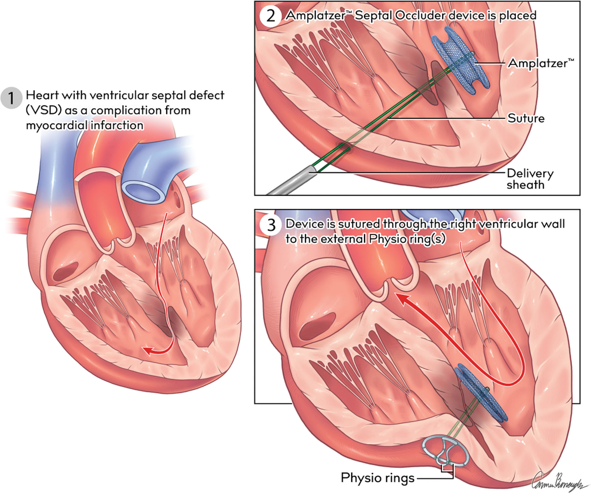
Post-infarction ventricular septal defect (post-MI VSD), also called ventricular septal rupture (VSR), is a tear in the muscular wall that separates the heart’s two lower chambers (the ventricles). It is a mechanical complication of acute myocardial infarction (heart attack): when a segment of the heart muscle dies from lack of blood flow, the weakened, necrotic septum can rupture, creating an abnormal hole between the high-pressure left ventricle and the lower-pressure right ventricle.
Through this hole, oxygen-rich blood is shunted from the left ventricle into the right ventricle (a left-to-right shunt). This overloads the right heart and lungs, reduces the blood the left ventricle can pump forward to the body, and frequently precipitates cardiogenic shock — a state in which the heart cannot maintain adequate circulation.
Post-MI VSD is rare but among the most lethal complications of a heart attack. It usually appears within the first days to two weeks after an MI, heralded by sudden deterioration and a new loud heart murmur. Because untreated rupture carries very high short-term mortality, it is a surgical emergency. Modern management combines rapid echocardiographic diagnosis, aggressive stabilization (including mechanical circulatory support), and definitive closure by surgical patch repair or, in selected patients, percutaneous (catheter-based) device closure. Outcomes have improved with faster reperfusion, but this remains a high-risk, time-critical condition requiring an experienced heart team.
2. Key Facts at a Glance
| Fact | Detail |
|---|---|
| Also known as | Ventricular septal rupture (VSR), post-MI VSD, ischemic ventricular septal rupture, myocardial septal rupture |
| Body system affected | Cardiovascular system — the interventricular septum, both ventricles, and pulmonary circulation |
| Common in | Older adults after a large, often first, myocardial infarction; more frequent in women, the elderly, and those without prior collateral circulation |
| Severity range | Serious to life-threatening — a surgical emergency; frequently causes cardiogenic shock |
| Key treatments | Emergency stabilization (IABP, mechanical support), surgical patch repair, percutaneous device closure, coronary revascularization |
| Outlook | Guarded; strongly dependent on shock status, defect size, and timing of repair, but markedly better than untreated rupture |
3. Alternative Names and Medical Terminology
- Ventricular septal rupture (VSR) — the most common formal term
- Post-infarction VSD / post-MI VSD
- Ischemic ventricular septal defect
- Acquired ventricular septal defect (to distinguish it from congenital VSD)
- Myocardial rupture, septal type — VSR is one of the “mechanical complications” of MI, alongside free-wall rupture and papillary muscle rupture
- Left-to-right shunt — describes the abnormal blood flow through the defect
Note that a congenital VSD is a hole present from birth, whereas post-MI VSD is acquired as a consequence of a heart attack; the two share anatomy but differ fundamentally in cause and management.
4. Relevant Heart, Lung or Vascular Anatomy
The heart has four chambers. The two lower pumping chambers — the left ventricle (LV) and right ventricle (RV) — are separated by the interventricular septum, a muscular wall with a small membranous portion near the top.
- The left ventricle generates high pressure to pump oxygenated blood to the body.
- The right ventricle operates at much lower pressure, sending blood to the lungs.
- The septum receives blood supply mainly from the left anterior descending (LAD) artery (which feeds the anterior and apical septum via septal perforators) and the posterior descending artery (PDA) from the right coronary or circumflex (feeding the inferior/basal septum).
When a coronary artery occlusion kills septal muscle, the dead tissue can tear. Anterior MIs (LAD territory) typically cause apical/anterior septal ruptures; inferior MIs cause basal/inferior-posterior ruptures, which are anatomically more complex and often harder to repair.
5. How the Disease Affects the Body
Post-MI VSD disrupts the heart’s normal one-way circulation. Because pressure in the left ventricle is far higher than in the right ventricle, blood is forced backward through the defect from left to right with each heartbeat. This left-to-right shunt has several damaging consequences that cascade quickly.
First, blood that should have gone forward to the body instead recirculates uselessly through the lungs, so systemic (forward) cardiac output falls and vital organs receive less oxygenated blood, driving cardiogenic shock.
Second, the right ventricle is suddenly overloaded with volume and pressure it is not built to handle. It dilates and can fail, causing right-heart failure with congestion of the veins, liver, and legs.
Third, the extra blood flooding the lungs raises pulmonary pressure, producing pulmonary congestion and edema (fluid on the lungs) and worsening breathlessness and low blood oxygen.
All of this occurs in a heart already weakened by the infarction, so contractile reserve is limited. The size of the shunt (the Qp:Qs ratio) largely determines severity: small defects may be tolerated, while large ones overwhelm the circulation within hours. The combination of a failing left ventricle, an overloaded right ventricle, and flooded lungs explains why untreated rupture is so often rapidly fatal.
6. Types and Classification
Post-MI VSDs are classified by location and morphology:
- Anterior (apical) VSR — follows anterior MI (LAD occlusion); the defect is usually a relatively simple, direct tract near the apex and is generally more accessible to repair.
- Inferior/posterior (basal) VSR — follows inferior MI; often complex, serpiginous (winding) tracts near the base of the septum, frequently involving the RV free wall and associated with worse outcomes.
By morphology, ruptures may be:
- Simple — a direct through-and-through channel at the same level on both sides.
- Complex — irregular, multiple, or serpiginous tracts with dissection between muscle layers.
Defects are also described by size and by the degree of shunting (Qp:Qs). Timing (early rupture within 24 hours vs. later rupture around 3–5 days) also carries prognostic weight.
7. Causes of the Disease
The fundamental cause is an acute myocardial infarction that produces full-thickness (transmural) death of septal muscle, which then tears. Contributing mechanisms include:
- Complete coronary occlusion without adequate collateral blood flow, causing a large, well-demarcated infarct.
- First myocardial infarction with no prior ischemic “preconditioning” or collateral vessels to limit tissue death.
- Delayed or absent reperfusion — hearts not opened promptly (by angioplasty or clot-busting drugs) are at higher risk.
- Transmural necrosis with tissue softening and inflammation during the first days after MI, when the necrotic septum is weakest.
VSR is therefore not caused by a birth defect or infection but by the mechanical failure of infarcted tissue.
8. How the Disease Develops
After a coronary artery blocks, the supplied septum begins to die within hours. Over the following days the necrotic muscle undergoes inflammation and softening, reaching maximum weakness around days 3 to 5, when the pressure of the beating left ventricle can split the wall and create the rupture. Rupture can also occur very early (within 24 hours) from an abrupt tear in a large infarct, or later (up to about two weeks). In the modern era of prompt reperfusion, ruptures tend to happen earlier than in past decades.
Once the hole forms, the left-to-right shunt develops immediately. A small tear may enlarge over hours to days as necrotic edges give way, so a briefly stable patient can deteriorate suddenly. The volume load causes progressive right ventricular failure, pulmonary overcirculation, and falling forward output — the pathway to cardiogenic shock and multi-organ failure if the defect is not closed.
9. Risk Factors
Non-modifiable / patient factors:
- Advanced age (typically older adults)
- Female sex (women are over-represented)
- First myocardial infarction with no prior collateral development
- Anterior MI location (large LAD-territory infarcts)
MI-related and modifiable factors:
- Delayed or failed reperfusion of the acute heart attack
- Complete, prolonged coronary occlusion with a large infarct
- Hypertension at the time of MI
- Single-vessel disease without collaterals (paradoxically, well-developed collaterals are protective)
- Chronic kidney disease and other comorbidity burden increase risk and worsen outcomes
The single most powerful protective factor is rapid restoration of coronary blood flow during the acute MI.
10. Genetic and Family-History Factors
Post-MI VSD is not an inherited or genetic condition. It is an acquired mechanical complication of a heart attack, so it is not passed from parent to child and does not follow a familial pattern in itself.
However, coronary artery disease — the underlying cause of the heart attack — does have hereditary components. A family history of premature coronary disease, along with inherited tendencies toward high cholesterol (familial hypercholesterolemia), diabetes, or high blood pressure, raises a person’s lifetime risk of MI and therefore, indirectly, of its complications. Managing these inherited cardiovascular risks helps prevent the heart attacks that can lead to VSR.
11. Who Is Most at Risk?
The highest-risk group is an older adult (often over 65–70), frequently a woman, presenting with a first, large anterior myocardial infarction that was not reperfused promptly. Additional high-risk profiles include:
- Patients with delayed hospital presentation after MI symptoms
- Those with hypertension and no prior angina or collateral circulation
- Patients with chronic kidney disease or multiple comorbidities
- Individuals in regions or situations with limited access to timely angioplasty or thrombolysis
Patients who receive prompt primary angioplasty are at much lower risk.
12. Prevalence and Epidemiology
Post-MI VSD is rare. In the modern reperfusion era it complicates well under 1% of acute myocardial infarctions — a substantial decline from the pre-thrombolytic era, when it was several times more common. This drop reflects widespread, faster primary angioplasty and clot-busting therapy, which limit infarct size.
Most ruptures occur within the first week after MI, and in contemporary practice often within the first few days. Because it is uncommon, individual hospitals may see only a handful of cases per year, which is one reason patients benefit from referral to high-volume cardiac surgery centres. Despite being rare, VSR accounts for a disproportionate share of mechanical-complication deaths after MI, underscoring its severity.
13. Signs and Symptoms
The hallmark of post-MI VSD is sudden deterioration in a patient recovering from a recent heart attack, accompanied by a new heart murmur. Typical features include:
- A new, loud, harsh holosystolic (pansystolic) murmur at the lower left sternal border, sometimes with a palpable thrill
- Abrupt worsening breathlessness and inability to lie flat
- Rapid low blood pressure and signs of shock — cool, clammy skin, mottling, weak pulses
- Signs of right-heart failure — distended neck veins, liver congestion, leg swelling
- Reduced urine output, confusion, or lethargy as perfusion drops
- Pulmonary congestion — lung crackles, low oxygen, frothy sputum
The speed of onset distinguishes VSR from the gradual decline of ordinary heart failure. Any sudden collapse with a new murmur after MI must prompt immediate evaluation for a mechanical complication.
14. Early-Stage Symptoms
In the earliest phase — sometimes the very first hours after rupture — a patient may report a sudden change in how they feel: renewed or increasing shortness of breath, a sense of chest tightness, palpitations, or unusual fatigue and weakness. Clinicians may detect a new murmur before the patient feels dramatically worse.
Because a small defect can enlarge quickly, even mild new symptoms after a recent MI should be treated as a warning sign rather than dismissed. Early recognition, before shock sets in, offers the best chance to stabilize and plan repair.
15. Advanced-Stage Symptoms
As the shunt enlarges and the circulation fails, symptoms become severe:
- Cardiogenic shock — profound weakness, cold and mottled extremities, thready pulse, and dangerously low blood pressure
- Severe pulmonary edema — gasping breathlessness, inability to lie flat, frothy or pink sputum
- Right-heart failure — markedly distended neck veins, tense abdomen from liver congestion, and swelling of the legs
- Falling urine output and rising confusion from poor perfusion of kidneys and brain
- Multi-organ dysfunction and, without intervention, circulatory collapse
At this stage the patient requires intensive care, mechanical circulatory support, and urgent decisions about definitive closure.
16. Symptoms in Women, Men and Older Adults
- Women are disproportionately affected by post-MI VSD and may present with less typical initial MI symptoms (fatigue, breathlessness, back or jaw discomfort), which can delay the original diagnosis and reperfusion.
- Men more often report classic crushing chest pain during the index MI, but once rupture occurs the presentation of VSR is similar.
- Older adults may have muted symptoms — confusion, weakness, or falls rather than dramatic chest pain — and often have comorbidities (kidney disease, frailty) that both raise risk and complicate treatment. A new murmur or unexplained deterioration in an elderly post-MI patient should trigger prompt echocardiography.
17. Emergency Warning Signs
Call emergency services immediately for a person recovering from a recent heart attack who develops:
- Sudden severe shortness of breath or gasping
- Fainting, collapse, or profound weakness
- Cold, clammy, mottled, or bluish skin
- A racing or very weak pulse with low blood pressure
- Coughing up pink, frothy fluid
These indicate possible rupture and cardiogenic shock — a life-threatening emergency requiring immediate hospital care.
18. When to Seek Medical Help
Any patient who has had a heart attack in the past two weeks and notices new or worsening breathlessness, a return of chest pain, palpitations, dizziness, or a sudden drop in energy should seek urgent medical attention. Do not wait to see if symptoms pass. If a new heart murmur is detected during follow-up, or if deterioration is abrupt, emergency evaluation with echocardiography is warranted to exclude a mechanical complication such as VSR.
19. Disease Stages, Grades and Severity
Severity is judged not by a formal “stage” system but by several clinical and anatomical factors:
- Hemodynamic status — the single most important factor; patients in cardiogenic shock do far worse than those who remain stable.
- Shunt size (Qp:Qs ratio) — larger shunts indicate greater severity.
- Defect location and morphology — inferior/posterior and complex defects are higher-risk than simple anterior ones.
- Biventricular function — combined right and left failure worsens prognosis.
- End-organ status — kidney injury, liver congestion, and lactic acidosis mark advanced severity.
Clinicians integrate these to gauge risk, often using surgical risk scores as adjuncts.
20. Disease Progression
Left unclosed, post-MI VSD tends to progress rapidly. The initial tear can enlarge over hours to days as necrotic edges break down, increasing the shunt and accelerating decline. A patient who appears deceptively stable at first can collapse into refractory cardiogenic shock, pulmonary edema, and multi-organ failure within a short time.
The highest-risk window is the first days after rupture. Patients who survive this period — usually because the defect is smaller, the shunt is limited, or effective support and repair are provided — may stabilize. But spontaneous healing of the defect is uncommon, and progression toward hemodynamic failure is the natural tendency without definitive closure.
21. Possible Complications
- Cardiogenic shock and multi-organ failure — the principal cause of death
- Right ventricular failure from acute volume/pressure overload
- Pulmonary edema and respiratory failure
- Acute kidney injury and hepatic congestion from low output
- Arrhythmias and conduction disturbances (heart block)
- Recurrent or residual shunt after repair, sometimes requiring reintervention
- Concurrent mechanical complications — free-wall rupture, papillary muscle rupture, or left ventricular aneurysm
- Surgical complications — bleeding, low-output syndrome, difficulty weaning from bypass
22. Related and Associated Medical Conditions
Post-MI VSD arises within coronary artery disease and shares company with other post-infarction problems:
- Cardiogenic shock — frequently coexists and drives outcomes
- Left ventricular aneurysm and ischemic cardiomyopathy — other consequences of large infarcts
- Papillary muscle rupture (acute mitral regurgitation) and free-wall rupture — the other mechanical complications of MI
- Heart failure, arrhythmias, and conduction block
- Hypertension, diabetes, chronic kidney disease — comorbidities that raise MI risk and complicate care
23. Screening and Early Detection
There is no population screening for post-MI VSD because it is an acute, unpredictable complication. Instead, “early detection” means vigilant monitoring of every patient during and after an acute MI:
- Careful, repeated cardiac auscultation to catch a new murmur
- Prompt echocardiography at any sign of hemodynamic change
- Continuous monitoring in a coronary care unit during the high-risk first week
- Awareness among staff that sudden deterioration plus a new murmur equals possible rupture until proven otherwise
The best “prevention through detection” is rapid reperfusion of the index MI, which limits infarct size and lowers rupture risk.
24. How the Disease Is Diagnosed
Diagnosis rests on combining a suggestive clinical picture with echocardiographic confirmation. The pathway typically unfolds as follows:
- Clinical suspicion — a recent-MI patient develops sudden deterioration, a new harsh pansystolic murmur, and signs of shock or heart failure.
- Transthoracic echocardiography (TTE) — the first-line and definitive test. It shows the septal defect, and color Doppler demonstrates the left-to-right shunt, its location, size, and severity, while also assessing ventricular function and excluding other mechanical complications.
- Transesophageal echocardiography (TEE) — used when transthoracic images are inadequate or to define complex or basal defects and guide repair.
- Coronary angiography — defines coronary anatomy to plan revascularization, unless the patient is too unstable to delay surgery.
- Right-heart catheterization — may confirm the shunt via an oxygen “step-up” and quantify Qp:Qs.
Echocardiography has largely replaced routine catheterization for diagnosis. Speed is essential, so that stabilization and definitive closure can be planned before irreversible shock develops.
25. Physical Examination and Medical History
The history is typically a recent (days-old) myocardial infarction followed by abrupt worsening. Key examination findings include:
- A new, loud, harsh holosystolic murmur at the lower left sternal border, often with a palpable thrill
- Signs of cardiogenic shock — hypotension, tachycardia, cool clammy skin
- Signs of right-heart failure — raised jugular venous pressure, hepatomegaly, peripheral edema
- Pulmonary crackles and respiratory distress
Distinguishing the VSR murmur from acute mitral regurgitation (papillary muscle rupture) at the bedside is difficult, which is why echocardiography is essential for confirmation.
26. Diagnostic Tests and Imaging
- Echocardiography (TTE and TEE) with color Doppler — the cornerstone; visualizes the defect and left-to-right shunt, quantifies severity, and assesses ventricular function.
- Electrocardiogram (ECG) — documents the infarct territory and detects arrhythmias or conduction block; does not diagnose VSR directly.
- Chest X-ray — may show pulmonary congestion and cardiac enlargement.
- Coronary angiography — maps coronary lesions to guide bypass or stenting.
- Right-heart catheterization — demonstrates oxygen step-up and measures shunt and pressures.
- Cardiac MRI or CT — occasionally used in stable patients for complex anatomy, though rarely feasible acutely.
27. Blood Tests, Biomarkers and Genetic Testing
- Cardiac troponin and CK-MB — elevated, confirming recent myocardial infarction.
- BNP/NT-proBNP — raised with heart failure and volume overload.
- Lactate and arterial blood gases — mark the severity of shock and hypoperfusion.
- Renal and liver function tests — detect end-organ injury from low output and congestion.
- Complete blood count and coagulation studies — surgical baseline and bleeding risk.
There is no genetic test for post-MI VSD; it is an acquired condition. Biomarkers gauge severity and guide intensive care rather than establish the diagnosis, which is made by echocardiography.
28. Understanding Test Results
- A visible septal defect with left-to-right color flow on echo confirms the diagnosis.
- A large shunt (high Qp:Qs) and a big defect indicate greater hemodynamic burden.
- Reduced left and/or right ventricular function on echo signals worse prognosis.
- A rising lactate, worsening kidney and liver numbers, and falling blood pressure reflect progressing shock.
- Oxygen step-up on right-heart catheterization objectively confirms and quantifies the shunt.
Results are interpreted together with the clinical picture by the heart team to decide timing and method of repair. Patients should ask their doctors to explain the defect size, shunt severity, and shock status, as these drive the treatment plan and prognosis.
29. Differential Diagnosis
The most important condition to distinguish is acute mitral regurgitation from papillary muscle rupture, which also causes a new murmur and shock after MI. Other considerations include:
- Free-wall rupture with tamponade
- Recurrent or extension of myocardial infarction
- Acute severe pulmonary edema from pump failure alone
- Right ventricular infarction
- Pulmonary embolism or other causes of sudden shock
Echocardiography reliably separates these, which is why it is performed urgently whenever a post-MI patient deteriorates.
30. Specialist and Multidisciplinary Evaluation
Post-MI VSD demands an urgent, coordinated “heart team”: an interventional cardiologist (angiography, revascularization, possible percutaneous closure), a cardiac surgeon (definitive patch repair), a cardiac intensivist (shock and mechanical support), a cardiac anesthesiologist and perfusionist, an imaging/echo specialist to define anatomy and guide intervention, and nursing and rehabilitation support. Decisions about timing, method, and support are made jointly and rapidly. Patients benefit from referral to a high-volume centre experienced in mechanical complications. Explore options through our hospitals directory and cardiac specialists.
31. Treatment Goals
- Restore adequate forward circulation and reverse cardiogenic shock
- Close the defect to eliminate the left-to-right shunt
- Support the failing ventricles and lungs during stabilization
- Revascularize significant coronary disease where appropriate
- Prevent and treat complications — organ failure, arrhythmia, residual shunt
- Preserve heart muscle and long-term ventricular function
- Return the patient to the best achievable quality of life through recovery and rehabilitation
32. When Is Treatment Required?
Post-MI VSD almost always requires treatment, and usually urgently. Because the defect tends to enlarge and precipitate shock, definitive closure is indicated in nearly all patients who are candidates. The main clinical question is timing and method, not whether to treat.
- Patients in cardiogenic shock need immediate stabilization and, in most cases, prompt closure.
- Even hemodynamically stable patients need close monitoring and planned repair, since apparent stability can be short-lived.
- Purely conservative management is reserved for patients considered unsalvageable or who decline intervention, and it carries very high mortality.
33. Active Monitoring and Watchful Waiting
True “watchful waiting” plays a limited role. In carefully selected, hemodynamically stable patients, some teams deliberately delay surgery for a period to allow the necrotic septal edges to firm up with scar tissue — because operating on friable, freshly infarcted muscle makes secure suturing difficult and raises the risk of patch dehiscence. During any such delay the patient is kept under intensive monitoring with mechanical support ready, and closure proceeds immediately if deterioration occurs. This strategy is a calculated balance of surgical risk versus rupture risk, not passive observation, and is only appropriate for stable patients.
34. Medications
Medications support but do not cure post-MI VSD; they buy time for definitive closure. Commonly used agents include:
- Vasodilators (e.g., nitroprusside, nitrates) — reduce systemic vascular resistance and afterload, which decreases the left-to-right shunt (used cautiously to avoid hypotension)
- Inotropes and vasopressors (e.g., dobutamine, dopamine, norepinephrine) — support blood pressure and cardiac output in shock
- Diuretics — relieve pulmonary congestion, used carefully given low output
- Antiplatelet and anticoagulant therapy — part of standard MI care and around intervention
- Statins, beta-blockers, ACE inhibitors/ARBs — introduced or resumed once stable, for long-term coronary and heart-failure management
The mainstay of acute care, however, is mechanical circulatory support and closure, not drugs alone.
35. Minimally Invasive Treatments
For post-MI VSD, the principal “minimally invasive” option is percutaneous (transcatheter) device closure, discussed in the next section, which avoids open-heart surgery in selected patients. In addition, minimally invasive circulatory support devices — inserted through blood vessels rather than open surgery — are used to stabilize patients, including the intra-aortic balloon pump (IABP) and percutaneous ventricular assist devices. Explore related approaches in our minimally invasive cardiac surgery overview. True minimally invasive surgical repair is uncommon because most cases require full sternotomy for secure, definitive closure.
36. Catheter-Based and Endovascular Treatments
Percutaneous device closure uses a catheter to deploy an occluder device across the defect, sealing it without open surgery and avoiding operating on fragile infarcted tissue. It is best suited to anatomically favorable, simple defects with adequate tissue rims, patients too high-risk for immediate surgery (as a bridge or definitive therapy), and residual or recurrent defects after surgery. Limitations include complex, large, or basal defects lacking secure margins and the risk of residual shunting or device instability in soft tissue. Catheter-based mechanical support (IABP, percutaneous assist devices) is also central to stabilization. See our endovascular stenting and angioplasty pages for related coronary techniques.
37. Surgical Treatment Options
Surgical patch repair remains the definitive, gold-standard treatment for most post-MI VSDs. It is performed through a sternotomy on cardiopulmonary bypass: the surgeon opens the infarcted region, identifies the defect, and closes it with a synthetic (Dacron or pericardial) patch, anchoring sutures in healthy tissue away from the friable necrotic edges.
Widely used approaches include:
- Infarct exclusion (endoventricular patch) technique — a patch is sewn to healthy endocardium to exclude the entire infarcted septum, avoiding tension on dead muscle; the most favored modern method for durable closure.
- Direct patch closure / infarctectomy — removal of necrotic tissue and patch closure, in selected anatomies.
- Concomitant coronary artery bypass grafting (CABG) — often done at the same time to revascularize significant lesions.
A central challenge is that freshly infarcted septum holds sutures poorly, so early surgery carries a higher risk of patch dehiscence and residual shunt — weighed against the danger of waiting. Operative mortality is significant, especially in shock or complex/inferior defects, but surgery offers the best chance of survival. See our coronary artery bypass grafting and congenital heart procedures resources and surgery section.
38. Advanced and Emerging Treatments
- Percutaneous occluder devices with improved designs tailored to post-infarction anatomy, expanding transcatheter closure to more patients.
- Temporary mechanical circulatory support — percutaneous ventricular assist devices and veno-arterial extracorporeal membrane oxygenation (VA-ECMO) used to stabilize shock, allow organs to recover, and let the septum scar before definitive repair (a “delay and repair” strategy).
- Hybrid procedures combining surgical and catheter techniques (see hybrid cardiac procedures).
- Staged approaches in which support and, sometimes, device closure bridge patients to surgery or recovery.
- Ongoing refinement of surgical patch techniques and biomaterials to improve durability.
These advances aim chiefly to improve the survival of patients in shock, the group with the poorest outcomes.
39. Treatment Options Compared
- Surgical patch repair — most durable and the standard of care; higher upfront risk with early surgery on friable tissue or in shock, but best long-term closure.
- Percutaneous device closure — less invasive; suits high-risk patients, simple defects, or residual leaks; risks residual shunt and is unsuitable for many complex or basal defects.
- Mechanical support (IABP, pVAD, ECMO) — not a cure but a vital bridge to stabilize and support recovery.
- Medical therapy alone — supportive only, when intervention is impossible; very high mortality.
Many patients receive a combination — support first, then closure, with revascularization as needed.
40. How Doctors Choose the Right Treatment
The heart team weighs:
- Hemodynamic status — shock usually mandates urgent stabilization and prompt closure.
- Defect location and morphology — simple anterior defects may suit device closure; complex/basal defects usually need surgery.
- Timing since rupture — early surgery on soft tissue vs. delaying to allow scarring in stable patients.
- Coronary anatomy — need for concurrent bypass.
- Overall condition, age, comorbidities, and patient wishes.
- Local expertise and availability of surgical and transcatheter options.
The choice is individualized and made rapidly, balancing the risk of the procedure against the risk of waiting.
41. Benefits and Risks of Treatment
Benefits:
- Closing the shunt can reverse shock, relieve congestion, and dramatically improve survival compared with no repair.
- Concurrent revascularization protects remaining heart muscle.
Risks:
- Surgery carries significant operative mortality, especially in shock, plus risks of bleeding, low-output syndrome, arrhythmia, and residual/recurrent shunt from patch dehiscence in friable tissue.
- Device closure risks residual shunting, device embolization, or incomplete seal in unsuitable anatomy.
- All approaches carry the general risks of anesthesia, infection, kidney injury, and stroke in a critically ill population.
Despite the risks, intervention offers far better odds than untreated rupture, which is usually fatal.
42. What Happens If the Disease Is Left Untreated?
Untreated post-MI VSD has a very poor prognosis. The defect tends to enlarge, the shunt worsens, and most patients progress to refractory cardiogenic shock, pulmonary edema, and multi-organ failure. The majority of untreated patients die within days to a few weeks, and survival beyond a month without closure is uncommon, particularly when shock is present. Even patients who initially seem stable are at high risk of sudden deterioration. This grim natural history is precisely why prompt evaluation and definitive closure are strongly recommended whenever the patient is a candidate.
43. Treatment Success and Expected Outcomes
Outcomes vary widely with the patient’s condition:
- Hemodynamically stable patients with favorable, simple defects who undergo timely repair have substantially better survival and often good functional recovery.
- Patients in cardiogenic shock face high operative mortality, though surgery still offers their best chance.
- Inferior/posterior and complex defects generally do worse than anterior/simple ones.
- Residual or recurrent shunt after repair occurs in a meaningful minority and may need reintervention.
Overall, while post-MI VSD carries one of the higher mortality rates in cardiac surgery, successful closure with recovery of ventricular function allows many survivors to return to meaningful activity. Outcomes are best at experienced, high-volume centres.
44. Prognosis and Long-Term Outlook
The prognosis of post-MI VSD is guarded and highly individual, shaped above all by whether cardiogenic shock is present and how quickly definitive treatment is achieved. The first weeks are by far the most dangerous; patients who survive surgery and the initial critical illness generally have a much better longer-term outlook.
Key prognostic factors include hemodynamic stability, defect size and location, biventricular function, timing and success of closure, and comorbidities such as kidney disease. Survivors who achieve complete closure with preserved heart function can enjoy years of reasonable quality of life, supported by guideline-based therapy and cardiac rehabilitation. Because the underlying problem is coronary disease, long-term risk-factor control is essential. Overall, VSR remains serious, but modern reperfusion, mechanical support, and surgical techniques have improved the outlook compared with previous decades, especially for those treated early at expert centres.
45. Recovery and Rehabilitation
Recovery begins in the intensive care unit, where the patient is supported after closure — often with continued mechanical support, ventilation, and careful management of the heart, kidneys, and lungs. As stability returns, the patient moves to a ward and then to a structured cardiac rehabilitation programme of gradual supervised exercise, heart-healthy education, medication optimization, and psychological support. Because these patients have had both a large MI and major surgery, recovery can be prolonged and is tailored to individual progress, with wound care and monitoring for residual shunt or heart failure. Full recovery may take weeks to months.
46. Follow-Up Tests and Long-Term Monitoring
After discharge, follow-up typically includes:
- Repeat echocardiography to confirm the defect remains closed (no residual shunt) and to assess ventricular function
- Clinical review for heart-failure symptoms and medication adjustment
- ECGs to monitor rhythm and conduction
- Blood tests for kidney function and heart-failure markers as needed
- Coronary follow-up to ensure revascularization remains effective and risk factors are controlled
Any new murmur, breathlessness, or fluid retention should prompt earlier review to detect a recurrent shunt or worsening heart failure.
47. Managing Recurrence or Disease Progression
The main “recurrence” concern is a residual or recurrent shunt through a patch leak or an enlarging defect, which can present with returning breathlessness, a murmur, or heart failure. Management options include repeat surgery or percutaneous device closure of the residual defect. Ongoing progression of the underlying ischemic heart disease and heart failure is managed with guideline-based medications, device therapy where indicated, and continued risk-factor control. Lifelong follow-up at a cardiology centre helps catch and address problems early.
48. Living with the Disease
Survivors live with the aftermath of both a major heart attack and major cardiac surgery. Day-to-day life focuses on taking medications reliably, attending cardiac rehabilitation and building activity gradually, eating a heart-healthy diet, stopping smoking, and limiting alcohol, watching for warning signs of heart failure or residual shunt, keeping follow-up appointments, and seeking emotional support. With good support, many survivors regain a meaningful and active quality of life.
49. Diet and Nutrition Guidelines
Because the root cause is coronary disease and the heart is weakened, a heart-healthy, heart-failure-conscious diet is advised:
- Emphasize vegetables, fruits, whole grains, legumes, fish, and healthy fats (a Mediterranean-style pattern)
- Limit saturated and trans fats, refined sugars, and processed foods
- Reduce salt (sodium) to help control fluid retention and blood pressure
- Monitor fluid intake if advised for heart failure
- Control portion sizes and weight, and manage diabetes and cholesterol through diet
A dietitian can tailor guidance to individual needs, kidney function, and medications.
50. Exercise and Physical-Activity Guidelines
In the acute and early recovery phase, activity is strictly limited and medically supervised. As the patient stabilizes, supervised cardiac rehabilitation introduces gradual, progressive aerobic exercise (such as walking and stationary cycling) at intensities matched to heart function.
- Start low and slow, guided by the rehab team
- Avoid heavy lifting and straining during sternal healing (typically several weeks)
- Progress activity as tolerated, guided by symptoms and follow-up testing
- Long-term, regular moderate activity supports heart health, but any exercise plan should be individualized with the cardiology team, since ventricular function varies.
51. Medications, Activities and Habits to Avoid
- Do not smoke — smoking accelerates coronary disease and impairs healing.
- Avoid heavy lifting, pushing, or straining during sternal recovery.
- Do not stop prescribed cardiac medications (antiplatelets, statins, beta-blockers, etc.) without medical advice.
- Avoid NSAIDs (such as ibuprofen) unless approved, as they can worsen fluid retention and heart failure.
- Limit alcohol and excess salt and fluids if heart failure is present.
- Avoid strenuous or competitive exertion until cleared by your cardiologist.
52. Preventing the Disease or Reducing Its Risks
Because post-MI VSD is a complication of heart attack, prevention centers on preventing and rapidly treating MI:
- Seek emergency care immediately for chest pain or heart-attack symptoms — fast reperfusion (primary angioplasty or clot-busting drugs) is the single best way to reduce rupture risk.
- Control coronary risk factors — blood pressure, cholesterol, diabetes, and stop smoking.
- Adopt a heart-healthy lifestyle — diet, activity, and weight management.
- Take prescribed preventive medications faithfully.
There is no way to specifically prevent rupture once a large MI has occurred except through prompt, effective reperfusion and close monitoring.
53. Pregnancy and the Disease
Post-MI VSD is overwhelmingly a condition of older adults, so it very rarely arises in pregnancy, and heart attacks during pregnancy are uncommon. Should a mechanical complication occur in a pregnant patient, care requires a specialized multidisciplinary team (cardiology, cardiac surgery, maternal-fetal medicine) to protect both mother and baby. Survivors considering future pregnancy should obtain pre-pregnancy counseling to assess heart function and risk.
54. Disease in Children and Young Adults
Post-infarction VSD is essentially a disease of older adults and is very rare in children and young adults, because heart attacks are uncommon at young ages. A hole in the septum found in a child is almost always a congenital ventricular septal defect, which is a different condition present from birth — see our congenital heart disease resources and ventricular septal defects page. On the rare occasion a young adult has a heart attack (for example from a coronary anomaly, dissection, or clotting disorder), the same principles of urgent recognition and repair apply.
55. Disease in Older Adults
Older adults are the typical patients with post-MI VSD and face the highest risk. They often have muted or atypical symptoms, more comorbidities (kidney disease, diabetes, frailty), and higher operative risk. Treatment decisions must balance the benefits of closure against the patient’s overall resilience and wishes. Percutaneous device closure may be considered for very high-risk elderly patients who cannot tolerate surgery. Careful, individualized care by an experienced heart team, with attention to quality of life, is essential in this group.
56. Emotional Health and Patient Support
Surviving a heart attack complicated by a life-threatening rupture and major surgery is emotionally as well as physically demanding. Patients and families commonly experience anxiety, depression, fear of recurrence, and post-intensive-care stress. Helpful support includes cardiac rehabilitation (which addresses psychological recovery), counseling or psychological support, peer support groups, open communication with the care team, and family involvement in recovery. Addressing emotional health improves adherence, recovery, and quality of life.
57. Preparing for Your Specialist Appointment
Post-MI VSD is usually diagnosed as an emergency, but for follow-up or second-opinion visits, prepare by:
- Bringing all records — MI details, echocardiograms, catheterization and surgical reports, and a current medication list
- Noting current symptoms (breathlessness, swelling, chest pain, energy levels)
- Listing questions and concerns in advance
- Bringing a family member for support and to help remember information
- Being ready to discuss lifestyle, risk factors, and goals of care
58. Questions to Ask Your Doctor
- How large is my septal defect, and how big is the shunt?
- Am I in cardiogenic shock, and how stable is my circulation?
- Is surgery or a catheter-based device the better option for me, and why?
- What is the best timing for closure — now or after a period of stabilization?
- Will I need coronary bypass or stents at the same time?
- What are the risks of the procedure in my specific situation?
- What is the chance of a residual or recurrent shunt afterward?
- What mechanical support might I need before or after repair?
- What does recovery and cardiac rehabilitation involve for me?
- What is my long-term outlook, and how will we monitor my heart?
59. Cost of Diagnosis and Treatment
Costs vary widely by country, hospital, complexity, need for mechanical support, and length of intensive care. The figures below are broad approximations for an emergency surgical repair episode and should be confirmed directly with providers.
| Region | Approximate cost (USD) | Notes |
|---|---|---|
| United States | $80,000 – $250,000+ | High ICU/support costs; varies by complications |
| United Kingdom / Western Europe | $40,000 – $120,000 | Public/private options differ |
| India | $8,000 – $30,000 | Often ~60–90% less than US; JCI hospitals available |
| Turkey | $12,000 – $35,000 | Growing cardiac-surgery hub |
| Thailand | $15,000 – $40,000 | Established medical-tourism centres |
| Singapore | $30,000 – $80,000 | High-end regional expertise |
These are approximate ranges for planning only. Emergency cases with prolonged ICU stays, ECMO, or reoperation cost substantially more. See our destinations guide for medical-travel options.
60. Factors Affecting Treatment Cost
- Severity and shock status — ICU stay and mechanical support (IABP, ECMO) add heavily to cost
- Type of procedure — surgery vs. percutaneous device, and any concurrent bypass
- Complications and reoperation for residual shunt
- Hospital and surgeon — accreditation, volume, and reputation
- Country and healthcare system — a major driver of price differences
- Length of hospital and rehabilitation stay, imaging, devices, and medications
- For international patients: travel, accommodation, and coordination fees
61. Choosing the Right Specialist
Look for a cardiac surgeon and interventional cardiologist experienced specifically in mechanical complications of MI and post-infarction VSD repair. Helpful criteria:
- Board certification and subspecialty training in cardiac surgery/interventional cardiology
- High personal and institutional case volume for VSR and complex cardiac surgery
- Access to a full heart team and mechanical circulatory support
- Transparent outcomes data and willingness to discuss risks
- Good communication and support for second opinions
Browse experienced cardiac specialists through our directory.
62. Choosing the Right Hospital or Treatment Centre
Because VSR is rare and high-risk, hospital selection matters greatly. Prioritize centres offering high cardiac-surgery volume and experience with mechanical complications, advanced mechanical support (IABP, percutaneous VAD, ECMO), 24/7 interventional cardiology and cardiac surgery, accreditation such as JCI for international patients, a dedicated cardiac ICU and heart team, and published outcomes. Explore accredited facilities via our hospitals directory and treatments overview.
63. Getting a Second Medical Opinion
Given the seriousness and the nuanced decisions about timing and method of closure, a second opinion can be valuable when time allows — for example, in a stable patient or when planning between initial stabilization and surgery. A second opinion can confirm the diagnosis, clarify whether surgery or device closure is preferable, and identify the most experienced centre. In true emergencies, however, decisions must be made rapidly by the on-site heart team. Our contact page can help you arrange an expert review.
64. Treatment Abroad and Medical-Travel Considerations
Post-MI VSD is typically an acute emergency treated where the heart attack occurs, so it is not usually a planned medical-tourism procedure. However, medical travel may be relevant for stabilized survivors needing complex reoperation, residual-shunt closure, or specialized care not available locally. Considerations include fitness to travel (only when stable and cleared), choosing a JCI-accredited, high-volume centre with VSR experience, continuity of care (sharing records and arranging home follow-up), and logistics (travel, accommodation, interpreters, cost). Countries such as India, Turkey, Thailand, and Singapore offer expert cardiac surgery at substantially lower cost. See our destinations guide.
65. Frequently Asked Questions
Is post-MI VSD the same as a congenital hole in the heart? No. Congenital VSD is present from birth; post-MI VSD is an acquired rupture caused by a heart attack, though both are holes in the septum.
How soon after a heart attack does it happen? Usually within the first days to two weeks, most often in the first week, and in the modern era often within the first few days.
Is it an emergency? Yes. It frequently causes cardiogenic shock and is a surgical emergency requiring urgent evaluation and, in most cases, closure.
Can it be fixed without open surgery? Sometimes. Percutaneous device closure is possible for selected, favorable defects or high-risk patients, but surgical patch repair remains the standard for most.
Why is surgery so risky? Freshly infarcted septal tissue is soft and holds stitches poorly, and many patients are critically ill in shock, which raises operative risk.
What is the survival rate? It varies widely — much better for stable patients with simple defects treated promptly, and poorer for those in shock. Untreated rupture is usually fatal.
Can the defect come back after repair? A residual or recurrent shunt can occur through a patch leak and may need reintervention.
What is the intra-aortic balloon pump for? It is a temporary support device that reduces the shunt and supports circulation to stabilize the patient before definitive closure.
66. Patient Stories and Treatment Experiences
The following are representative, anonymized examples for illustration and do not depict specific individuals.
-
Margaret, United Kingdom: After a large anterior heart attack, she became suddenly breathless on day four and a new murmur was heard. Echo confirmed a septal rupture; she was stabilized with a balloon pump, underwent successful patch repair, and completed cardiac rehabilitation.
-
Rajiv, India: His inferior MI was complicated by a complex basal rupture. The heart team used mechanical support for several days before an infarct-exclusion repair with concurrent bypass. He recovered steadily and returned to light work.
-
Elena, Spain: Too high-risk for immediate surgery, she had a favorable defect closed with a percutaneous device as a bridge, illustrating how treatment is individualized.
67. Latest Research and Clinical Trials
Research focuses on improving survival for patients in cardiogenic shock, the group with the poorest outcomes. Active areas include:
- Optimal timing of surgery — immediate operation versus stabilizing and delaying to allow septal scarring
- Mechanical circulatory support — percutaneous assist devices and VA-ECMO to bridge patients to repair or recovery
- Improved percutaneous closure devices for post-infarction anatomy
- Hybrid strategies and better patch materials to reduce residual shunt
Patients interested in trials should ask their heart team or major cardiac centres. (No specific study data are cited here; consult current guidelines and your specialists.)
68. Related Diseases and Conditions
- Left ventricular aneurysm — another consequence of large infarcts
- Isolated coronary artery disease — the underlying cause
- Complex coronary artery disease
- Ischemic cardiomyopathy — heart failure after infarction
- Congenital ventricular septal defects — the congenital counterpart
- Coronary artery disease overview
69. Related Treatments and Procedures
- Coronary artery bypass grafting — often performed alongside VSD repair
- Congenital heart procedures — septal closure techniques
- Minimally invasive cardiac surgery
- Hybrid cardiac procedures — combined surgical and catheter approaches
- Angioplasty — to treat the underlying coronary blockage
70. Medical Glossary
- Ventricular septal rupture (VSR): A tear in the wall between the ventricles after a heart attack.
- Interventricular septum: The muscular wall separating the left and right ventricles.
- Left-to-right shunt: Abnormal blood flow from the high-pressure left ventricle to the right ventricle through the defect.
- Cardiogenic shock: A state in which the heart cannot pump enough blood to meet the body’s needs.
- Qp:Qs ratio: A measure of shunt size comparing pulmonary to systemic blood flow.
- Intra-aortic balloon pump (IABP): A temporary support device that improves circulation and reduces the shunt.
- VA-ECMO: Veno-arterial extracorporeal membrane oxygenation, a form of heart-lung support.
- Infarct exclusion: A surgical technique using a patch to seal off the infarcted septum.
- Patch dehiscence: Breakdown or loosening of the surgical patch, causing a residual leak.
- Percutaneous device closure: Sealing the defect with a catheter-delivered occluder, without open surgery.
- Reperfusion: Restoring blood flow to the heart during a heart attack, by angioplasty or clot-busting drugs.
- Holosystolic (pansystolic) murmur: A murmur heard throughout the heart’s contraction, typical of VSR.
71. Medical Review, Editorial Policy and Last Updated Date
Last updated: 11 July 2026.
This article is reviewed for accuracy against established cardiology and cardiac-surgery guidance, including principles from the ACC/AHA, ESC, STS, and NHS. Our editorial process aims to present balanced, up-to-date, and clearly sourced information, using ranges and qualifiers rather than false precision.
Disclaimer: This content is for general educational purposes only and is not a substitute for professional medical advice, diagnosis, or treatment. Post-infarction ventricular septal defect is a medical emergency. Always seek the guidance of a qualified physician or emergency services regarding any medical condition or symptoms.
72. Clinical Guidelines and Medical References
For authoritative, current guidance, consult:
- American College of Cardiology (ACC) / American Heart Association (AHA) — guidelines on management of ST-elevation myocardial infarction and its mechanical complications
- European Society of Cardiology (ESC) — acute coronary syndrome and heart-failure guidelines
- Society of Thoracic Surgeons (STS) — cardiac surgery standards and outcomes data
- National Health Service (NHS, UK) and World Health Organization (WHO) — patient information and cardiovascular health resources
- Standard cardiology and cardiac-surgery textbooks
These bodies publish periodically updated recommendations; your specialist can advise on the latest guidance relevant to your case.
73. Book an Appointment or Request a Second Opinion
If you or a loved one has been diagnosed with post-infarction ventricular septal defect, or you want an expert review of your options, our team can help connect you with experienced cardiac surgeons and accredited heart centres worldwide.
- Book an appointment: /contact/
- Request a second opinion or ask a question: visit our contact page
- Explore hospitals, specialist doctors, and treatment destinations
Early, expert care makes a real difference. Reach out today to discuss your diagnosis and the best path forward.