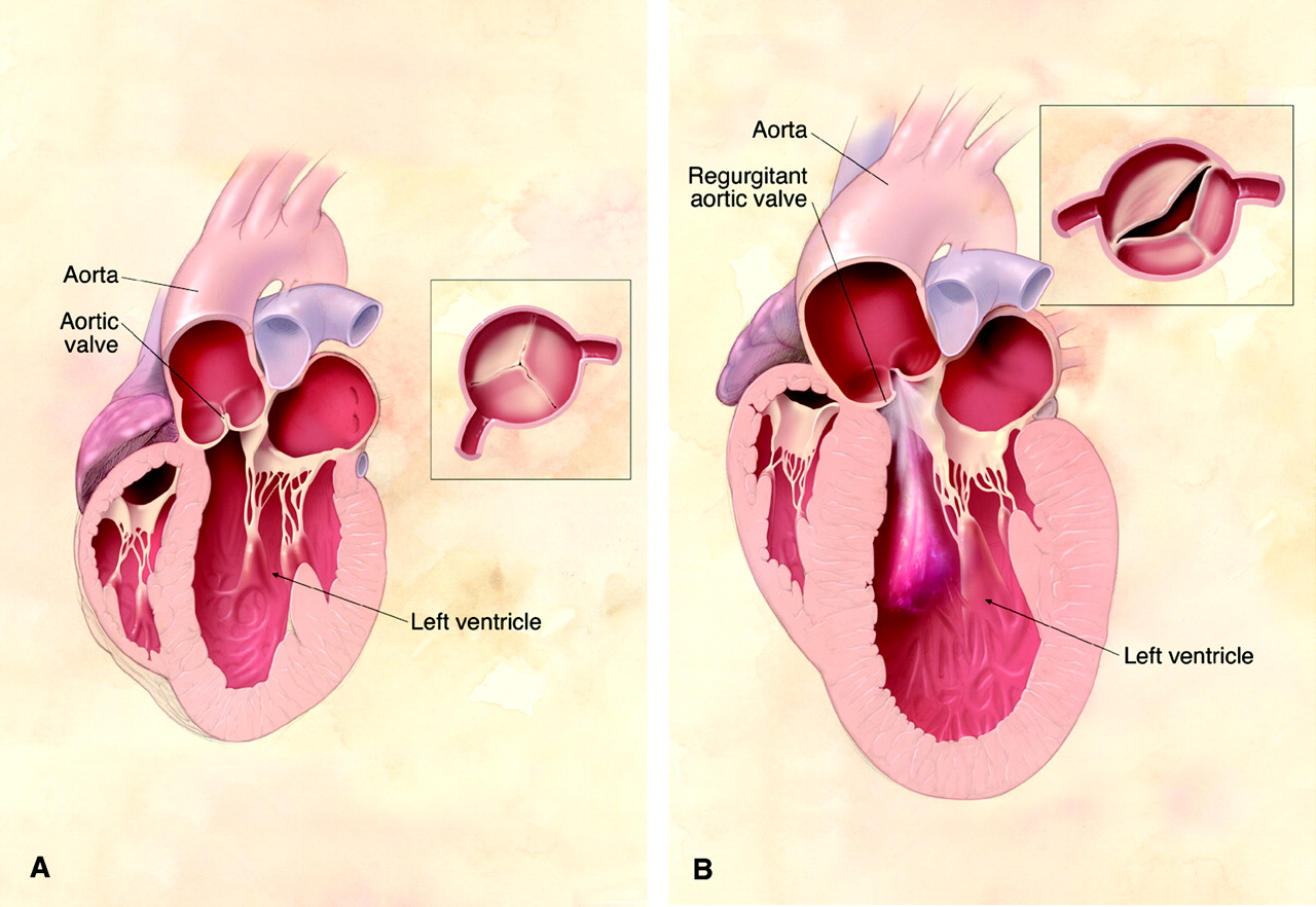
1. Disease Overview
Aortic valve disease is a condition in which the aortic valve — the one-way door between the heart’s main pumping chamber (the left ventricle) and the body’s largest artery (the aorta) — no longer works properly. Every heartbeat, this valve opens to let oxygen-rich blood surge out to the body, then snaps shut to stop it flowing backward.
There are two main problems that can affect it. In aortic stenosis, the valve becomes stiff, thickened or narrowed, so the heart must work much harder to push blood through a smaller opening. In aortic regurgitation (also called aortic insufficiency), the valve fails to close tightly, allowing blood to leak back into the ventricle after each beat. Some patients have a mix of both.
Aortic valve disease may be present from birth — most often as a bicuspid aortic valve, where the valve has two leaflets instead of the usual three — or it may develop over decades because of age-related calcium build-up, rheumatic fever, or infection. Mild disease often causes no symptoms for years and is picked up as a heart murmur. When it becomes severe, it can cause chest pain, blackouts and heart failure, and it becomes one of the most treatable serious heart conditions, with excellent results from valve replacement or repair.
2. Key Facts at a Glance
| Also known as | Aortic stenosis, aortic regurgitation, aortic insufficiency, aortic valve stenosis/incompetence |
| Body system affected | Cardiovascular system (left heart and aorta) |
| Common in | Older adults (calcific stenosis); people born with a bicuspid valve; those with a history of rheumatic fever |
| Severity range | Mild and symptom-free to severe, life-threatening |
| Key treatments | Monitoring, medications, TAVR/TAVI, surgical valve replacement (SAVR), valve repair, Ross procedure, balloon valvuloplasty |
| Outlook | Excellent after timely valve replacement; poor if severe symptomatic disease is left untreated |
3. Alternative Names and Medical Terminology
- Aortic stenosis (AS) — narrowing of the valve opening
- Aortic regurgitation (AR) / aortic insufficiency (AI) — leaking valve
- Aortic valve incompetence — older term for regurgitation
- Calcific / degenerative aortic valve disease — age-related form
- Bicuspid aortic valve (BAV) — congenital two-leaflet valve
- Mixed aortic valve disease — combined stenosis and regurgitation
- Aortic sclerosis — early thickening without significant obstruction
4. Relevant Heart, Lung or Vascular Anatomy
The aortic valve sits at the junction between the left ventricle and the aorta. In its normal form it has three thin, flexible flaps (cusps or leaflets) — the right, left and non-coronary cusps — arranged in a crown shape within the aortic root. Just above two of these cusps lie the openings of the coronary arteries, which feed the heart muscle itself.
When the ventricle contracts, the leaflets open fully to create a wide passage; when it relaxes, they fall back together and seal so no blood leaks backward. The valve works in concert with the left ventricular outflow tract below it and the ascending aorta above. Because the same tissue and blood flow influence the aortic root, aortic valve disease — especially with a bicuspid valve — is often accompanied by widening of the ascending aorta.
5. How the Disease Affects the Body
Both forms of aortic valve disease place an abnormal load on the left ventricle, and the heart adapts in different ways.
In aortic stenosis, the narrowed opening forces the left ventricle to generate very high pressure to eject blood. Over time the ventricle wall thickens (hypertrophy) to cope. This thickened muscle is stiff, needs more oxygen, and eventually cannot keep up. The result is reduced blood flow to the body and brain, high pressures backing up into the lungs, and an increased risk of angina, fainting and heart failure.
In aortic regurgitation, blood that leaked backward is added to the incoming blood on the next beat, so the ventricle must handle a larger volume. It responds by enlarging (dilating) and, for a while, copes well. But sustained volume overload eventually weakens the muscle, reducing its pumping efficiency irreversibly if not corrected in time.
In both cases, the heart’s ability to deliver oxygen-rich blood is compromised. The consequences ripple outward — fatigue, breathlessness, fluid retention, and reduced exercise capacity — and, when severe, they raise the risk of sudden cardiac death.
6. Types and Classification
- By valve problem — stenosis (narrowing), regurgitation (leaking), or mixed disease.
- By cause — degenerative/calcific, congenital (bicuspid or unicuspid), rheumatic, and infective (endocarditis-related).
- By valve anatomy — tricuspid (normal three-leaflet), bicuspid, or rarely unicuspid/quadricuspid.
- By severity — mild, moderate or severe, graded on echocardiography.
- By flow/gradient (in stenosis) — high-gradient severe AS, and low-flow low-gradient AS (which may occur with a normal or reduced ejection fraction).
- By onset (in regurgitation) — acute (sudden, e.g. endocarditis or dissection) or chronic.
7. Causes of the Disease
- Age-related calcification — the leading cause of aortic stenosis in high-income countries; calcium gradually stiffens the leaflets.
- Bicuspid aortic valve — a common congenital abnormality that wears out faster, causing stenosis or regurgitation, often in middle age.
- Rheumatic heart disease — scarring after rheumatic fever; still a major cause worldwide, especially in lower-income regions.
- Infective endocarditis — infection that destroys or perforates leaflets, causing acute regurgitation.
- Aortic root dilatation or dissection — stretching or tearing that stops leaflets meeting, causing regurgitation.
- Connective-tissue disorders — such as Marfan or Ehlers–Danlos syndrome.
8. How the Disease Develops
Aortic valve disease usually develops slowly over years. In calcific stenosis, the process resembles atherosclerosis: minor injury to the leaflet surface triggers inflammation, lipid deposition and, ultimately, active calcium formation within the valve tissue. What begins as harmless thickening (aortic sclerosis) can, over a decade or more, progress to significant narrowing. A bicuspid valve experiences abnormal mechanical stress with every beat, accelerating this same calcification so that severe stenosis often appears one to two decades earlier than in people with a normal valve.
In chronic regurgitation, the leaflets either become scarred and retracted (rheumatic disease) or fail to meet because the aortic root has enlarged. The leak starts small and the heart compensates by dilating, so patients can remain symptom-free for many years. During this silent compensated phase the ventricle steadily enlarges; eventually its walls thin and its contraction weakens.
Progression rates vary widely between individuals. Once symptoms appear in severe disease, however, deterioration tends to accelerate, which is why timing of treatment matters so much.
9. Risk Factors
Non-modifiable:
- Older age
- Being born with a bicuspid or unicuspid valve
- Male sex (slightly higher risk of stenosis)
- History of rheumatic fever
- Family history and certain genetic conditions
Modifiable / associated:
- High blood pressure
- High cholesterol
- Diabetes
- Smoking
- Chronic kidney disease
- Poor dental and oral health (increases endocarditis risk)
10. Genetic and Family-History Factors
Genetics plays a clear role in some forms of aortic valve disease. Bicuspid aortic valve clusters in families and is one of the most common inherited heart abnormalities; first-degree relatives of an affected person have a meaningfully higher chance of having a bicuspid valve or an enlarged aorta, so screening of close family members is often recommended.
Connective-tissue disorders such as Marfan syndrome, Loeys–Dietz syndrome and Ehlers–Danlos syndrome carry inherited mutations that weaken the aortic wall and valve, predisposing to regurgitation and aneurysm. Some cases of familial calcific valve disease are also being linked to specific genetic variants. A detailed family history helps identify who may benefit from earlier imaging.
11. Who Is Most at Risk?
- Adults over 65–70 — for calcific/degenerative stenosis.
- People born with a bicuspid valve — often present in their 40s–60s.
- Anyone with a history of rheumatic fever — particularly from regions where it remains common.
- People with Marfan or related connective-tissue syndromes.
- Those with untreated infections or intravenous drug use — higher endocarditis risk.
- Patients with hypertension, high cholesterol, diabetes or kidney disease.
12. Prevalence and Epidemiology
Aortic valve disease is among the most common valve conditions worldwide. Aortic stenosis becomes increasingly frequent with age and is found in a significant minority of people over 75, with severe disease in a smaller but substantial fraction of the very elderly. Bicuspid aortic valve is present in roughly 1–2% of the general population, making it one of the most common congenital heart abnormalities.
In high-income countries, degenerative calcific disease predominates, whereas in many lower- and middle-income regions rheumatic aortic disease remains a leading cause and affects younger adults. As populations age globally, the number of people needing aortic valve treatment is rising steeply. These figures are broad estimates and vary considerably by country and age group.
13. Signs and Symptoms
Many people with mild or moderate aortic valve disease feel completely normal, and the first clue is a heart murmur heard by a doctor. As the disease becomes severe, characteristic symptoms emerge.
The classic triad for severe aortic stenosis is:
- Chest pain or tightness (angina), especially on exertion
- Fainting or near-fainting (syncope), often during activity
- Breathlessness and heart failure symptoms — shortness of breath on exertion, then at rest, and swelling of the legs
Other common symptoms across both stenosis and regurgitation include:
- Fatigue and reduced exercise tolerance
- Palpitations or an awareness of a forceful heartbeat (common in regurgitation)
- Dizziness or light-headedness
- Difficulty lying flat, or waking at night breathless
Symptoms often appear gradually, and older patients may unconsciously slow down to avoid them, masking how severe the disease has become. The onset of any of these symptoms in someone with known severe disease is an important signal that treatment is needed.
14. Early-Stage Symptoms
In the early stages most patients have no symptoms at all, and the condition is silent. When subtle signs do appear, they may include:
- Mild breathlessness or fatigue during vigorous exercise
- A feeling of reduced stamina compared with the past
- Occasional palpitations
- A heart murmur found incidentally on a routine check-up
15. Advanced-Stage Symptoms
- Angina — chest tightness on exertion
- Syncope or near-fainting, especially with activity
- Marked breathlessness on mild exertion or at rest
- Orthopnoea (breathless lying flat) and paroxysmal nocturnal dyspnoea (waking gasping)
- Swollen ankles and legs, abdominal bloating (fluid retention)
- Extreme fatigue and, occasionally, sudden collapse
16. Symptoms in Women, Men and Older Adults
The core symptoms are similar across sexes, but presentation can differ. Women with aortic stenosis may report more breathlessness and fatigue and are sometimes diagnosed later; they may also have relatively smaller valve and ventricle dimensions, which can complicate assessment. Men may present somewhat earlier and with more calcification.
Older adults frequently attribute early symptoms to “just getting old” and reduce their activity to compensate, so severe disease can go unrecognised. In the elderly, the first presentation may be heart failure, falls related to fainting, or confusion. Because symptoms can be atypical or masked, a low threshold for echocardiography is important in older patients with a murmur.
17. Emergency Warning Signs
Seek emergency care immediately (call local emergency services) if there is:
- Fainting or collapse
- Severe or crushing chest pain
- Sudden, severe breathlessness or inability to breathe when lying down
- Coughing up pink, frothy sputum (a sign of acute heart failure)
- A tearing chest or back pain (possible aortic dissection with acute regurgitation)
18. When to Seek Medical Help
Arrange a prompt medical review if you have a known heart murmur or valve problem and notice new breathlessness, chest discomfort, dizziness, fainting or reduced exercise tolerance. Anyone told they have aortic valve disease should keep scheduled monitoring appointments even when feeling well. New or worsening symptoms in someone with severe stenosis or regurgitation should never be ignored, as timely treatment is often life-saving.
19. Disease Stages, Grades and Severity
Cardiologists grade aortic valve disease mainly by echocardiography and by whether symptoms are present. A widely used framework describes:
- Stage A (at risk) — e.g. a bicuspid valve or aortic sclerosis, no obstruction yet.
- Stage B (progressive) — mild to moderate stenosis or regurgitation.
- Stage C (severe, asymptomatic) — severe disease but no symptoms, sometimes split by whether heart function is preserved (C1) or reduced (C2).
- Stage D (severe, symptomatic) — severe disease with symptoms.
Severity of stenosis is judged from the valve area, the peak jet velocity and the mean pressure gradient across the valve. Severity of regurgitation is graded from the width and volume of the backward jet and the effect on ventricle size and function.
20. Disease Progression
Aortic valve disease tends to progress gradually but relentlessly. Stenosis typically worsens at a variable pace — some valves narrow slowly over many years, others faster, especially bicuspid or heavily calcified valves. Once severe stenosis becomes symptomatic, the outlook without treatment declines sharply over the following months to a couple of years.
Chronic regurgitation may remain stable and well tolerated for a long time, but continued volume overload eventually enlarges and weakens the ventricle. If left too long, this damage can become irreversible even after the valve is fixed, which is why regular imaging to catch progression early is essential.
21. Possible Complications
- Heart failure — the most common serious consequence
- Arrhythmias, including atrial fibrillation and dangerous ventricular rhythms
- Sudden cardiac death, particularly with severe symptomatic stenosis
- Infective endocarditis — infection of the diseased or replaced valve
- Pulmonary hypertension and right-heart strain
- Aortic aneurysm or dissection — especially with bicuspid valves
- Stroke or systemic embolism from calcium debris or clots
- Bleeding tendency (a rare association of severe stenosis)
22. Related and Associated Medical Conditions
Aortic valve disease often coexists with coronary artery disease, since both share risk factors and age; many patients need coronary assessment before valve surgery. Bicuspid valve is strongly linked with ascending aortic aneurysm and, occasionally, coarctation of the aorta. Other associations include hypertension, atrial fibrillation, mitral valve disease (particularly in rheumatic disease affecting multiple valves), chronic kidney disease, and heart failure. Endocarditis can affect the aortic valve as a primary or complicating condition.
23. Screening and Early Detection
There is no population-wide screening programme, but aortic valve disease is often detected early through the routine detection of a heart murmur during physical examination. Anyone with a murmur, unexplained breathlessness, or a family history of bicuspid valve should have an echocardiogram. First-degree relatives of a person with a bicuspid aortic valve are frequently advised to be screened by echocardiography, since the condition and associated aortic enlargement run in families. Good dental hygiene and prompt treatment of streptococcal throat infections help prevent rheumatic and infective valve damage.
24. How the Disease Is Diagnosed
Diagnosis usually begins when a doctor hears a heart murmur or when a patient reports breathlessness, chest pain, dizziness or fainting. The evaluation then follows a logical pathway.
First, a careful history and physical examination identify the type and possible severity of the valve problem — the character, timing and location of the murmur give important clues, as do the pulse and blood pressure. The single most important test is a transthoracic echocardiogram (echo), an ultrasound of the heart that shows the valve structure, measures how narrow or leaky it is, and assesses the size and pumping strength of the left ventricle. It confirms the diagnosis, grades severity and is repeated over time to track progression.
Supporting tests include an ECG (looking for thickened muscle or rhythm problems), a chest X-ray, and blood tests. When more detail is needed — for example before a procedure — a transoesophageal echo, cardiac CT (to measure valve calcium and size the aorta and valve for TAVR), cardiac MRI, or cardiac catheterisation with coronary angiography may be performed. Together these build a complete picture that guides treatment timing and choice.
25. Physical Examination and Medical History
The history focuses on symptoms (breathlessness, chest pain, fainting), exercise tolerance, past rheumatic fever, known murmurs, and family history of valve or aortic disease. On examination, the doctor listens for characteristic murmurs — a harsh ejection systolic murmur radiating to the neck in stenosis, or an early diastolic murmur in regurgitation. Other signs include a slow-rising, weak pulse in stenosis, or a bounding, “collapsing” pulse with wide pulse pressure in regurgitation. Signs of heart failure, such as raised neck veins, lung crackles and leg swelling, are also assessed.
26. Diagnostic Tests and Imaging
- Transthoracic echocardiogram — the cornerstone test; measures valve area, gradients, regurgitation severity and ventricular function.
- Transoesophageal echo (TOE) — detailed views, especially for endocarditis or before intervention.
- ECG — shows left ventricular hypertrophy, strain, or arrhythmias.
- Chest X-ray — heart size, calcification, lung congestion.
- Cardiac CT — quantifies valve calcium, sizes the annulus for TAVR, and images the aorta.
- Cardiac MRI — accurate assessment of regurgitation and ventricular volumes.
- Cardiac catheterisation and coronary angiography — checks for coexisting coronary disease before surgery.
- Exercise (stress) testing — used cautiously to unmask symptoms in “asymptomatic” severe stenosis.
27. Blood Tests, Biomarkers and Genetic Testing
There is no blood test that diagnoses aortic valve disease directly, but several are useful. Natriuretic peptides (BNP or NT-proBNP) rise with heart strain and can help judge severity, timing of surgery and prognosis. Routine bloods assess kidney and liver function, blood count and electrolytes, which matter for procedural planning and anticoagulation. Blood cultures are essential when endocarditis is suspected. Genetic testing and family screening may be advised where a bicuspid valve, Marfan syndrome or other connective-tissue disorder is suspected.
28. Understanding Test Results
Echo results are usually reported as mild, moderate or severe. For stenosis, severe disease is generally indicated by a small valve area, a high peak velocity and a high mean gradient; sometimes flow is low, producing a “low-gradient” severe stenosis that needs extra tests to confirm. For regurgitation, severity reflects the size of the leak and its impact on ventricle dimensions. Ejection fraction describes the ventricle’s pumping strength; a falling value is an important warning sign. Doctors interpret all these numbers together with symptoms — no single figure decides treatment.
29. Differential Diagnosis
Conditions that can mimic or coexist with aortic valve disease include:
- Mitral valve disease — another cause of murmur and breathlessness
- Hypertrophic cardiomyopathy — can cause an outflow murmur and fainting
- Coronary artery disease / angina — overlapping chest pain
- Heart failure from other causes
- Anaemia or thyroid disease — can produce flow murmurs and fatigue
- Pulmonary conditions — as a cause of breathlessness
- Aortic aneurysm or dissection — for acute regurgitation
30. Specialist and Multidisciplinary Evaluation
Modern valve care is delivered by a Heart Valve Team (or Heart Team). This typically includes a cardiologist, a cardiac surgeon, an interventional (structural) cardiologist, a cardiac imaging specialist, and a cardiac anaesthetist, often supported by valve-clinic nurses and, for older patients, geriatric and frailty input. The team reviews imaging, symptoms, overall health and personal preferences to recommend the best-timed and best-suited treatment — whether monitoring, surgery or a catheter-based procedure. You can find experienced valve specialists through our doctors directory and accredited hospitals.
31. Treatment Goals
- Relieve symptoms such as breathlessness, chest pain and fainting
- Protect and preserve the heart muscle before irreversible damage occurs
- Prevent complications — heart failure, arrhythmia and sudden death
- Restore normal or near-normal blood flow through the valve
- Improve quality of life and life expectancy
- Individualise timing so intervention happens at the right moment — not too early, not too late
32. When Is Treatment Required?
Intervention is generally recommended for severe aortic valve disease once symptoms appear, as symptomatic severe stenosis or regurgitation carries a poor outlook without treatment. It is also considered in severe disease without symptoms when the heart shows signs of strain — for example, a falling ejection fraction, an enlarging ventricle, an abnormal exercise test, or when heart surgery is being done for another reason. Mild and moderate disease usually needs monitoring rather than intervention. The decision balances valve severity, symptoms, heart function, age, other illnesses and personal wishes.
33. Active Monitoring and Watchful Waiting
Many people with mild or moderate — or even severe but symptom-free — aortic valve disease are managed by regular surveillance rather than immediate treatment. This involves periodic clinic reviews and echocardiograms, with the interval depending on severity: milder disease may be checked every couple of years, while severe asymptomatic disease is watched closely, often every 6–12 months. The aim is to intervene at the optimal moment — as soon as symptoms develop or the heart begins to show early strain. Patients are taught which symptoms to report promptly between visits.
34. Medications
There is no medication that cures aortic valve disease or reliably slows valve narrowing. Drugs are used to manage symptoms, control associated conditions and support the heart:
- Diuretics — reduce fluid overload and breathlessness (used cautiously in stenosis)
- Blood-pressure medicines (e.g. ACE inhibitors, ARBs) — especially helpful in regurgitation
- Beta-blockers or rate-control drugs — for arrhythmias such as atrial fibrillation
- Anticoagulants — for atrial fibrillation or after mechanical valve replacement
- Statins and standard cardiovascular risk control
- Antibiotics — to treat endocarditis and, in selected patients, to prevent it around dental procedures
Medication is a bridge and a support, but severe symptomatic disease ultimately requires valve replacement or repair.
35. Minimally Invasive Treatments
Where suitable, aortic valve replacement can be done through smaller incisions rather than a full breastbone split. Minimally invasive aortic valve surgery uses a partial upper sternotomy or a small right-chest incision, offering less scarring, reduced blood loss, less pain and a quicker recovery for appropriately selected patients. These techniques require an experienced surgical team and careful patient selection. Learn more about minimally invasive cardiac surgery.
36. Catheter-Based and Endovascular Treatments
- TAVR / TAVI (Transcatheter Aortic Valve Replacement / Implantation) — a new valve is threaded to the heart through an artery (usually in the groin) and expanded inside the diseased valve, without open surgery. It is now a mainstream option for many patients with severe stenosis, particularly older or higher-risk patients, and increasingly for selected intermediate- and lower-risk patients.
- Balloon aortic valvuloplasty — a balloon is inflated to stretch a narrowed valve; effects are usually temporary, so it is mainly used as a bridge or in children/young adults with non-calcified valves.
- Valve-in-valve TAVR — placing a transcatheter valve inside a previously implanted tissue valve that has worn out.
Explore aortic valve procedures for more detail.
37. Surgical Treatment Options
Open-heart surgery remains a cornerstone of treatment, especially for younger patients, those with additional heart problems, or anatomy unsuitable for a catheter approach.
- Surgical Aortic Valve Replacement (SAVR) — the diseased valve is removed and replaced with either a mechanical valve (very durable but requires lifelong blood-thinning medication) or a tissue (bioprosthetic) valve (no long-term anticoagulation but a limited lifespan). The choice depends largely on age, lifestyle and bleeding risk.
- Aortic valve repair — in selected cases of regurgitation, the patient’s own valve can be repaired rather than replaced, preserving natural tissue.
- The Ross procedure — the diseased aortic valve is replaced with the patient’s own pulmonary valve, and a donor valve is placed in the pulmonary position. Mainly used in younger patients and children, it avoids anticoagulation and allows the new aortic valve to grow.
- Combined surgery — the valve may be replaced at the same time as coronary bypass, aortic root/ascending aorta repair, or another valve procedure.
Surgical valve replacement has a long, proven track record with excellent outcomes in experienced centres. See our overview of heart surgery and aortic valve procedures.
38. Advanced and Emerging Treatments
Innovation in this field is rapid. Advances include newer-generation transcatheter valves with better durability and lower complication rates, refined techniques to treat aortic regurgitation by catheter (historically harder than stenosis), and valve-in-valve approaches that let a failed tissue valve be replaced without repeat open surgery. Research also focuses on tissue-engineered and longer-lasting bioprosthetic valves, improved imaging and computer-based (“digital twin”) procedure planning, and drugs aimed at slowing valve calcification. Many of these are available in leading centres or through clinical trials.
39. Treatment Options Compared
- Monitoring — best for mild/moderate or asymptomatic disease; no procedural risk, but requires regular follow-up.
- Medications — ease symptoms and treat associated conditions; do not fix the valve.
- TAVR/TAVI — no open surgery, faster recovery, ideal for older or higher-risk patients; long-term durability data still maturing and not suitable for all anatomies.
- SAVR (surgical replacement) — durable, well-proven, allows repair of other problems; involves open-heart surgery and longer recovery.
- Ross procedure — excellent for young patients, avoids anticoagulation; complex operation done in specialist centres.
- Balloon valvuloplasty — quick but temporary; mainly a bridge or for young non-calcified valves.
40. How Doctors Choose the Right Treatment
The Heart Team weighs many factors: the type and severity of valve disease, symptoms, the strength of the heart, and the patient’s age, frailty and other illnesses. Anatomy matters — cardiac CT shows whether a valve is suitable for TAVR. Younger patients and those needing surgery for other problems often favour SAVR or the Ross procedure; older or higher-risk patients often favour TAVR. Patient preference — including attitudes toward anticoagulation, recovery time and future re-intervention — is central. The goal is a shared, individualised decision.
41. Benefits and Risks of Treatment
Benefits: relief of breathlessness, chest pain and fainting; a lower risk of heart failure and sudden death; improved quality of life; and, in severe symptomatic disease, a substantial gain in life expectancy.
Risks vary by procedure but can include:
- Bleeding, infection and reactions to anaesthesia
- Stroke
- Heart rhythm problems, sometimes needing a pacemaker (more common after TAVR)
- Vascular access complications (TAVR) or wound issues (surgery)
- Leak around a new valve (paravalvular leak)
- Valve deterioration over time, needing re-intervention
In experienced, high-volume centres these risks are low, and the benefits for severe symptomatic disease clearly outweigh them.
42. What Happens If the Disease Is Left Untreated?
Untreated severe symptomatic aortic stenosis carries a serious prognosis — once the classic symptoms of angina, fainting or heart failure appear, survival without valve replacement is often measured in months to a few years, and there is a real risk of sudden death. Untreated severe regurgitation allows progressive, eventually irreversible enlargement and weakening of the left ventricle, leading to heart failure. Mild or moderate disease may remain stable for years, but severe disease that is left untreated tends to end in disabling heart failure or death. Timely treatment dramatically changes this outlook.
43. Treatment Success and Expected Outcomes
Aortic valve treatment is one of the great success stories of modern cardiology. In experienced centres, both surgical replacement and TAVR have high success rates and low procedural mortality in appropriately selected patients. Most people experience marked relief of symptoms — better breathing, more energy and freedom from fainting — often within weeks. Tissue and mechanical valves both function well for many years, though tissue valves may eventually need replacement. Outcomes are best when treatment is carried out before the heart is severely and irreversibly damaged, underscoring the value of timely referral.
44. Prognosis and Long-Term Outlook
The long-term outlook after successful treatment is generally very good. Most patients return to normal or near-normal activities and enjoy a life expectancy that approaches that of people without valve disease, particularly when the heart function was preserved beforehand. Mechanical valves are highly durable but require lifelong anticoagulation and careful monitoring; tissue valves avoid long-term blood thinners but may wear out over 10–20 years, potentially needing a repeat procedure — increasingly done by valve-in-valve TAVR.
Prognosis is less favourable when treatment is delayed until the ventricle is badly weakened, or in patients who are very frail or have multiple other serious illnesses. Lifelong follow-up, good blood-pressure control, dental hygiene and prompt treatment of infections all help protect the long-term result. With modern care, the great majority of treated patients do well for many years.
45. Recovery and Rehabilitation
Recovery depends on the procedure. After TAVR, many patients go home within a few days and resume light activity quickly. After open surgery, the hospital stay is typically around a week, with full recovery over 6–12 weeks as the breastbone heals; heavy lifting and driving are restricted for several weeks. Cardiac rehabilitation — a supervised programme of gradual exercise, education and support — is strongly encouraged and improves fitness, confidence and outcomes. Wound care, pain control, medication adherence and attending follow-up appointments are all part of a smooth recovery.
46. Follow-Up Tests and Long-Term Monitoring
Lifelong follow-up is important. Patients are usually reviewed with periodic clinical checks and echocardiograms to confirm the valve is working well and to watch for late problems such as valve deterioration or leak. Those on anticoagulation for a mechanical valve need regular blood monitoring (INR). People with unrepaired mild-to-moderate disease or a bicuspid valve also have scheduled echoes to track progression and monitor the aorta. Any new symptoms should prompt an earlier review.
47. Managing Recurrence or Disease Progression
Aortic valve disease can progress even after treatment. A tissue valve may degenerate over years, and a native valve under surveillance may worsen. Ongoing monitoring is designed to catch this early. When a previously implanted tissue valve fails, a valve-in-valve TAVR often allows treatment without repeat open surgery. Good control of blood pressure and cardiovascular risk factors, dental hygiene to prevent endocarditis, and prompt reporting of new symptoms all help manage progression and protect the result over the long term.
48. Living with the Disease
Many people live full, active lives with aortic valve disease, especially after successful treatment. Day-to-day, it helps to stay physically active within recommended limits, take medications reliably, attend follow-up appointments, and maintain excellent dental hygiene to reduce endocarditis risk. Knowing your own warning symptoms and acting on them quickly is key. Carrying details of your valve type and any anticoagulation, avoiding smoking, controlling blood pressure and eating heart-healthily all support long-term wellbeing.
49. Diet and Nutrition Guidelines
- Follow a heart-healthy diet rich in vegetables, fruit, whole grains, legumes and fish.
- Limit salt to reduce fluid retention and control blood pressure.
- Reduce saturated fat and processed foods to help manage cholesterol.
- Keep a healthy weight.
- Limit alcohol.
- If taking warfarin, keep vitamin K intake (green leafy vegetables) steady rather than fluctuating, and discuss diet with your clinic.
- Stay well hydrated but follow any fluid limits advised for heart failure.
50. Exercise and Physical-Activity Guidelines
Regular moderate activity is beneficial and encouraged for most patients, but the right level depends on severity. Those with mild disease can usually exercise normally. People with severe stenosis, especially before treatment, should avoid intense or competitive exertion because of the risk of fainting or sudden problems, and should follow individualised advice. After successful treatment, most patients can gradually return to an active lifestyle, ideally guided by cardiac rehabilitation. Always agree an exercise plan with your cardiologist.
51. Medications, Activities and Habits to Avoid
- Avoid strenuous or competitive sport with untreated severe stenosis.
- Do not stop anticoagulation (mechanical valve) without medical advice.
- Use caution with medications that lower blood pressure abruptly in severe stenosis — take only as prescribed.
- Avoid smoking entirely.
- Limit excess alcohol.
- Do not neglect dental care or ignore infections (endocarditis risk).
- Avoid abrupt, heavy exertion (such as sudden heavy lifting) if advised.
52. Preventing the Disease or Reducing Its Risks
Not all aortic valve disease is preventable — a bicuspid valve is present from birth. However, risks can be reduced by:
- Preventing and promptly treating strep throat to avoid rheumatic fever
- Maintaining good dental and oral hygiene to lower endocarditis risk
- Controlling blood pressure, cholesterol and diabetes
- Not smoking and keeping a healthy weight
- Regular check-ups, especially if you have a family history of valve or aortic disease
53. Pregnancy and the Disease
Pregnancy places extra demands on the heart, so women with aortic valve disease should ideally have pre-pregnancy counselling with a cardiologist. Mild disease is often well tolerated, but severe stenosis can be dangerous and may need treatment before conceiving. Women on warfarin for a mechanical valve require specialist planning, as anticoagulation must be managed carefully in pregnancy. Care is best delivered by a joint cardiology and obstetric team, with close monitoring throughout pregnancy and delivery.
54. Disease in Children and Young Adults
In the young, aortic valve disease is usually congenital — most often a bicuspid or unicuspid valve — or a consequence of rheumatic fever. Children may need balloon valvuloplasty or surgical repair; the Ross procedure is particularly valuable because the pulmonary autograft can grow with the child and avoids lifelong blood thinners. Young patients need lifelong follow-up, as valve problems and any associated aortic enlargement can progress over time. Care is provided through specialised congenital heart procedures programmes.
55. Disease in Older Adults
In older adults, calcific degenerative stenosis is the dominant form. Age, frailty and other illnesses influence both symptoms and treatment choice. TAVR has transformed care for this group, offering effective treatment with faster recovery for many who were once considered too high-risk for surgery. A comprehensive assessment — including frailty, kidney function and cognition — helps the Heart Team recommend treatment that improves both quality and length of life while matching the patient’s overall health and goals.
56. Emotional Health and Patient Support
A diagnosis of heart valve disease, and the prospect of surgery or a procedure, can cause anxiety, low mood or fear. These feelings are common and understandable. Support helps — talking with your care team, connecting with patient support groups or others who have had valve treatment, and involving family. Cardiac rehabilitation also supports emotional recovery and confidence. If anxiety or depression is significant, ask your doctor about counselling or other help; addressing emotional health improves overall recovery.
57. Preparing for Your Specialist Appointment
- Write down your symptoms, when they occur and what triggers them.
- List all medications, doses and allergies.
- Note your medical history, including rheumatic fever, murmurs and family heart/aortic disease.
- Bring previous echo reports, ECGs and imaging if available.
- Prepare your questions in advance (see next section).
- Consider bringing a family member to help remember information.
58. Questions to Ask Your Doctor
- What type of aortic valve disease do I have, and how severe is it?
- Is my valve narrowed (stenosis), leaking (regurgitation), or both?
- Do I have a bicuspid valve, and does it affect my aorta?
- How is my heart’s pumping function?
- Do I need treatment now, or can we monitor it?
- Which treatment is best for me — surgery, TAVR, or another option?
- If I need a replacement valve, should it be mechanical or tissue, and why?
- What are the risks and benefits, and what recovery should I expect?
- Will I need long-term blood-thinning medication?
- What symptoms should prompt me to seek urgent help?
59. Cost of Diagnosis and Treatment
Costs vary enormously by country, hospital and procedure. The figures below are broad approximate ranges for aortic valve replacement and should be confirmed with each provider.
| Region | Approx. cost of aortic valve replacement (USD) |
|---|---|
| United States | $80,000 – $200,000+ |
| United Kingdom (private) | $35,000 – $80,000 |
| Singapore | $25,000 – $55,000 |
| Thailand | $18,000 – $40,000 |
| Turkey | $12,000 – $30,000 |
| India | $6,000 – $20,000 |
Medical-tourism destinations such as India, Turkey, Thailand and Singapore often provide high-quality valve surgery and TAVR at roughly 50–90% less than US/UK prices. TAVR is generally more expensive than surgical replacement because of the cost of the transcatheter valve. Explore leading destinations and hospitals for current quotes.
60. Factors Affecting Treatment Cost
- Type of procedure — TAVR is usually costlier than surgical replacement due to device price.
- Valve type — mechanical vs tissue vs transcatheter.
- Additional procedures — e.g. coronary bypass or aortic surgery at the same time.
- Hospital reputation, accreditation and surgeon experience.
- Country and city.
- Length of hospital stay and level of care needed.
- Pre-operative tests, complications and rehabilitation.
- Travel, accommodation and follow-up for international patients.
61. Choosing the Right Specialist
Look for a cardiologist and cardiac surgeon (or structural/interventional cardiologist) with specific expertise in aortic valve disease and access to a full Heart Valve Team. Helpful markers include high procedure volume, published or audited outcomes, board certification, and experience with both SAVR and TAVR so that advice is not biased toward a single technique. Good communication and willingness to explain options matter too. Browse experienced valve specialists in our doctors directory.
62. Choosing the Right Hospital or Treatment Centre
Choose a centre with a dedicated heart valve or structural heart programme, ideally with international accreditation such as JCI. Key factors include:
- A multidisciplinary Heart Team and hybrid operating facilities
- High case volumes and strong, transparent outcome data
- Availability of both surgical and transcatheter options
- Advanced imaging (CT, TOE, MRI)
- Comprehensive cardiac rehabilitation and follow-up
- Clear pricing and international-patient support
See our recommended hospitals and destinations.
63. Getting a Second Medical Opinion
A second opinion is valuable and appropriate before major valve treatment. It can confirm the diagnosis and severity, clarify whether treatment is needed now, and compare options such as surgery versus TAVR — particularly where a case is borderline or complex. Bring your echo reports and imaging so the reviewing team can assess them directly. A good specialist will welcome a second opinion. You can request a second opinion through our network of experienced valve centres.
64. Treatment Abroad and Medical-Travel Considerations
Many patients travel for aortic valve treatment to access high-quality care at lower cost or shorter waiting times. When planning treatment abroad, consider:
- Accreditation and outcomes of the hospital (e.g. JCI)
- Surgeon and centre experience with your specific procedure
- Clear, itemised cost estimates
- Travel fitness and timing — long flights soon after heart surgery need medical clearance
- Arrangements for follow-up and rehabilitation back home
- Language, coordination and aftercare support
Our destinations pages highlight leading medical-tourism centres for cardiac valve care.
65. Frequently Asked Questions
Is aortic valve disease serious? Mild disease is often harmless and simply monitored, but severe symptomatic disease is serious and needs treatment. The good news is that it is highly treatable.
Can aortic valve disease be cured with medication? No. Medicines ease symptoms and treat related conditions but cannot fix a narrowed or leaking valve. Severe disease needs repair or replacement.
What is the difference between TAVR and open surgery? TAVR places a new valve through a catheter without open-heart surgery and suits many older or higher-risk patients; surgical replacement is open-heart surgery, very durable, and often preferred for younger patients or when other heart problems must be fixed too.
How long do replacement valves last? Tissue valves typically last around 10–20 years; mechanical valves can last a lifetime but require lifelong blood thinners.
I have a bicuspid valve but feel fine — what should I do? Attend regular monitoring, as the valve and aorta need watching over time, and close relatives may benefit from screening.
Can I exercise with aortic stenosis? Usually yes if it is mild, but strenuous exertion should be avoided with severe stenosis until treated. Always follow your cardiologist’s advice.
Will I need blood thinners after treatment? Mechanical valves require lifelong anticoagulation; tissue valves and TAVR usually do not, though short-term medication may be used.
66. Patient Stories and Treatment Experiences
The following are representative, anonymised examples for illustration only.
Ramesh, India (72): After months of breathlessness and one fainting episode, Ramesh was found to have severe aortic stenosis. He underwent TAVR and went home within a few days, describing his energy as “back to what it was ten years ago.”
Sofia, Spain (45): Born with a bicuspid valve, Sofia developed significant regurgitation in her forties. She had surgical valve replacement and a mild aortic enlargement repaired at the same time, returning to gentle running after cardiac rehabilitation.
David, UK (58): Diagnosed with severe stenosis, David travelled for treatment to reduce waiting time. His surgical replacement went smoothly and he now attends yearly follow-up, feeling well and active.
67. Latest Research and Clinical Trials
Research is advancing quickly. Studies continue to extend TAVR to younger and lower-risk patients and to improve valve durability and reduce the need for a pacemaker. Trials are developing transcatheter solutions for aortic regurgitation, which has been harder to treat by catheter than stenosis. Other work explores tissue-engineered valves, better imaging and AI-assisted procedure planning, and drugs that might slow valve calcification. Leading centres often offer participation in clinical trials; ask your specialist whether any are suitable for you. (This is a general summary, not a reference to specific published studies.)
68. Related Diseases and Conditions
- Mitral Valve Disease
- Combined and Multi-Valvular Disease
- Tricuspid Valve Disease
- Coronary Artery Disease
- Aortic Root Dilatation
- Infective Endocarditis
69. Related Treatments and Procedures
- Aortic Valve Procedures
- Minimally Invasive Cardiac Surgery
- Hybrid Cardiac Procedures
- Congenital Heart Procedures
- Coronary Artery Bypass Grafting
70. Medical Glossary
- Aortic valve — the valve between the left ventricle and the aorta.
- Aortic stenosis — narrowing of the aortic valve opening.
- Aortic regurgitation — leaking of the aortic valve.
- Bicuspid aortic valve — a valve with two leaflets instead of three.
- Left ventricle — the heart’s main pumping chamber.
- Hypertrophy — thickening of the heart muscle.
- TAVR / TAVI — replacing the valve via a catheter, without open surgery.
- SAVR — surgical (open) aortic valve replacement.
- Ross procedure — using the patient’s own pulmonary valve to replace the aortic valve.
- Balloon valvuloplasty — widening a narrowed valve with a balloon.
- Bioprosthetic (tissue) valve — a replacement valve made from animal or human tissue.
- Mechanical valve — a durable artificial valve needing lifelong anticoagulation.
- Echocardiogram — an ultrasound scan of the heart.
- Ejection fraction — a measure of the heart’s pumping strength.
- Endocarditis — infection of a heart valve.
71. Medical Review, Editorial Policy and Last Updated Date
Last updated: 11 July 2026.
This article is written for patient education and is reviewed by qualified medical professionals for accuracy against current cardiology guidance from bodies such as the ACC/AHA, the European Society of Cardiology (ESC) and the NHS. We aim for clear, balanced, evidence-based information and update our content periodically.
Disclaimer: This content is for general educational purposes only and is not a substitute for professional medical advice, diagnosis or treatment. Always consult a qualified healthcare provider about your individual condition and before making any treatment decision.
72. Clinical Guidelines and Medical References
This article draws on general, widely accepted medical knowledge and the published guidelines of major professional bodies, including:
- American College of Cardiology (ACC) / American Heart Association (AHA) — guidelines for the management of valvular heart disease
- European Society of Cardiology (ESC) / EACTS — valvular heart disease guidelines
- Society of Thoracic Surgeons (STS) — surgical outcome standards
- NHS and World Health Organization (WHO) — patient information and rheumatic heart disease resources
These are cited as reputable sources of standard-of-care guidance rather than specific studies.
73. Book an Appointment or Request a Second Opinion
If you or a loved one has been diagnosed with aortic valve disease, expert help is available. Our network connects you with experienced valve specialists and accredited hospitals worldwide for diagnosis, treatment and second opinions.
- Book an appointment: Get started here
- Request a second opinion or ask a question: Contact us
- Explore top hospitals, doctors and destinations for aortic valve care.
Take the next step toward better heart health today.