1. Disease Overview
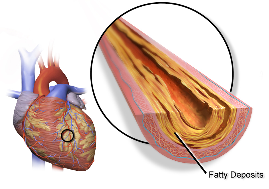
Recurrent coronary artery disease (recurrent CAD) is the return of reduced blood flow to the heart muscle after a patient has already been treated for coronary artery disease with either a percutaneous coronary intervention (PCI, angioplasty and stenting) or coronary artery bypass grafting (CABG). Instead of a completely new problem, it represents the reappearance or progression of ischemia in a heart that has already undergone revascularization.
Recurrence can arise from several distinct mechanisms: in-stent restenosis (gradual re-narrowing inside a previously placed stent), stent thrombosis (sudden clot formation within a stent), bypass graft failure (early graft closure or later vein-graft disease), and progression of atherosclerosis in native coronary arteries that were not treated or in new locations. Because the coronary anatomy is now altered by scar tissue, stents, and grafts, recurrent CAD is often technically more complex to diagnose and treat than the original disease.
Patients may notice returning angina (chest pain or pressure), breathlessness, or reduced exercise tolerance, sometimes years after they felt fully recovered. Modern cardiology offers effective repeat treatments, but careful evaluation is needed to select the safest and most durable option, whether that is intensified medication, redo angioplasty, or redo surgery.
2. Key Facts at a Glance
| Fact | Detail |
|---|---|
| Also known as | Recurrent CAD, restenosis, recurrent ischemia, graft failure, recurrent angina |
| Body system affected | Cardiovascular system (coronary arteries, bypass grafts, stents) |
| Common in | Patients with prior PCI or CABG, diabetics, smokers, those with poorly controlled risk factors |
| Severity range | Mild (stable recurrent angina) to life-threatening (acute stent thrombosis, graft occlusion causing heart attack) |
| Key treatments | Optimized medical therapy, repeat angioplasty/stenting, redo CABG, risk-factor control |
| Outlook | Generally good with timely re-intervention and aggressive secondary prevention |
3. Alternative Names and Medical Terminology
- Recurrent CAD / recurrent ischemic heart disease
- In-stent restenosis (ISR) — re-narrowing within a stent
- Stent thrombosis (ST) — clot inside a stent
- Bypass graft failure / graft occlusion
- Saphenous vein graft (SVG) disease / vein-graft degeneration
- Recurrent angina or post-revascularization angina
- De novo disease progression — new blockages in native arteries
- Target lesion revascularization (TLR) and target vessel revascularization (TVR) — clinical terms describing repeat treatment of a previously treated segment
4. Relevant Heart, Lung or Vascular Anatomy
The coronary arteries supply oxygen-rich blood to the heart muscle. The main vessels are the left main, which divides into the left anterior descending (LAD) and left circumflex (LCx), and the right coronary artery (RCA). After revascularization, the anatomy is modified:
- Stents are metal-mesh scaffolds implanted inside native arteries.
- Bypass grafts are new conduits: the left internal mammary artery (LIMA), radial artery, and saphenous vein grafts (SVGs) rerouting blood around blockages.
Recurrent disease may occur inside a stent, at the anastomosis (where a graft joins the artery), within the body of a vein graft, or in previously untreated native segments. Arterial grafts (LIMA) tend to remain durable for decades, whereas vein grafts are more prone to degeneration.
5. How the Disease Affects the Body
Recurrent CAD reduces or interrupts blood flow to the myocardium, producing ischemia — a mismatch between the oxygen the heart muscle needs and what it receives. When a stent re-narrows or a graft fails, the territory it supplied becomes under-perfused, especially during exertion when demand rises.
The consequences depend on how quickly and how severely flow drops. Gradual re-narrowing (in-stent restenosis or vein-graft disease) tends to cause progressive, exertional angina and declining exercise capacity, giving the heart time to develop some collateral circulation. Sudden occlusion (stent thrombosis or acute graft closure) causes an acute coronary syndrome — unstable angina or a myocardial infarction (heart attack) — because the muscle is abruptly deprived of blood before collaterals can compensate.
Repeated or prolonged ischemia can permanently damage heart muscle, weaken the pumping function, and lead to heart failure or dangerous arrhythmias. Because recurrent CAD often affects a heart already scarred from earlier disease, the reserve to tolerate a new ischemic insult may be reduced, which is why prompt recognition and treatment matter.
6. Types and Classification
- In-stent restenosis (ISR): focal (short segment) or diffuse (long segment or multiple areas); more common with bare-metal stents than drug-eluting stents.
- Stent thrombosis: classified by timing — acute (within 24 hours), subacute (1–30 days), late (30 days–1 year), and very late (beyond 1 year).
- Bypass graft failure: early (thrombotic/technical, within 30 days), intermediate, and late (vein-graft atherosclerosis, typically after several years).
- Native-vessel disease progression: new or worsening atherosclerotic lesions in untreated segments.
- Recurrent coronary spasm: vasospastic (variant/Prinzmetal) angina causing recurrent ischemia without fixed obstruction.
7. Causes of the Disease
- Neointimal hyperplasia — excessive scar-tissue growth inside a stent (main cause of restenosis).
- Neoatherosclerosis — new plaque forming inside older stents.
- Incomplete stent expansion or under-sizing — mechanical factors predisposing to restenosis or thrombosis.
- Premature stopping of antiplatelet drugs — a major trigger for stent thrombosis.
- Vein-graft degeneration — accelerated atherosclerosis in saphenous vein grafts.
- Ongoing atherosclerosis driven by uncontrolled cholesterol, diabetes, smoking, and hypertension.
- Coronary vasospasm.
8. How the Disease Develops
After a stent is placed, the vessel wall heals by growing a layer of tissue over the metal struts. Normally this is thin, but in some patients — particularly those with diabetes, small vessels, or long stents — the healing response is exaggerated, producing neointimal hyperplasia that gradually narrows the lumen over months. Years later, fresh cholesterol plaque (neoatherosclerosis) can develop inside the stent and rupture, causing an acute event.
Stent thrombosis develops when the stent surface is not yet fully covered by healthy lining and platelets clump on the exposed metal, especially if antiplatelet medication is stopped early or the stent was poorly expanded.
Bypass grafts follow their own timeline. Early failure usually reflects clotting or technical issues at the join. Over years, saphenous vein grafts undergo intimal thickening, lipid accumulation, and atherosclerosis far faster than native arteries, so vein-graft disease is a common cause of late recurrence. Meanwhile, the underlying atherosclerotic process continues in native arteries whenever risk factors remain uncontrolled, generating new blockages independent of the original treatment.
9. Risk Factors
Modifiable:
- Continued smoking
- Poorly controlled diabetes, hypertension, and high LDL cholesterol
- Obesity and physical inactivity
- Non-adherence to antiplatelet or statin therapy
Non-modifiable / procedural:
- Prior PCI or CABG (by definition)
- Diabetes (strongest driver of restenosis)
- Small-caliber vessels, long or bifurcation lesions, chronic total occlusions
- Bare-metal (vs. drug-eluting) stents
- Multiple or overlapping stents
- Use of saphenous vein grafts
- Chronic kidney disease
10. Genetic and Family-History Factors
There is no single gene that causes recurrent CAD, but genetics influence the underlying atherosclerotic tendency that drives recurrence. A strong family history of premature coronary disease, familial hypercholesterolemia, elevated lipoprotein(a), and inherited predispositions to diabetes and hypertension all increase the risk that disease will return after treatment. Some patients also have genetic variation in how they metabolize antiplatelet drugs such as clopidogrel (for example, CYP2C19 loss-of-function variants), which can reduce drug effectiveness and raise the risk of stent thrombosis. Genetic testing is not routine but may guide antiplatelet choice in selected cases.
11. Who Is Most at Risk?
- Patients with diabetes, particularly insulin-dependent
- Current smokers and those who resume smoking after treatment
- People with poorly controlled cholesterol or who stop statins
- Patients who received multiple, long, or overlapping stents, or stents in small vessels
- Those treated with saphenous vein grafts rather than arterial grafts
- Patients with chronic kidney disease
- Anyone who stops antiplatelet medication prematurely
- Patients with prior in-stent restenosis (recurrence begets recurrence)
12. Prevalence and Epidemiology
Coronary artery disease is the leading cause of death worldwide, and millions of PCI and CABG procedures are performed each year, so recurrent disease is common. With modern drug-eluting stents, in-stent restenosis affects a minority of patients — typically a low single-digit to low double-digit percentage over several years, considerably less than with older bare-metal stents. Stent thrombosis is uncommon but serious. Saphenous vein grafts show meaningful attrition over a decade, whereas arterial grafts (LIMA) remain patent in the large majority of patients long term. Rates are approximate and vary widely with diabetes, lesion complexity, and risk-factor control.
13. Signs and Symptoms
The hallmark of recurrent CAD is the return of symptoms the patient may have hoped were gone for good. Common features include:
- Angina — chest pressure, tightness, heaviness, or burning, often provoked by exertion or emotional stress and relieved by rest or nitroglycerin.
- Shortness of breath, especially on exertion.
- Reduced exercise tolerance — being unable to do activities that were comfortable after the first procedure.
- Radiating discomfort to the left arm, neck, jaw, or back.
- Fatigue, lightheadedness, or palpitations.
Because these patients have been symptomatic before, they often recognize their “typical” angina returning. Importantly, symptom patterns can differ from the original presentation: in-stent restenosis usually produces gradually worsening stable angina, while stent thrombosis or acute graft closure typically presents abruptly as a heart attack with severe, unrelenting chest pain, sweating, and nausea. Some patients — particularly those with diabetes — have silent ischemia with few or atypical symptoms, detected only on testing.
14. Early-Stage Symptoms
Early recurrence often begins subtly:
- A gradual decline in exercise capacity — needing to stop and rest more often.
- Mild, exertional chest discomfort or breathlessness that resolves with rest.
- Angina occurring at a lower level of activity than before.
- Vague fatigue or a sense that “something isn’t right.”
These early symptoms of restenosis or graft narrowing can be easy to dismiss, so any change in a previously stable pattern should prompt evaluation.
15. Advanced-Stage Symptoms
- Angina at rest or with minimal exertion (unstable pattern)
- Prolonged, severe chest pain unrelieved by nitroglycerin (possible acute occlusion)
- Marked breathlessness, inability to lie flat, or leg swelling (heart-failure features)
- Fainting or near-fainting
- Signs of a myocardial infarction — crushing chest pain, cold sweat, nausea, radiating pain
Advanced symptoms indicate significant flow limitation or an acute coronary event and require urgent care.
16. Symptoms in Women, Men and Older Adults
- Women more often present with atypical symptoms — unusual fatigue, breathlessness, nausea, jaw or back discomfort, or indigestion-like sensations rather than classic crushing chest pain. This can delay diagnosis.
- Men more commonly report the “classic” exertional central chest pressure radiating to the arm.
- Older adults frequently have blunted or silent symptoms, presenting instead with breathlessness, confusion, weakness, or falls. Diabetics of any sex may have reduced pain perception (silent ischemia).
Awareness of these differences is essential so recurrent ischemia is not overlooked in higher-risk groups.
17. Emergency Warning Signs
Call emergency services immediately if you experience:
- Severe or crushing chest pain lasting more than a few minutes or not relieved by rest/nitroglycerin
- Chest pain with sweating, nausea, or vomiting
- Sudden severe breathlessness
- Pain spreading to the arm, jaw, neck, or back with faintness
- Loss of consciousness
These may signal stent thrombosis, acute graft closure, or a heart attack — every minute matters.
18. When to Seek Medical Help
Seek prompt (non-emergency) medical review if your angina returns, changes character, occurs more easily, or is less responsive to medication than before. Any new or worsening breathlessness, declining exercise tolerance, or unexplained fatigue after a prior PCI or CABG warrants evaluation. Do not wait for symptoms to become severe — early assessment allows less invasive treatment.
19. Disease Stages, Grades and Severity
Severity is described using several tools:
- Canadian Cardiovascular Society (CCS) angina classes I–IV grade how much activity provokes symptoms.
- Angiographic severity — degree of stenosis (e.g., mild <50%, moderate 50–70%, severe >70%), lesion length, and number of vessels involved.
- Functional significance — measured by fractional flow reserve (FFR) or instantaneous wave-free ratio (iFR) during angiography.
- Acuity — stable recurrent angina versus acute coronary syndrome (unstable angina, NSTEMI, STEMI).
Grading guides whether treatment can be elective or must be urgent.
20. Disease Progression
Untreated recurrent CAD tends to worsen. In-stent restenosis typically progresses over months from mild exertional angina to symptoms at lower workloads. Vein-graft disease advances over years and can develop friable, clot-prone plaque. Native-vessel atherosclerosis continues silently until a new lesion becomes flow-limiting or ruptures. At any point, a stable narrowing can destabilize and cause an acute coronary syndrome. Repeated ischemic episodes can progressively injure the myocardium, reducing pump function and increasing the risk of heart failure and arrhythmia over time.
21. Possible Complications
- Myocardial infarction (heart attack) — especially with stent thrombosis or acute graft occlusion
- Heart failure from accumulated muscle damage
- Dangerous arrhythmias and risk of sudden cardiac death
- Repeat procedures with their own procedural risks
- Progressive, difficult-to-treat anatomy (calcified, previously stented, or diffusely diseased vessels)
- Reduced quality of life from limiting angina
- Complications of long-term medication (e.g., bleeding on antiplatelet therapy)
22. Related and Associated Medical Conditions
Recurrent CAD rarely occurs in isolation. Commonly associated conditions include diabetes mellitus, hypertension, dyslipidemia, chronic kidney disease, peripheral arterial disease, atrial fibrillation, and heart failure. Many patients also have obesity and obstructive sleep apnea. Because atherosclerosis is a systemic disease, those with recurrent coronary disease are also at higher risk of stroke and carotid artery disease. Managing these coexisting conditions is central to reducing further recurrence.
23. Screening and Early Detection
Patients with prior revascularization are followed for early recurrence rather than screened from scratch. Strategies include symptom review, resting and exercise ECGs, and stress imaging (stress echocardiography, nuclear perfusion, or stress MRI) when symptoms return or when there is a high-risk profile. Routine stress testing in asymptomatic patients is generally not recommended unless they are in high-risk occupations or have concerning features. Ongoing risk-factor monitoring — lipids, HbA1c, blood pressure — is a key part of early detection of the biological drivers of recurrence.
24. How the Disease Is Diagnosed
Diagnosis of recurrent CAD combines the patient’s history with objective testing to confirm ischemia and locate its source in a now-complex anatomy. The pathway usually begins with a clinical assessment of returning symptoms and a resting ECG, followed by functional (stress) testing to demonstrate reversible ischemia and estimate its extent.
When ischemia is confirmed or when an acute coronary syndrome is suspected, invasive coronary angiography remains the definitive test. It visualizes native arteries, previously placed stents, and all bypass grafts, allowing the cardiologist to distinguish in-stent restenosis, stent thrombosis, graft failure, and new native disease. Graft angiography requires selectively cannulating each conduit, which can be technically demanding.
Additional intracoronary tools sharpen the diagnosis: intravascular ultrasound (IVUS) and optical coherence tomography (OCT) show whether a stent is under-expanded, has neointimal growth, or has developed neoatherosclerosis, while FFR/iFR confirm whether a narrowing is truly flow-limiting. Non-invasively, coronary CT angiography can assess graft patency in selected patients, though heavily stented segments are harder to evaluate. The goal is to identify the exact mechanism so the most durable treatment can be chosen.
25. Physical Examination and Medical History
A detailed history focuses on the original diagnosis and procedures (type and number of stents, which grafts were used and when), current symptom pattern, medication adherence (especially antiplatelets and statins), and risk-factor control. Examination may reveal signs of heart failure (raised jugular venous pressure, crackles, edema), peripheral vascular disease, hypertension, or arrhythmia. Often the physical exam is normal between episodes, so history and testing carry the most diagnostic weight.
26. Diagnostic Tests and Imaging
- Resting ECG — may show ischemic changes or prior infarction
- Exercise or pharmacologic stress testing with ECG
- Stress imaging — stress echocardiography, nuclear myocardial perfusion (SPECT/PET), or stress cardiac MRI to localize ischemia
- Coronary CT angiography — non-invasive assessment, useful for graft patency
- Invasive coronary and graft angiography — the gold standard
- IVUS / OCT — intracoronary imaging of stents and vessel walls
- FFR / iFR — functional significance of a lesion
- Echocardiography — assesses pump function and wall-motion abnormalities
27. Blood Tests, Biomarkers and Genetic Testing
- Cardiac troponin — confirms myocardial injury during acute events
- Lipid profile (LDL, HDL, triglycerides) and lipoprotein(a) — atherosclerosis drivers
- HbA1c and glucose — diabetes control
- Renal function — important before contrast angiography
- Complete blood count and coagulation before procedures
- hs-CRP — inflammatory marker in selected cases
- CYP2C19 genotype / platelet-function testing — occasionally used to guide antiplatelet selection in patients with recurrent stent thrombosis
28. Understanding Test Results
A positive stress test with reversible perfusion defects points to significant recurrent ischemia and its likely location. On angiography, a stenosis over ~70%, or a lower-grade lesion with an abnormal FFR (≤0.80), is generally considered flow-limiting. IVUS/OCT clarify whether the problem is neointimal tissue, neoatherosclerosis, or stent under-expansion — each of which is treated differently. A rise in troponin indicates acute muscle injury. Results are interpreted together with symptoms; a mild angiographic narrowing that is not causing ischemia may be safely managed medically.
29. Differential Diagnosis
Returning chest pain after revascularization is not always recurrent CAD. Alternatives to consider include coronary vasospasm, microvascular angina, musculoskeletal (chest-wall) pain, gastro-esophageal reflux, pericarditis, aortic dissection, pulmonary embolism, anxiety/panic, and anemia or thyroid disease unmasking angina. Distinguishing these from true recurrent ischemia relies on the character of symptoms, ECG, biomarkers, and functional testing, since treatment differs substantially.
30. Specialist and Multidisciplinary Evaluation
Complex recurrent CAD is best managed by a Heart Team comprising an interventional cardiologist, a cardiac surgeon, and a clinical (general) cardiologist, often with input from diabetology, nephrology, and imaging specialists. The team weighs the technical feasibility and durability of redo PCI versus redo CABG versus medical therapy, taking account of anatomy, prior grafts, comorbidities, and patient preference. This shared decision-making improves outcomes, particularly for patients with prior bypass surgery or diffuse disease. You can find experienced teams through our doctors and hospitals directories.
31. Treatment Goals
- Relieve angina and restore quality of life
- Prevent heart attack and death by treating flow-limiting or unstable lesions
- Preserve heart-muscle function and prevent heart failure
- Halt further disease progression through aggressive risk-factor control
- Maintain the durability of previous and new revascularization
- Minimize procedural risk by choosing the most appropriate re-intervention
32. When Is Treatment Required?
Treatment intensity depends on acuity and ischemic burden. Urgent revascularization is required for an acute coronary syndrome — stent thrombosis, acute graft occlusion, or new heart attack. Elective re-intervention is considered when there is significant, symptom-limiting angina despite medical therapy or a large area of proven ischemia. When narrowings are mild, non-flow-limiting, or asymptomatic, optimized medical therapy alone is usually appropriate, with re-intervention reserved for progression.
33. Active Monitoring and Watchful Waiting
Not every recurrence needs a procedure. Patients with mild symptoms, small ischemic burden, or non-flow-limiting lesions can be managed with guideline-directed medical therapy and close follow-up. Monitoring includes regular symptom review, periodic stress testing if symptoms change, and tight control of cholesterol, blood pressure, and diabetes. Watchful waiting is only safe when acute coronary syndrome and high-risk anatomy (such as left main disease) have been excluded.
34. Medications
- Antiplatelet therapy — aspirin plus a P2Y12 inhibitor (clopidogrel, ticagrelor, or prasugrel); crucial after repeat stenting and to prevent stent thrombosis
- High-intensity statins and, if needed, ezetimibe or PCSK9 inhibitors to lower LDL aggressively
- Beta-blockers — reduce angina and myocardial oxygen demand
- Nitrates — relieve angina and coronary spasm
- Calcium-channel blockers — for angina and vasospasm
- ACE inhibitors / ARBs — for hypertension, diabetes, and reduced ejection fraction
- Ranolazine — anti-anginal for refractory symptoms
- Glucose-lowering agents (SGLT2 inhibitors, GLP-1 agonists) with cardiovascular benefit
35. Minimally Invasive Treatments
Many recurrences are treated through minimally invasive, catheter-based methods that avoid open surgery. These are performed via a small puncture in the wrist (radial) or groin (femoral) artery and include repeat balloon angioplasty and stenting. For patients who ultimately need surgery, minimally invasive and off-pump techniques may sometimes be options depending on anatomy. Explore minimally invasive cardiac surgery and related treatments for more detail.
36. Catheter-Based and Endovascular Treatments
Percutaneous re-intervention is often the first-line treatment for recurrence:
- Repeat drug-eluting stent implantation for in-stent restenosis
- Drug-coated balloon (DCB) angioplasty — treats restenosis without adding another metal layer
- Rotational or orbital atherectomy and intravascular lithotripsy for heavily calcified or under-expanded stents
- Aspiration thrombectomy and stenting for stent thrombosis
- PCI of bypass grafts or the native vessel — often native-vessel PCI is preferred over treating a degenerated vein graft; embolic protection devices may be used during vein-graft intervention
See our angioplasty and endovascular stenting pages.
37. Surgical Treatment Options
Redo coronary artery bypass grafting (redo CABG) is considered when percutaneous options are unlikely to be durable — for example, extensive in-stent restenosis, multiple failed grafts, complex diffuse disease, or unprotected left main involvement not suited to PCI. Redo surgery is technically demanding: the chest has adhesions from the first operation, a patent LIMA graft must be protected, and there may be fewer conduits available. As a result, redo CABG generally carries a higher operative risk than a first-time operation.
Surgical strategies include using arterial conduits (internal mammary, radial) for their superior long-term patency, off-pump (beating-heart) techniques in selected patients to reduce complications, and hybrid procedures that combine a surgical LIMA-to-LAD graft with PCI of other vessels. In carefully chosen patients, minimally invasive approaches may be feasible.
For many patients with prior CABG and new symptoms, PCI is preferred over reoperation because of the added risk of redo surgery; the Heart Team individualizes the decision. Learn more about coronary artery bypass grafting and hybrid cardiac procedures. You can also review the range of options on our surgery page.
38. Advanced and Emerging Treatments
- Bioresorbable and next-generation drug-eluting stents with thinner struts and improved healing
- Improved drug-coated balloons to avoid additional stent layers
- Intravascular lithotripsy for calcified restenosis
- Refined intracoronary imaging (OCT/IVUS) to optimize stent expansion and reduce recurrence
- Chronic total occlusion (CTO) PCI techniques for previously untreatable occluded grafts or arteries
- Advanced lipid-lowering agents (PCSK9 inhibitors, inclisiran) and anti-inflammatory therapy research
- Robotic and hybrid revascularization to lower the burden of redo surgery
39. Treatment Options Compared
- Optimized medical therapy: least invasive; best for mild, non-flow-limiting, or stable disease; requires strict adherence.
- Repeat PCI (stent/DCB): minimally invasive, quick recovery, ideal for focal restenosis or discrete new lesions; risk of further recurrence in complex or diabetic patients.
- Redo CABG: most durable for extensive/diffuse disease and failed multiple grafts; higher upfront operative risk and longer recovery.
- Hybrid revascularization: combines the durability of a LIMA graft with the low invasiveness of PCI.
The right choice balances durability against procedural risk and anatomy.
40. How Doctors Choose the Right Treatment
Key decision factors include the mechanism of recurrence (restenosis vs. thrombosis vs. graft failure vs. new disease), lesion complexity and calcification, number and status of prior grafts, left ventricular function, diabetes and kidney function, surgical risk (age, frailty, comorbidities), and patient preference. Intracoronary imaging and FFR refine the plan. A Heart Team discussion is recommended for left main, multivessel, or prior-CABG patients to weigh redo PCI against redo surgery.
41. Benefits and Risks of Treatment
Benefits: relief of angina, improved exercise capacity and quality of life, reduced risk of heart attack, and preserved heart function.
Risks:
- PCI: bleeding or vessel injury, contrast-related kidney injury, further restenosis or stent thrombosis, rarely emergency surgery
- Redo CABG: higher risk of bleeding, stroke, infection, and prolonged recovery than first-time surgery
- Medication: bleeding on antiplatelets, statin side-effects
Benefits usually outweigh risks when significant ischemia is present, but risks rise with complex anatomy and comorbidity.
42. What Happens If the Disease Is Left Untreated?
Untreated flow-limiting recurrent CAD tends to cause worsening angina, declining exercise capacity, and a rising risk of myocardial infarction. Repeated ischemia can progressively weaken the heart, leading to heart failure and life-threatening arrhythmias, and increases the risk of sudden cardiac death. An unstable lesion or stent thrombosis can occlude abruptly at any time. Even when symptoms are mild, ignoring the underlying atherosclerotic process allows disease to advance in native vessels and grafts. Timely treatment and risk-factor control substantially improve the outlook.
43. Treatment Success and Expected Outcomes
Most patients treated for recurrent CAD gain significant symptom relief and improved function. Repeat PCI for focal restenosis has good immediate success rates, though the chance of a further recurrence is higher than after first-time stenting, especially in diabetics. Redo CABG provides durable relief but with greater upfront risk. Outcomes are best when the mechanism is correctly identified, the intervention is imaging-optimized, and secondary prevention (statins, antiplatelets, risk-factor control) is rigorously maintained. Results vary with individual anatomy and comorbidity.
44. Prognosis and Long-Term Outlook
The long-term outlook for recurrent CAD is generally favorable when it is recognized early and treated appropriately, but it depends heavily on several factors: the mechanism of recurrence, the extent of heart-muscle damage already present, left ventricular function, the presence of diabetes and kidney disease, and — crucially — the patient’s commitment to secondary prevention.
Patients who present with a large heart attack from stent thrombosis or acute graft occlusion have a more guarded prognosis than those with gradual, stable restenosis treated electively. Repeated re-interventions can be successful, but each recurrence signals an active atherosclerotic process that must be controlled to prevent the next one.
The most powerful determinants of long-term outcome are within the patient’s and care team’s control: complete smoking cessation, aggressive LDL lowering, tight diabetes and blood-pressure control, medication adherence, and cardiac rehabilitation. With these in place, many patients live active lives for many years after treatment. Regular follow-up ensures that any new recurrence is caught and managed before it becomes dangerous.
45. Recovery and Rehabilitation
Recovery after repeat PCI is usually rapid — most patients go home within a day and resume normal activity within a week. Recovery after redo CABG is longer, often several weeks to a few months, and tends to be slower than after first surgery. Cardiac rehabilitation — a supervised program of exercise, education, and risk-factor counseling — is strongly recommended after any revascularization and improves survival, symptoms, and confidence. Adherence to antiplatelet therapy during recovery is essential to protect newly placed stents.
46. Follow-Up Tests and Long-Term Monitoring
Follow-up includes regular clinical review, ECGs, and stress imaging when symptoms recur or change. Lipids, HbA1c, blood pressure, and renal function are monitored to guide medical therapy. Routine repeat angiography in asymptomatic patients is generally avoided. Patients are counseled to report any return of angina promptly. Lifelong surveillance of risk factors is the cornerstone of preventing further recurrence.
47. Managing Recurrence or Disease Progression
Because recurrent CAD reflects ongoing atherosclerosis, management is a long-term partnership. If disease recurs again, the Heart Team reassesses the mechanism and may escalate from medical therapy to repeat PCI or surgery. Equally important is intensifying prevention after each event — pushing LDL lower, confirming diabetes control, ensuring antiplatelet adherence, and reinforcing smoking cessation. Coordinated care and patient engagement reduce the frequency and severity of future recurrences.
48. Living with the Disease
Living with recurrent CAD means adopting a heart-healthy lifestyle for life and staying alert to symptoms. Most patients can work, travel, and enjoy activity, guided by their cardiologist. Key habits include taking medications reliably, attending follow-up, carrying nitroglycerin if prescribed, and knowing the warning signs of a heart attack. Emotional adjustment is normal; support from family, cardiac rehab, and patient groups helps. Many people find that structured lifestyle change gives them a renewed sense of control.
49. Diet and Nutrition Guidelines
- Follow a Mediterranean-style diet rich in vegetables, fruits, whole grains, legumes, nuts, and olive oil
- Choose oily fish and lean protein; limit red and processed meats
- Reduce saturated and trans fats, refined sugar, and salt
- Limit alcohol and avoid sugary drinks
- Emphasize fiber and control portion sizes to manage weight
- For diabetics, coordinate carbohydrate intake with glucose management
A dietitian can personalize the plan, especially for coexisting diabetes or kidney disease.
50. Exercise and Physical-Activity Guidelines
Regular aerobic activity — brisk walking, cycling, or swimming, aiming for around 150 minutes weekly — improves cardiovascular health, ideally started within a cardiac rehabilitation program after treatment. Add light resistance training as advised. Warm up and cool down, avoid extreme exertion in very cold weather, and stop and seek help if angina occurs during exercise. Activity should be individualized to heart function and symptoms; your cardiologist or rehab team will set safe targets.
51. Medications, Activities and Habits to Avoid
- Do not stop antiplatelet or statin medication without medical advice — premature cessation risks stent thrombosis
- Avoid smoking and second-hand smoke entirely
- Limit alcohol and avoid recreational stimulants (e.g., cocaine) that can trigger spasm and heart attack
- Avoid sudden extreme exertion without conditioning
- Use caution with NSAIDs and check for interactions with prescribed drugs
- Avoid high-salt, high-saturated-fat diets and unmanaged stress
52. Preventing the Disease or Reducing Its Risks
Preventing recurrence centers on aggressive secondary prevention: complete smoking cessation, high-intensity statin therapy to drive LDL well below target, tight control of diabetes and blood pressure, a heart-healthy diet, regular exercise, weight management, and strict medication adherence. Optimizing the first procedure with intracoronary imaging and choosing durable conduits also reduces recurrence. Attending cardiac rehabilitation and regular follow-up ties these measures together.
53. Pregnancy and the Disease
Recurrent CAD in women of childbearing age is uncommon but requires specialist, pre-conception counseling because pregnancy increases cardiac workload and some medications (statins, ACE inhibitors, certain antiplatelets) are unsafe in pregnancy. Women with prior revascularization who are pregnant or planning pregnancy should be managed jointly by a cardiologist and obstetric team in a high-risk pregnancy service, with a tailored medication and monitoring plan.
54. Disease in Children and Young Adults
Coronary artery disease requiring revascularization is rare in children and young adults, so recurrent CAD in this group is unusual. When it occurs, it is often linked to familial hypercholesterolemia, Kawasaki disease (coronary aneurysms), congenital coronary anomalies, or premature atherosclerosis. Young patients with recurrent disease need evaluation for these underlying causes and especially aggressive lifelong risk-factor management.
55. Disease in Older Adults
Older adults form the largest group with recurrent CAD and often have multiple comorbidities, frailty, calcified vessels, and atypical or silent symptoms. Treatment decisions weigh the benefits of revascularization against higher procedural risk, and redo surgery carries particular risk in this group. A less invasive PCI approach and careful medical optimization are frequently preferred, with attention to quality of life, kidney function, and bleeding risk from antiplatelet therapy.
56. Emotional Health and Patient Support
A recurrence after treatment can be discouraging and provoke anxiety, depression, or fear of another heart attack. These feelings are common and treatable. Cardiac rehabilitation, counseling, stress-management techniques, and support from family and patient groups all help. Addressing mental health is not optional — depression and anxiety worsen cardiac outcomes and medication adherence. Patients should feel free to raise emotional concerns with their care team.
57. Preparing for Your Specialist Appointment
- Bring records of your prior procedures — stent types, graft details, angiography reports, and discharge summaries
- Prepare a current medication list including doses
- Note your symptom pattern — what triggers it, how it has changed, and what relieves it
- List risk factors and recent test results (lipids, HbA1c)
- Write down your questions and bring a family member if helpful
- Note any medication side-effects or adherence difficulties
58. Questions to Ask Your Doctor
- What is causing my recurrent symptoms — restenosis, graft failure, or new disease?
- How significant is the narrowing, and is it limiting blood flow?
- What are my treatment options — medication, repeat angioplasty, or redo surgery?
- What are the risks and expected durability of each option for me?
- Is redo bypass surgery riskier than another stent in my case?
- What LDL, blood-pressure, and HbA1c targets should I aim for?
- How long must I stay on antiplatelet medication, and what if I need surgery?
- What symptoms should prompt emergency care?
- Am I a candidate for cardiac rehabilitation?
- What can I do to reduce the chance of another recurrence?
59. Cost of Diagnosis and Treatment
Costs are approximate and vary widely by country, hospital, and complexity. Medical-tourism destinations such as India, Turkey, and Thailand often cost 50–90% less than the US or UK.
| Procedure (approx.) | US / UK | India / Turkey / Thailand |
|---|---|---|
| Diagnostic angiography | $3,000–$8,000 | $500–$2,000 |
| Repeat PCI with drug-eluting stent | $15,000–$40,000+ | $3,000–$9,000 |
| Redo CABG surgery | $40,000–$120,000+ | $7,000–$15,000 |
| Cardiac rehabilitation program | $1,000–$5,000 | $300–$1,500 |
Figures are indicative only; obtain a personalized quote. See our destinations guide.
60. Factors Affecting Treatment Cost
- Type of procedure — medical therapy vs. PCI vs. redo surgery
- Number and type of stents or conduits used
- Complexity — calcified, multivessel, or prior-CABG anatomy
- Use of advanced tools (IVUS/OCT, atherectomy, embolic protection)
- Hospital accreditation, surgeon expertise, and country
- Length of stay, ICU time, and complications
- Pre- and post-operative testing and rehabilitation
- Insurance coverage and, for international patients, travel and accommodation
61. Choosing the Right Specialist
Look for a board-certified interventional cardiologist or cardiac surgeon with specific experience in complex and redo revascularization, including in-stent restenosis, chronic total occlusions, and graft interventions. High procedure volume, access to intracoronary imaging, and participation in a Heart Team are markers of quality. Ask about their outcomes for redo cases. Browse our doctors directory to compare specialists.
62. Choosing the Right Hospital or Treatment Centre
Choose a centre with cardiac-surgery back-up, a catheterization laboratory equipped for complex PCI (atherectomy, IVUS/OCT, embolic protection), and an experienced Heart Team. International accreditation such as JCI, high case volumes, transparent outcome data, and strong post-operative and rehabilitation services are important. For international patients, confirm interpreter services and coordinated aftercare. Explore accredited options in our hospitals directory.
63. Getting a Second Medical Opinion
Because the choice between repeat PCI, redo surgery, and medical therapy can be finely balanced — especially after prior bypass — a second opinion is valuable and reasonable. Another Heart Team may offer alternative techniques (hybrid procedures, CTO PCI, off-pump surgery) or a different risk assessment. Bring your angiography images and reports. Requesting a second opinion is a normal part of good care and does not offend your treating team. You can request a second opinion through our network.
64. Treatment Abroad and Medical-Travel Considerations
Many patients travel for recurrent CAD treatment to access expert redo-revascularization teams at lower cost, often with shorter waiting times. Key considerations: choose a JCI-accredited hospital with experienced complex-PCI and cardiac-surgery teams, share complete prior records in advance, plan for adequate recovery time before flying (longer after redo surgery), and arrange continuity of care and medication supply back home. Popular destinations include India, Turkey, Thailand, and Singapore. Our destinations and hospitals pages can help you plan.
65. Frequently Asked Questions
Q: Why did my blockage come back after a stent? A: Usually because of in-stent restenosis (scar-tissue re-growth), new plaque inside the stent, or progression of atherosclerosis elsewhere — often driven by diabetes, smoking, or uncontrolled cholesterol.
Q: Is a second stent or another bypass better? A: It depends on your anatomy, prior grafts, diabetes, and surgical risk. A Heart Team weighs the durability of surgery against the lower risk of repeat PCI.
Q: Is redo bypass surgery more dangerous? A: Generally it carries higher risk than first-time surgery because of scar tissue and fewer available conduits, so PCI is often preferred when feasible.
Q: Can I prevent another recurrence? A: Yes — stopping smoking, aggressive LDL lowering, controlling diabetes and blood pressure, and taking medications reliably substantially reduce the risk.
Q: Why must I keep taking antiplatelet drugs? A: They prevent clots from forming in your stents; stopping them early is a major cause of stent thrombosis and heart attack.
Q: Are my symptoms always from the heart? A: Not always — chest pain after revascularization can also be from reflux, muscles, or anxiety. Testing distinguishes true recurrent ischemia.
Q: How soon can I return to normal life? A: Within days after repeat PCI; several weeks to months after redo surgery, aided by cardiac rehabilitation.
66. Patient Stories and Treatment Experiences
These stories are representative illustrations, not real individual patients.
Rajesh, India: Three years after stenting, Rajesh’s exertional chest pain returned. Imaging showed in-stent restenosis, treated with a drug-coated balloon. With stricter diabetes and cholesterol control, he has stayed symptom-free since.
Margaret, UK: A decade after bypass surgery, Margaret developed breathlessness from a failing vein graft. Her Heart Team chose PCI of the native artery rather than redo surgery, and cardiac rehabilitation restored her confidence and activity.
Ahmed, UAE: After stopping his antiplatelet medication early, Ahmed suffered stent thrombosis and an urgent heart attack. Emergency angioplasty reopened the vessel; he now emphasizes never missing his medications.
67. Latest Research and Clinical Trials
Research is advancing on several fronts: drug-coated balloons that treat restenosis without adding stent layers, thin-strut and bioresorbable stent technologies to lower recurrence, and intracoronary imaging-guided PCI to optimize stent expansion. Intravascular lithotripsy is improving treatment of calcified and under-expanded stents. In prevention, PCSK9 inhibitors, inclisiran, and anti-inflammatory strategies are being studied to slow atherosclerosis progression. Techniques for chronic total occlusion PCI and hybrid revascularization continue to expand options for patients who previously had only surgery. Ask your specialist about relevant clinical trials.
68. Related Diseases and Conditions
- Isolated Coronary Artery Disease
- Complex Coronary Artery Disease
- Combined Valvular and Coronary Artery Disease
- Left Ventricular Aneurysm
- Ischemic Cardiomyopathy
- Heart Failure
69. Related Treatments and Procedures
- Coronary Artery Bypass Grafting
- Angioplasty
- Endovascular Stenting
- Minimally Invasive Cardiac Surgery
- Hybrid Cardiac Procedures
70. Medical Glossary
- In-stent restenosis (ISR): re-narrowing of an artery inside a previously placed stent.
- Stent thrombosis: sudden blood-clot formation within a stent, often causing a heart attack.
- Neointimal hyperplasia: excessive scar-tissue growth inside a stent.
- Neoatherosclerosis: new cholesterol plaque forming inside an older stent.
- Saphenous vein graft (SVG): a leg-vein conduit used in bypass surgery, prone to later degeneration.
- LIMA: left internal mammary artery, a durable arterial bypass graft.
- Redo CABG: repeat coronary artery bypass surgery.
- Target lesion revascularization (TLR): repeat treatment of a previously treated segment.
- Drug-eluting stent (DES): a stent coated with medication to reduce restenosis.
- Drug-coated balloon (DCB): a balloon delivering anti-restenosis drug without leaving a stent.
- FFR: fractional flow reserve, a measure of whether a narrowing limits blood flow.
- IVUS / OCT: intracoronary imaging methods to assess stents and vessel walls.
- Ischemia: insufficient blood supply to heart muscle.
- Acute coronary syndrome (ACS): sudden reduction of blood flow causing unstable angina or heart attack.
71. Medical Review, Editorial Policy and Last Updated Date
Last updated: 11 July 2026.
This article is reviewed for medical accuracy against current cardiology guidance from bodies such as the ACC/AHA, ESC, STS, and NHS. Our editorial policy emphasizes evidence-based, patient-friendly information written and reviewed by qualified medical professionals, with regular updates as practice evolves.
Disclaimer: This content is for educational purposes only and is not a substitute for professional medical advice, diagnosis, or treatment. Always consult a qualified healthcare provider about your individual condition.
72. Clinical Guidelines and Medical References
Guidance in this article aligns with general recommendations from reputable bodies, including:
- American College of Cardiology (ACC) and American Heart Association (AHA) — guidelines on coronary revascularization and chronic coronary disease
- European Society of Cardiology (ESC) — myocardial revascularization guidelines
- Society of Thoracic Surgeons (STS) — surgical outcomes standards
- National Institute for Health and Care Excellence (NICE) and NHS — patient guidance
- World Health Organization (WHO) — cardiovascular disease resources
Consult these organizations and your care team for detailed, up-to-date recommendations.
73. Book an Appointment or Request a Second Opinion
If you or a loved one has returning chest pain or breathlessness after a previous angioplasty or bypass, expert help is available. Our network connects you with experienced Heart Teams and accredited hospitals worldwide for evaluation of recurrent coronary artery disease.
- Book an appointment: /contact/
- Request a second opinion or ask a question: Contact us
Explore our hospitals, doctors, and destinations directories to plan your care with confidence.