1. Disease Overview
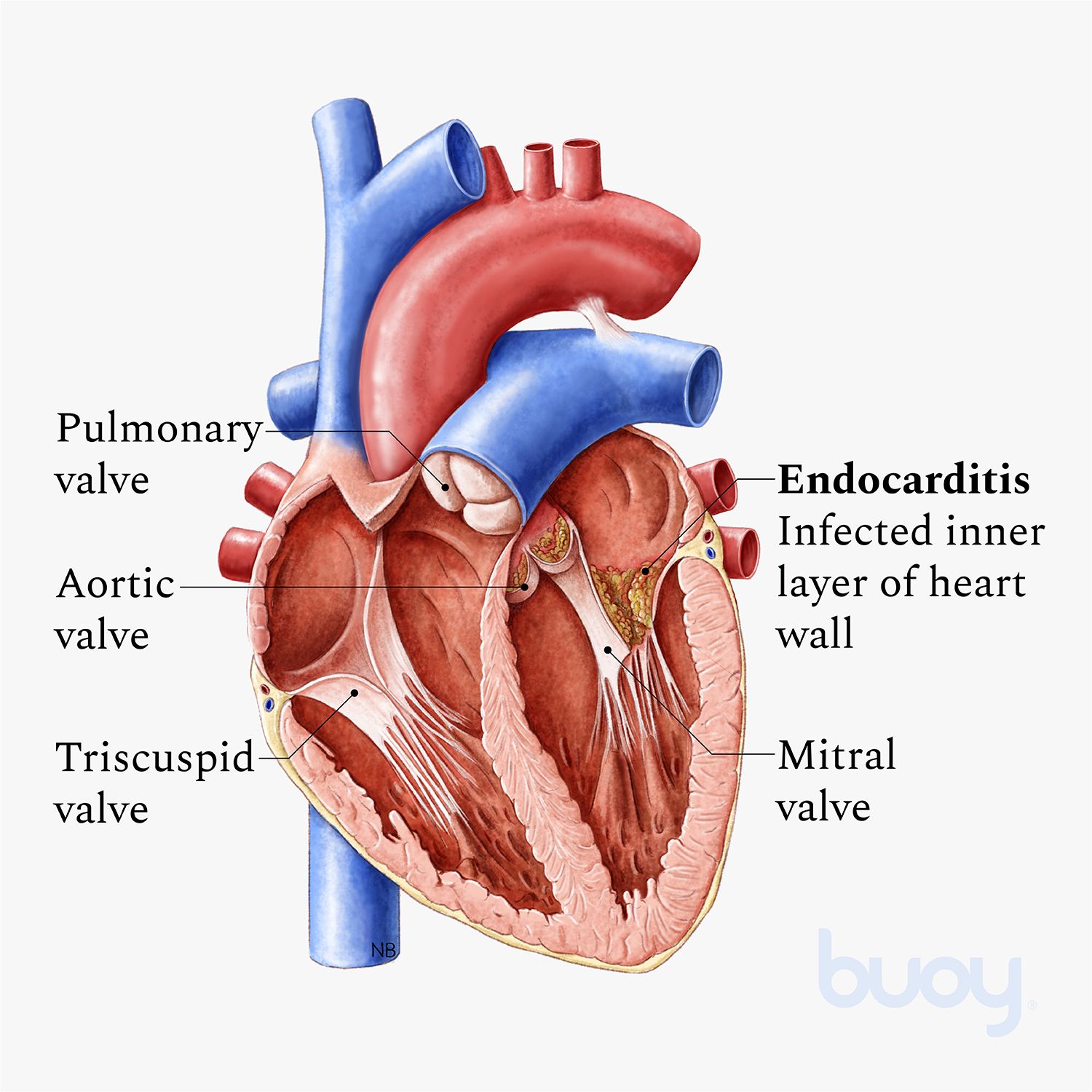
Infective endocarditis (IE) is a serious infection of the inner lining of the heart (the endocardium), most often affecting one or more of the heart valves. It develops when bacteria — or less commonly fungi — enter the bloodstream, settle on a valve or an area of damaged endocardium, and multiply to form clumps called vegetations. These vegetations are a mixture of microorganisms, platelets, fibrin and inflammatory cells.
Once established, the infection can destroy valve tissue, cause severe valve leakage (regurgitation), form abscesses in the heart muscle, and shed infected fragments (septic emboli) that travel to the brain, lungs, kidneys, spleen and limbs. Left untreated it is almost always fatal; even with modern treatment it remains a life-threatening condition requiring urgent, specialist care.
Endocarditis can arise on native (natural) valves, prosthetic (artificial) valves, pacemaker or defibrillator leads, and in people who inject drugs. Treatment combines several weeks of intravenous antibiotics with, in many cases, heart surgery to repair or replace the damaged valve. This page explains the causes, symptoms, diagnosis, treatment and long-term outlook, with attention to patients considering care abroad.
2. Key Facts at a Glance
| Fact | Detail |
|---|---|
| Also known as | IE, bacterial endocarditis, valve infection, subacute/acute endocarditis |
| Body system affected | Heart — endocardium and heart valves; secondary effects on brain, kidneys, lungs |
| Common in | People with damaged or prosthetic valves, injection-drug users, those with implanted cardiac devices, dialysis patients |
| Severity range | Serious to life-threatening; a medical emergency |
| Key treatments | Prolonged IV antibiotics; valve repair or replacement surgery when indicated |
| Outlook | Good with early diagnosis and treatment; poor if diagnosis is delayed or complications develop |
3. Alternative Names and Medical Terminology
- Infective endocarditis (IE) — the current preferred term.
- Bacterial endocarditis — older term, still widely used when bacteria are the cause.
- Subacute bacterial endocarditis (SBE) — a slower, smouldering form, often caused by Streptococcus viridans.
- Acute infective endocarditis — a rapidly progressive, aggressive form, often due to Staphylococcus aureus.
- Prosthetic valve endocarditis (PVE) — infection of an artificial valve.
- Native valve endocarditis (NVE) — infection of a natural valve.
- Cardiac device-related endocarditis — involving pacemaker or ICD leads.
- Common abbreviation: IE; the infected clumps are vegetations.
4. Relevant Heart, Lung or Vascular Anatomy
The heart has four chambers and four valves lined by a thin membrane, the endocardium. The four valves — aortic, mitral, tricuspid and pulmonary — open and close to keep blood flowing in one direction.
- The mitral and aortic valves (left side) are most commonly affected in the general population.
- The tricuspid valve (right side) is most often involved in people who inject drugs, as bacteria enter through veins.
- Valve leaflets have a poor blood supply, which limits the immune system’s ability to clear infection once bacteria attach.
- Nearby structures at risk include the valve annulus, the aortic root, the chordae tendineae and the conduction system, damage to which can cause heart block.
5. How the Disease Affects the Body
Infective endocarditis harms the body in three main ways. First, the infection destroys valve tissue. As vegetations grow and eat into a valve, the leaflets can tear, perforate or fail to close, producing severe regurgitation (backward leakage) that forces the heart to work harder and can quickly lead to heart failure — the most common reason patients with IE need surgery.
Second, infected fragments break off and become septic emboli that lodge in distant organs. A fragment reaching the brain can cause a stroke; one reaching the lungs (in right-sided IE) causes septic pulmonary emboli; fragments in the kidneys, spleen or limbs cause infarcts and abscesses. Emboli can also weaken artery walls, forming mycotic aneurysms that may rupture.
Third, the body mounts a systemic inflammatory and immune response. Ongoing infection releases bacteria and toxins into the blood, causing sepsis and organ dysfunction, while immune complexes damage the kidneys (glomerulonephritis) and produce the classic skin and eye signs. Infection may also burrow into the heart muscle to form an abscess, disrupting the conduction system.
6. Types and Classification
Infective endocarditis is classified in several overlapping ways:
- By valve type: native valve endocarditis (NVE) versus prosthetic valve endocarditis (PVE). PVE is further split into early (within 12 months of surgery, often hospital-acquired) and late (after 12 months).
- By side of the heart: left-sided (mitral/aortic — most common) versus right-sided (tricuspid/pulmonary — typical of injection-drug use).
- By speed of onset: acute (aggressive, days) versus subacute/chronic (indolent, weeks to months).
- By setting: community-acquired, healthcare-associated, and injection-drug-use-associated.
- By causative organism: streptococcal, staphylococcal, enterococcal, fungal, or culture-negative endocarditis.
- Device-related endocarditis involving pacemaker/ICD leads is a distinct, growing category.
7. Causes of the Disease
Infective endocarditis is caused by microorganisms entering the bloodstream and colonising the heart. Common causes include:
- Staphylococcus aureus — the leading cause overall in many countries; aggressive and often affects previously normal valves.
- Streptococcus viridans — from the mouth; classic cause of subacute endocarditis, often after dental work.
- Enterococci — from the gastrointestinal or genitourinary tract, especially in older adults.
- Staphylococcus epidermidis (coagulase-negative staph) — a major cause of prosthetic valve infection.
- The HACEK group and other fastidious organisms — less common bacteria that are slow to grow in culture.
- Fungi (Candida, Aspergillus) — rare but severe, seen in immunocompromised patients, IV-drug users and those with prosthetic valves.
Bacteria enter through dental or gum disease, skin infections, intravenous lines, contaminated needles, surgery, or invasive procedures.
8. How the Disease Develops
Infective endocarditis usually begins where the endocardium is already abnormal or damaged. Turbulent blood flow across a diseased or prosthetic valve injures the smooth lining, and a small clot of platelets and fibrin forms over the injured area — a sterile lesion called nonbacterial thrombotic endocarditis.
When bacteria enter the bloodstream (bacteraemia) — for example after dental work, a skin infection, or injection of drugs — they can stick to this sticky platelet-fibrin patch. Some organisms, notably Staphylococcus aureus, are able to attach even to healthy, undamaged valves.
Once attached, bacteria multiply and are covered by more platelets and fibrin, forming a protective vegetation. Deep inside, the bacteria are shielded from white blood cells and only partly reached by antibiotics, which is why treatment must be intravenous and prolonged. As the vegetation grows it destroys the valve, sheds emboli and may invade surrounding tissue to form an abscess — a cycle that continues until the heart fails or a catastrophic embolic event occurs.
9. Risk Factors
Cardiac risk factors:
- Previous infective endocarditis (strongest single risk factor)
- Prosthetic heart valves or valve repair with prosthetic material
- Damaged or leaking valves (rheumatic, degenerative, congenital)
- Certain unrepaired congenital heart defects
- Hypertrophic cardiomyopathy; mitral valve prolapse with regurgitation
- Implanted pacemakers and defibrillators
Non-cardiac and behavioural risk factors:
- Injection drug use (needle-borne bacteria)
- Poor dental hygiene and gum disease
- Long-term intravenous catheters or dialysis access
- Recent invasive or surgical procedures
- Weakened immune system, diabetes, or chronic illness
10. Genetic and Family-History Factors
Infective endocarditis is not an inherited disease and does not run in families in the way genetic disorders do. However, some underlying conditions that predispose to IE do have a genetic or congenital basis. These include inherited connective-tissue disorders (such as Marfan syndrome) that cause valve or aortic problems, bicuspid aortic valve (a common congenital variant present from birth), and various congenital heart defects.
People born with these structural abnormalities carry a lifelong, higher risk of endocarditis and may need extra vigilance and, in specific cases, antibiotic prophylaxis. If you or a close relative has a congenital heart condition, mention this to your cardiologist so appropriate preventive advice can be given.
11. Who Is Most at Risk?
Those at greatest risk of infective endocarditis include:
- People with a prosthetic heart valve or previous valve surgery.
- Anyone with a prior episode of endocarditis.
- People who inject drugs, who are especially prone to right-sided (tricuspid) infection.
- Patients with structural heart disease — congenital defects, rheumatic valve disease, degenerative valve disease.
- People with implanted cardiac devices (pacemakers, ICDs).
- Dialysis patients and those with long-term central venous catheters.
- Older adults, in whom degenerative valve disease and healthcare exposure are common.
- Immunocompromised and diabetic patients.
12. Prevalence and Epidemiology
Infective endocarditis is relatively uncommon but far from rare, and its incidence has been slowly rising in many high-income countries. It affects roughly a few people per 100,000 population each year, though rates are higher in specific groups such as injection-drug users and dialysis patients.
The disease profile has shifted over recent decades. In wealthier nations, rheumatic disease has declined while degenerative valve disease, prosthetic valves, cardiac devices and injection-drug use have become more important, and Staphylococcus aureus has overtaken viridans streptococci as the leading organism. In lower-income countries, rheumatic heart disease in younger patients remains a major cause. Men are affected somewhat more often than women, and the average patient age has risen. These figures are approximate and vary by setting.
13. Signs and Symptoms
Infective endocarditis can develop suddenly and severely (acute) or slowly over weeks (subacute), so the picture varies widely. Common features include:
- Fever and chills — the most common symptom, sometimes low-grade in subacute cases.
- A new or changed heart murmur — a key clinical clue.
- Fatigue, weakness, night sweats and unexplained weight loss.
- Aching joints and muscles.
- Shortness of breath, especially if valve leakage causes heart failure.
More specific signs reflect emboli and immune reactions: petechiae (tiny red-purple spots on the skin, mouth or eyes), splinter haemorrhages under the nails, Janeway lesions (painless palm/sole spots), Osler nodes (painful finger nodules) and Roth spots in the eye. Because symptoms are often vague, a persistent unexplained fever in an at-risk person should always prompt evaluation.
14. Early-Stage Symptoms
In the subacute form, early symptoms are often mild and non-specific, which is why diagnosis is frequently delayed. Watch for:
- Persistent low-grade fever lasting days to weeks.
- Unusual tiredness, weakness and loss of energy.
- Night sweats and mild weight loss.
- Vague muscle and joint aches.
- Poor appetite and a general “unwell” feeling.
In at-risk people — those with prosthetic valves, prior endocarditis, or injection-drug use — any of these early, unexplained symptoms should trigger prompt medical review and blood cultures.
15. Advanced-Stage Symptoms
As the disease progresses and complications develop, more dramatic symptoms appear:
- Breathlessness, ankle swelling and inability to lie flat — signs of heart failure from valve destruction.
- Sudden neurological symptoms — weakness, slurred speech, confusion or loss of consciousness from a stroke.
- Sharp chest or flank pain from emboli to the lungs, kidneys or spleen.
- Blood in the urine and reduced urine output from kidney involvement.
- High fever with rigors and signs of sepsis — rapid heart rate, low blood pressure, drowsiness.
- Visible skin and nail signs (petechiae, splinter haemorrhages, Osler nodes).
These features signal a medical emergency requiring immediate hospital care.
16. Symptoms in Women, Men and Older Adults
- Men are affected more often than women overall, particularly with injection-drug-related, right-sided disease.
- Women may experience more non-specific and constitutional symptoms and can face diagnostic delay; outcomes are broadly similar when treated promptly.
- Older adults often present atypically — with confusion, loss of appetite, falls, general decline or low-grade fever rather than classic signs. Degenerative valve disease, cancer screening bacteraemia and healthcare procedures make IE an important consideration in any older patient with unexplained fever or a new murmur.
- In all groups, a new heart murmur with fever deserves urgent evaluation.
17. Emergency Warning Signs
Call emergency services or go to hospital immediately if a person with known heart-valve disease, a prosthetic valve, or a history of injection-drug use develops:
- Sudden weakness, facial droop, slurred speech or confusion (possible stroke).
- Severe breathlessness or inability to lie flat.
- High fever with shaking chills, drowsiness or collapse (possible sepsis).
- Sudden severe chest, back or abdominal pain.
- A cold, painful, pulseless limb (arterial embolism).
18. When to Seek Medical Help
See a doctor promptly if you have a fever lasting more than a few days that has no obvious cause, especially if you have a heart-valve problem, a prosthetic valve, a previous episode of endocarditis, an implanted cardiac device, or you inject drugs. Do not start antibiotics on your own before blood cultures are taken, as this can mask the infection and make diagnosis much harder. Early assessment with blood tests and an echocardiogram can be life-saving.
19. Disease Stages, Grades and Severity
Infective endocarditis is not staged like cancer, but its severity and diagnostic certainty are graded in structured ways.
- Modified Duke Criteria classify cases as definite, possible, or rejected endocarditis, based on combinations of major criteria (positive blood cultures with typical organisms; echocardiographic evidence of vegetation, abscess or new valve leak) and minor criteria (fever, predisposing heart condition, vascular and immune phenomena, suggestive microbiology).
- Severity is judged by the presence of complications: heart failure, perivalvular abscess, large mobile vegetations, systemic emboli, persistent infection despite antibiotics, and prosthetic-valve involvement.
- Cases with these complicated features carry a higher risk and more often require urgent surgery.
20. Disease Progression
Untreated, infective endocarditis follows a relentless downhill course. The vegetation enlarges, progressively destroying the valve and worsening regurgitation until the heart can no longer cope, producing acute heart failure, while fragments repeatedly embolise to the brain and other organs. Infection may spread into the heart muscle, forming an abscess that causes heart block, or loosen a prosthetic valve (dehiscence). The speed depends on the organism: Staphylococcus aureus can destroy a valve within days, whereas viridans streptococcal disease may smoulder for weeks. Prompt antibiotics and, where needed, surgery halt this progression.
21. Possible Complications
- Heart failure — the leading complication and most common reason for surgery.
- Stroke and systemic emboli — to brain, spleen, kidneys, limbs and gut.
- Perivalvular abscess and heart block from conduction damage.
- Valve destruction, perforation or prosthetic dehiscence.
- Septic pulmonary emboli (right-sided disease) and mycotic aneurysms that may rupture.
- Kidney injury, and persistent or recurrent infection with sepsis and multi-organ failure.
22. Related and Associated Medical Conditions
Infective endocarditis is closely linked with several other conditions, both as causes and consequences:
- Valvular heart disease — mitral valve disease and aortic valve disease both predispose to and result from IE.
- Congenital heart disease and bicuspid aortic valve.
- Rheumatic heart disease, still important worldwide.
- Heart failure — a frequent complication.
- Disorders of heart rhythm, especially heart block from abscess.
- Sepsis, stroke and kidney disease as systemic consequences.
- Diabetes, chronic kidney disease and immunosuppression as contributing comorbidities.
23. Screening and Early Detection
There is no population screening programme for infective endocarditis because it is uncommon and cannot be predicted by a simple test. Instead, early detection relies on clinical vigilance in high-risk people. Anyone with a prosthetic valve, prior endocarditis, congenital heart disease or injection-drug use should be aware of the warning signs and seek early blood cultures for any unexplained persistent fever.
Good dental care and prompt treatment of infections are the practical “screening” equivalents — reducing the bacteraemia that seeds the heart. Clinicians maintain a low threshold to order blood cultures and an echocardiogram whenever endocarditis is plausible.
24. How the Disease Is Diagnosed
Diagnosing infective endocarditis relies on combining clinical findings, blood cultures and cardiac imaging, formalised in the Modified Duke Criteria. No single test is definitive; the diagnosis is built from a pattern of evidence.
The two cornerstones are:
- Blood cultures — usually three sets drawn from different sites before antibiotics are started. Repeatedly positive cultures growing a typical organism are a major diagnostic criterion and identify the bacteria so that antibiotics can be targeted.
- Echocardiography — a transthoracic echocardiogram (TTE) is the first imaging test, but a transoesophageal echocardiogram (TOE/TEE), in which a probe is passed into the gullet behind the heart, is far more sensitive for vegetations, abscesses and prosthetic valve infection.
Supporting tests include inflammatory markers, an ECG (to detect conduction problems suggesting abscess), and, in difficult cases, cardiac CT, MRI, or PET-CT. A cardiologist, microbiologist, cardiac surgeon and infection specialist typically review the findings together as an “Endocarditis Team” to confirm the diagnosis and plan treatment.
25. Physical Examination and Medical History
The doctor will ask about fever, night sweats, weight loss, recent dental or surgical procedures, injection-drug use, prosthetic valves and previous endocarditis. On examination they look for:
- A new or changed heart murmur — a central clue.
- Fever and signs of sepsis.
- Peripheral signs — splinter haemorrhages under the nails, petechiae, Osler nodes (painful finger nodules), Janeway lesions (painless palm/sole spots) and Roth spots in the eyes.
- Splenomegaly (enlarged spleen) and signs of heart failure.
- Evidence of emboli — neurological deficits, cold limbs or flank tenderness.
26. Diagnostic Tests and Imaging
- Transthoracic echocardiogram (TTE) — initial, non-invasive scan of valve structure and function.
- Transoesophageal echocardiogram (TOE/TEE) — the gold standard for vegetations, abscesses and prosthetic valves.
- Electrocardiogram (ECG) — checks for conduction block from abscess and rhythm disturbance.
- Chest X-ray — looks for heart failure and septic lung emboli.
- Cardiac CT, MRI and PET-CT — define abscesses and valve anatomy and help in prosthetic-valve or device infection.
- Brain/abdominal imaging — to identify embolic strokes, organ infarcts and mycotic aneurysms.
27. Blood Tests, Biomarkers and Genetic Testing
- Blood cultures — the single most important test; multiple sets before antibiotics identify the organism and guide therapy.
- Full blood count — often shows anaemia and a raised white-cell count.
- Inflammatory markers — CRP and ESR are usually elevated and help track treatment response.
- Kidney and liver function tests — assess organ involvement and guide antibiotic dosing.
- Urinalysis — may reveal blood or protein from kidney involvement.
- Specialised serology or PCR — for culture-negative endocarditis. There is no genetic test for IE itself.
28. Understanding Test Results
- Persistently positive blood cultures with a typical organism strongly support the diagnosis and tell the team which antibiotic to use.
- A vegetation, abscess or new valve leak on echocardiography is major evidence of endocarditis.
- Negative initial cultures do not exclude IE — the patient may have received antibiotics, or the organism may be hard to grow (culture-negative endocarditis).
- Rising CRP/ESR suggests ongoing infection; falling levels indicate a response to treatment.
- New conduction abnormality on ECG raises concern for a root abscess and often prompts surgery.
- Results are always interpreted together, using the Duke framework, rather than in isolation.
29. Differential Diagnosis
Because its symptoms are non-specific, infective endocarditis can be confused with many conditions, including:
- Other causes of prolonged fever of unknown origin.
- Sepsis or bacteraemia from another source.
- Rheumatic fever and autoimmune conditions such as lupus (which can cause non-infective valve lesions).
- Cancers, especially lymphoma, and atrial myxoma (a heart tumour that can mimic vegetations).
- Vasculitis and connective-tissue disease.
- Non-bacterial thrombotic (marantic) endocarditis associated with cancer.
- Antiphospholipid syndrome.
30. Specialist and Multidisciplinary Evaluation
Modern guidelines recommend that infective endocarditis be managed by a dedicated “Endocarditis Team” at a centre with cardiac surgery on site. This team typically includes a cardiologist, cardiac surgeon, infectious-diseases specialist and clinical microbiologist, plus imaging, neurology and, where relevant, addiction specialists. Together they confirm the diagnosis, monitor antibiotics, decide whether and when surgery is needed, and manage complications such as stroke — an approach shown to improve outcomes. Find experienced doctors and specialist hospitals through our directory.
31. Treatment Goals
The main goals of treating infective endocarditis are to:
- Eradicate the infection completely with targeted, prolonged antibiotics.
- Prevent or treat complications — heart failure, emboli, abscess and sepsis.
- Preserve or restore valve function, repairing or replacing the valve when destroyed.
- Remove infected material when antibiotics alone cannot control the infection.
- Prevent recurrence by treating the underlying source of bacteraemia.
- Restore the best possible quality of life and long-term heart health.
32. When Is Treatment Required?
Infective endocarditis always requires urgent treatment — it is never a “watch and wait” condition once diagnosed. Intravenous antibiotics should begin as soon as blood cultures have been taken. The key decisions are which antibiotics and, critically, whether surgery is needed and how soon. Surgery is considered when there is heart failure from valve destruction, uncontrolled or persistent infection (including abscess), or a high risk of embolism from large mobile vegetations. In some situations surgery is performed as an emergency (within 24 hours), in others urgently (within days), and in others electively after a course of antibiotics.
33. Active Monitoring and Watchful Waiting
True “watchful waiting” has no role in confirmed infective endocarditis, because the infection progresses without treatment. However, patients who respond well to antibiotics and do not have surgical indications are managed with close in-hospital monitoring rather than an operation. This involves repeated clinical assessment, serial echocardiograms, tracking of inflammatory markers and blood cultures, and continuous vigilance for emerging complications. The team continually reassesses whether the patient can complete antibiotics without surgery or whether the situation is changing and an operation has become necessary.
34. Medications
The foundation of treatment is prolonged intravenous (IV) antibiotic therapy, typically for 4–6 weeks, matched to the organism and its sensitivities. Key points include:
- Empirical antibiotics are started immediately after cultures are taken, then narrowed once the organism is identified.
- Bactericidal, high-dose IV combinations are used because bacteria deep within vegetations are hard to kill.
- Prosthetic-valve infection usually needs longer courses and often an additional agent such as rifampicin plus gentamicin.
- Antifungal drugs are needed for fungal endocarditis, usually combined with surgery.
- Some stable patients complete part of the course with outpatient IV antibiotics (OPAT) or a partial oral regimen under specialist supervision.
- Supportive medicines treat heart failure, fever and pain; anticoagulation is used cautiously.
Explore related treatments in our directory.
35. Minimally Invasive Treatments
For most patients, the “minimally invasive” element of care is non-surgical management with antibiotics and, where suitable, outpatient IV antibiotic therapy to avoid a prolonged hospital stay. When surgery is required, some experienced centres can perform valve operations through smaller incisions — such as a mini-thoracotomy or partial sternotomy — rather than a full breastbone opening. These minimally invasive cardiac approaches may reduce blood loss, pain and recovery time, but suitability depends on the extent of infection, the valve involved and the presence of abscess; extensive destruction usually requires conventional open surgery.
36. Catheter-Based and Endovascular Treatments
Traditional catheter-based valve procedures play a limited role in active infective endocarditis, and infected valves are generally not treated by percutaneous valve implantation. However, catheter techniques are used in specific situations:
- Percutaneous lead extraction — removing infected pacemaker or defibrillator leads.
- Aspiration/debulking devices — in selected right-sided (tricuspid) endocarditis, specialised catheter systems can remove large vegetations in patients too high-risk for surgery.
- Endovascular treatment of mycotic aneurysms — coiling or stenting to seal an infected, weakened artery.
- Drainage of abscesses and management of embolic complications.
37. Surgical Treatment Options
Surgery is needed in a substantial proportion of patients and can be life-saving. The main indications are heart failure from a destroyed valve, uncontrolled infection (abscess or persistent positive cultures), and prevention of embolism from large, mobile vegetations. The core operations are:
- Valve repair — preferred when feasible, particularly for the mitral valve, preserving the patient’s own tissue by removing infected areas and reconstructing the valve.
- Valve replacement — when the valve is too damaged to repair, using a mechanical or biological (tissue) prosthesis. This is common for the aortic valve.
- Radical debridement — removing all infected tissue, draining abscesses and reconstructing the valve annulus or aortic root, sometimes with a homograft (human donor valve).
- Device/lead removal — complete extraction of infected pacemaker or ICD systems.
The surgeon removes vegetations and infected tissue, repairs or replaces the valve, and drains any abscess; an infected prosthesis is usually replaced. Timing is crucial — delaying surgery in a deteriorating patient worsens outcomes, while operating on an actively infected valve carries its own risks. The decision is made by the multidisciplinary Endocarditis Team. Browse our surgery and procedures sections for more detail.
38. Advanced and Emerging Treatments
- Rapid molecular diagnostics — PCR and next-generation sequencing to identify hard-to-culture organisms quickly, improving culture-negative diagnosis.
- Advanced imaging — routine use of PET-CT and cardiac CT to detect prosthetic-valve infection, hidden abscesses and silent emboli.
- Percutaneous vegetation aspiration systems for high-risk right-sided disease.
- Shorter and partial-oral antibiotic regimens for selected, stable patients, reducing hospital time.
- Tissue-engineered and antibiotic-impregnated valves and grafts to reduce reinfection.
- Bacteriophage therapy and novel antimicrobials under investigation for resistant organisms.
39. Treatment Options Compared
- Antibiotics alone — appropriate for uncomplicated cases that respond well, with no heart failure, abscess or high embolic risk. Avoids surgery but requires weeks of IV therapy and close monitoring.
- Antibiotics plus surgery — necessary when there is valve destruction with heart failure, abscess, persistent infection or dangerous vegetations. Offers the best chance of cure in complicated disease but carries operative risk.
- Valve repair vs replacement — repair preserves natural tissue and avoids long-term anticoagulation but is not always possible; replacement is definitive but introduces a prosthesis that itself can become infected.
- Mechanical vs tissue valve — mechanical valves last longer but need lifelong blood thinners; tissue valves avoid anticoagulation but may wear out sooner.
The right choice depends on the individual and is made by the specialist team.
40. How Doctors Choose the Right Treatment
Treatment is tailored to each patient. Key factors doctors weigh include:
- The causative organism and its antibiotic sensitivity.
- Native versus prosthetic valve, and which valve is involved.
- Presence of heart failure, abscess, or persistent infection.
- Size and mobility of vegetations and any embolic events already suffered.
- The patient’s overall health, age and surgical risk.
- Whether the infection is controlled by antibiotics.
- Neurological status — recent stroke affects the timing of surgery.
- Patient preferences regarding valve type and lifestyle.
41. Benefits and Risks of Treatment
Benefits:
- Cure of a life-threatening infection and prevention of death.
- Restoration of valve function and relief of heart failure.
- Prevention of stroke and further emboli.
- Return to normal or near-normal activity for most successfully treated patients.
Risks:
- Antibiotics — allergic reactions, kidney toxicity, IV-line infections and antibiotic resistance.
- Surgery — bleeding, stroke, infection, heart rhythm problems and the general risks of major heart surgery, which are higher during active infection.
- Prosthetic valves — risk of reinfection, clotting and need for anticoagulation.
- Risk of recurrent endocarditis, particularly if the underlying cause persists.
42. What Happens If the Disease Is Left Untreated?
Untreated infective endocarditis is almost universally fatal. Without antibiotics, the infection continues to destroy the valve, causing progressive heart failure; repeatedly sheds emboli, causing strokes and organ infarction; and burrows into the heart to form abscesses and heart block, with overwhelming sepsis eventually following. Even a short delay worsens the outlook, so any at-risk person with an unexplained persistent fever must be assessed promptly — early treatment transforms an otherwise lethal disease into a curable one for most patients.
43. Treatment Success and Expected Outcomes
With early diagnosis and appropriate treatment, many patients are cured of infective endocarditis. Outcomes depend strongly on the organism, the valve involved, the presence of complications and how quickly treatment began.
- Viridans streptococcal, native-valve disease treated promptly generally has the best outcomes.
- Staphylococcus aureus, prosthetic-valve, fungal, and complicated disease carry higher risks.
- Combined medical-plus-surgical treatment improves survival in complicated cases.
- Most survivors regain good function, though some are left with a repaired or prosthetic valve requiring lifelong follow-up.
In-hospital mortality remains significant for complicated disease, which is why specialist, high-volume care matters. Figures vary widely and should be discussed individually with your team.
44. Prognosis and Long-Term Outlook
The long-term outlook depends on how much heart damage occurred, whether surgery was needed, and the underlying cause. Patients treated early, with a favourable organism and no major complications, often make a full recovery and return to normal life, though they carry a lifelong increased risk of a future episode and must take prevention seriously.
Those left with a prosthetic valve, significant valve dysfunction, or a history of stroke need ongoing cardiology follow-up. Prosthetic-valve and fungal endocarditis have a more guarded prognosis and higher relapse rates. Recurrence is a real concern — particularly for people who continue to inject drugs — so addressing the root cause is essential. Regular monitoring, good dental hygiene and prompt attention to future fevers give patients the best chance of a good long-term outcome. Overall survival has improved with modern team-based care, but IE remains a serious condition.
45. Recovery and Rehabilitation
Recovery is often prolonged because treatment itself lasts weeks. Key elements include:
- Completing the full IV antibiotic course, sometimes at home via OPAT.
- Gradual mobilisation and rebuilding of strength and stamina.
- Cardiac rehabilitation after valve surgery — supervised exercise, education and lifestyle support.
- Wound care and recovery from surgery, typically several weeks to a few months.
- Attention to nutrition, dental health and any contributing addiction.
- Emotional support, as many patients feel exhausted after such a serious illness.
46. Follow-Up Tests and Long-Term Monitoring
After treatment, follow-up aims to confirm cure and detect any relapse or valve problem. It typically includes:
- A repeat (baseline) echocardiogram at the end of treatment to document valve function.
- Clinical review, blood tests and inflammatory markers in the months after discharge.
- Ongoing cardiology follow-up, especially for those with prosthetic or repaired valves.
- Prompt blood cultures for any new fever, as relapse is most likely in the first months.
- Regular dental checks and reinforcement of prevention.
47. Managing Recurrence or Disease Progression
Recurrent endocarditis may be a relapse (same organism, usually within weeks) or a new infection (later, often a different organism). Management focuses on preventing recurrence by eliminating the underlying source — treating gum disease, removing infected catheters or devices, and supporting people to stop injecting drugs. Any new unexplained fever in a previous IE patient should be treated as possible endocarditis, with blood cultures before antibiotics and an early echocardiogram. Prosthetic-valve relapse often needs repeat surgery, planned by the specialist team.
48. Living with the Disease
Life after infective endocarditis centres on preventing another episode and looking after a heart that may now have a repaired or prosthetic valve. Practical steps include:
- Carrying a patient alert card noting your valve condition and IE history.
- Maintaining excellent dental hygiene and regular dental visits.
- Telling every dentist and doctor about your history so antibiotic prophylaxis can be considered when appropriate.
- Cleaning and caring for any skin wounds promptly.
- Taking prescribed medicines, including anticoagulants if you have a mechanical valve, exactly as directed.
- Seeking early help for any fever, and avoiding self-medication with antibiotics.
49. Diet and Nutrition Guidelines
There is no special “endocarditis diet”, but good nutrition supports recovery and heart health:
- Eat a balanced, heart-healthy diet rich in vegetables, fruit, whole grains and lean protein.
- Ensure adequate protein and calories during recovery to help tissue healing after severe infection.
- If you have heart failure or a prosthetic valve, follow any advice on limiting salt and fluid.
- If you take warfarin, keep your intake of vitamin-K-rich foods (green leafy vegetables) consistent rather than fluctuating.
- Stay well hydrated and limit alcohol.
- Maintaining good nutrition also strengthens the immune system against future infection.
50. Exercise and Physical-Activity Guidelines
During active infection, rest is important and strenuous activity should be avoided. As you recover:
- Increase activity gradually, guided by your care team.
- Attend cardiac rehabilitation after valve surgery for a structured, supervised programme.
- Aim for regular moderate activity — such as walking — once cleared, to rebuild fitness.
- Avoid heavy lifting and strenuous upper-body exercise while a surgical breastbone wound heals (usually several weeks).
- Those with residual valve disease or heart failure should follow individualised activity advice from their cardiologist.
51. Medications, Activities and Habits to Avoid
- Do not stop antibiotics early — completing the full course is essential to cure.
- Do not take antibiotics for a fever before blood cultures — this can hide the diagnosis.
- Avoid injection drug use, the single most preventable and dangerous risk factor.
- Avoid poor dental hygiene and non-sterile procedures such as unregulated tattooing or piercing.
- If on warfarin, keep a steady diet and check before starting interacting drugs.
- Do not ignore new fevers, breathlessness or neurological symptoms.
52. Preventing the Disease or Reducing Its Risks
Prevention focuses on reducing bacteraemia and protecting vulnerable valves:
- Maintain excellent oral and dental hygiene with regular check-ups — the most important everyday measure.
- Antibiotic prophylaxis before certain dental and invasive procedures for high-risk patients (prosthetic valves, previous endocarditis, specific congenital disease). Guidelines (ACC/AHA, ESC, NICE) differ, so follow your specialist’s advice.
- Never inject drugs; access support services if you do.
- Care meticulously for IV lines and catheters and treat skin infections promptly.
- Keep vaccinations up to date and manage conditions such as diabetes.
53. Pregnancy and the Disease
Infective endocarditis in pregnancy is uncommon but very serious, threatening both mother and baby. Women with a prosthetic valve, congenital heart disease or previous endocarditis should have pre-pregnancy counselling and be cared for by a joint cardiac-obstetric team. Antibiotic choice must consider fetal safety, and the timing of any necessary surgery is complex. Women on warfarin for a mechanical valve need careful anticoagulation planning throughout pregnancy. Any pregnant woman with a heart-valve condition and unexplained fever should be assessed urgently.
54. Disease in Children and Young Adults
In children and young adults, infective endocarditis usually occurs against a background of congenital heart disease, previous cardiac surgery, or, in adolescents and young adults, injection-drug use. Presentation may be non-specific, so a high index of suspicion is needed in a child with a heart defect and persistent fever. Rheumatic heart disease remains an important underlying cause in many countries. Management principles are similar to adults — prolonged IV antibiotics and surgery when indicated — but doses, valve choices and long-term planning are tailored by paediatric cardiac specialists. See our congenital heart procedures section.
55. Disease in Older Adults
Older adults now make up a growing proportion of endocarditis patients. Contributing factors include degenerative (calcific) valve disease, prosthetic valves, cardiac devices, healthcare procedures and bacteraemia from the gut (for example, from colon conditions, which raises concern for Streptococcus gallolyticus). Presentation is often atypical — confusion, weakness, poor appetite or falls rather than classic signs. Older patients may be higher-risk surgical candidates, so treatment decisions carefully balance the benefits of surgery against frailty and other illnesses, ideally through the multidisciplinary team.
56. Emotional Health and Patient Support
A diagnosis of infective endocarditis — a sudden, severe, weeks-long illness often followed by major surgery — can be frightening and exhausting. Many patients experience anxiety, low mood or fear of recurrence. Helpful support includes:
- Clear information from your care team about progress and outlook.
- Cardiac rehabilitation, which supports emotional as well as physical recovery.
- Counselling or psychological support where needed, including addiction services for those recovering from drug use.
- Peer-support groups and involving family and carers.
- Reassurance that, with treatment, most patients recover and return to normal life.
57. Preparing for Your Specialist Appointment
To make the most of your appointment:
- Write down all your symptoms, when they started and how they have changed.
- List all medicines, allergies and supplements you take.
- Note your medical history — valve problems, previous endocarditis, prosthetic valves, devices, recent dental or surgical procedures.
- Bring previous test results, echocardiograms and hospital records if available.
- Be honest about injection-drug use — it is vital for correct treatment.
- Bring a companion and a written list of questions.
58. Questions to Ask Your Doctor
- What type of endocarditis do I have, and which organism is causing it?
- Which valve is affected, and how badly is it damaged?
- Will antibiotics alone cure me, or will I need surgery?
- How long will my antibiotic treatment last, and can any be given at home?
- If I need surgery, will my valve be repaired or replaced, and what type of valve?
- What complications should I watch for, and what are my warning signs?
- What is my risk of another episode, and how can I prevent it?
- Do I need antibiotics before dental or surgical procedures in future?
- When can I return to work, exercise and normal activities?
- What follow-up tests and appointments will I need?
59. Cost of Diagnosis and Treatment
Costs vary enormously by country, hospital and complexity. The table gives approximate, indicative ranges for a full episode (diagnosis, hospital stay, IV antibiotics, and where needed valve surgery). Medical-tourism destinations are often substantially less expensive.
| Region | Approx. cost (medical therapy) | Approx. cost with valve surgery |
|---|---|---|
| United States | $30,000–$80,000+ | $120,000–$250,000+ |
| United Kingdom / Western Europe | $20,000–$50,000 | $50,000–$120,000 |
| India | $4,000–$10,000 | $8,000–$20,000 |
| Turkey | $5,000–$12,000 | $12,000–$25,000 |
| Thailand / Singapore | $6,000–$15,000 | $15,000–$40,000 |
Figures are approximate and for general guidance only; obtain a written quote from the treating hospital.
60. Factors Affecting Treatment Cost
- Length of hospital stay — often several weeks for IV antibiotics.
- Whether surgery is required, and its complexity (repair vs replacement, single vs multiple valves).
- Type of prosthetic valve used and any antifungal therapy.
- Intensive-care needs and management of complications such as stroke or kidney failure.
- Diagnostic tests — repeated echocardiograms, CT, MRI, PET-CT.
- Country and hospital chosen, and whether follow-up and rehabilitation are included.
61. Choosing the Right Specialist
Look for a cardiologist or cardiac surgeon experienced in valve disease and endocarditis, ideally practising within a formal Endocarditis Team. Helpful considerations include:
- Board certification and specific experience with valve surgery and infection.
- Access to a multidisciplinary team (cardiology, surgery, infectious diseases, microbiology, imaging).
- Good volume and published outcomes for valve procedures.
- Availability of advanced imaging and on-site cardiac surgery.
- Clear communication and willingness to answer your questions.
Browse experienced doctors in our directory.
62. Choosing the Right Hospital or Treatment Centre
Endocarditis is best treated at a specialist heart centre with cardiac surgery on site and a dedicated endocarditis service. When choosing a hospital, consider:
- International accreditation such as JCI, indicating recognised quality and safety standards.
- High case volumes and good outcomes for valve surgery.
- Availability of a 24/7 cardiac surgical and intensive-care service.
- Full imaging and microbiology facilities.
- Experience with international patients, if travelling abroad.
Explore leading hospitals and destinations in our directory.
63. Getting a Second Medical Opinion
Because decisions in endocarditis — especially whether and when to operate — are complex and consequential, a second opinion can be valuable, particularly for prosthetic-valve infection, borderline surgical indications, or a poor response to treatment. A second specialist can confirm the diagnosis, review imaging and cultures, and discuss alternatives. In an acute emergency, however, treatment must not be delayed. For stable patients and long-term planning, seeking another expert view is reasonable and often reassuring. Request a second opinion through our team.
64. Treatment Abroad and Medical-Travel Considerations
Many patients travel abroad for valve surgery following endocarditis, drawn by high-quality care at lower cost in countries such as India, Turkey, Thailand and Singapore. Important considerations include:
- Active infection usually requires urgent local treatment — travel suits planned valve surgery after infection is controlled.
- Choose JCI-accredited hospitals with experienced cardiac teams.
- Plan for a lengthy stay, as recovery takes weeks.
- Arrange clear handover of records, cultures and imaging between teams.
- Confirm follow-up and rehabilitation after returning home, and consider travel insurance and fitness to fly.
See our destinations guide for more.
65. Frequently Asked Questions
Is infective endocarditis contagious? No. It is an infection of your own heart valves and cannot be caught from or spread to other people.
Can it be cured? Yes — with prompt, appropriate antibiotics (and surgery when needed), many patients are cured, though they carry a lifelong higher risk of a future episode.
Why do I need weeks of antibiotics? Bacteria hide deep within the vegetation where the immune system and drugs struggle to reach them, so prolonged, high-dose IV treatment is required.
Will I definitely need surgery? Not always. Surgery is needed for heart failure, abscess, uncontrolled infection or high embolic risk; many uncomplicated cases are cured with antibiotics alone.
How did I get it? Bacteria entered your bloodstream — often from the mouth, skin, an IV line or injecting drugs — and settled on a damaged or prosthetic valve.
Do I need antibiotics before dental work in future? If you are high-risk, you may need prophylactic antibiotics; follow your specialist’s advice. Recurrence is also possible, so prevention and good dental hygiene are essential.
66. Patient Stories and Treatment Experiences
The following are representative, anonymised examples for illustration only.
-
Rajesh, India — After weeks of low-grade fever and fatigue following a dental infection, Rajesh was found to have streptococcal endocarditis on his mitral valve. Six weeks of IV antibiotics cured the infection, and a valve repair restored his heart function. He now attends regular dental check-ups.
-
Maria, Spain — Maria developed Staphylococcus aureus endocarditis on a prosthetic aortic valve. Her Endocarditis Team recommended urgent surgery to replace the infected valve, followed by prolonged antibiotics. She recovered well and returned to work after cardiac rehabilitation.
-
James, United Kingdom — James, who had a history of injecting drugs, presented with fever and breathlessness from tricuspid-valve endocarditis. Alongside antibiotics and addiction support, he recovered and has remained drug-free, greatly reducing his risk of recurrence.
67. Latest Research and Clinical Trials
Research in infective endocarditis is advancing on several fronts. Rapid molecular and sequencing techniques are improving the diagnosis of culture-negative disease. Trials of partial oral antibiotic therapy for selected, stable patients suggest that some may safely complete treatment without prolonged IV therapy, shortening hospital stays. Advanced imaging with PET-CT and cardiac CT is increasingly used to detect prosthetic-valve infection and silent emboli. Researchers are also studying optimal timing of surgery, percutaneous devices to remove right-sided vegetations, and antibiotic-impregnated or engineered valve materials to reduce reinfection. Prevention research continues to refine antibiotic-prophylaxis guidelines. Patients interested in trials should ask their specialist about reputable studies at accredited centres.
68. Related Diseases and Conditions
- Mitral Valve Disease — a common site and consequence of endocarditis.
- Aortic Valve Disease — frequently involved in left-sided IE.
- Tricuspid Valve Disease — the typical site in injection-drug-related IE.
- Heart Failure — the leading complication of valve destruction.
- Disorders of Heart Rhythm — can result from abscess and heart block.
- Congenital Heart Disease — an important predisposing condition.
69. Related Treatments and Procedures
- Aortic Valve Procedures — repair or replacement of an infected aortic valve.
- Mitral Valve Procedures — repair or replacement of an infected mitral valve.
- Minimally Invasive Cardiac Surgery — smaller-incision valve surgery in selected patients.
- Congenital Heart Procedures — for endocarditis on congenital defects.
- Endovascular Stenting — treatment of mycotic aneurysms.
70. Medical Glossary
- Endocardium — the thin inner lining of the heart chambers and valves.
- Vegetation — a clump of bacteria, platelets and fibrin on an infected valve.
- Bacteraemia — bacteria circulating in the bloodstream.
- Septic embolus — an infected fragment that breaks off and lodges in a distant organ.
- Regurgitation — backward leakage of blood through a damaged valve.
- Abscess — a walled-off pocket of infection in the heart tissue.
- Prosthetic valve — an artificial mechanical or tissue valve (versus a natural, native valve).
- Modified Duke Criteria — the standard framework for diagnosing IE.
- Transoesophageal echocardiogram (TOE/TEE) — an ultrasound probe passed into the gullet for detailed heart imaging.
- Mycotic aneurysm — an infected, weakened bulge in an artery wall.
- Debridement — surgical removal of infected or dead tissue.
- Homograft — a valve from a human donor.
- Prophylaxis — preventive antibiotics given before high-risk procedures.
71. Medical Review, Editorial Policy and Last Updated Date
Last updated: 11 July 2026.
This article was written for patient education and reviewed for general accuracy against recognised cardiology guidance, including that of the ACC/AHA, the European Society of Cardiology (ESC), the NHS and NICE. Our editorial policy is to present balanced, evidence-based information using clear language, to avoid fabricated statistics, and to update content periodically.
Disclaimer: This information is for general education only and is not a substitute for professional medical advice, diagnosis or treatment. Always consult a qualified healthcare provider about your individual situation. If you have symptoms of a serious infection or a cardiac emergency, seek medical help immediately.
72. Clinical Guidelines and Medical References
This content reflects widely accepted guidance and textbook knowledge, including:
- American College of Cardiology / American Heart Association (ACC/AHA) guidelines on valvular heart disease and endocarditis prevention.
- European Society of Cardiology (ESC) guidelines for the management of infective endocarditis.
- National Institute for Health and Care Excellence (NICE) guidance on antibiotic prophylaxis.
- NHS and World Health Organization (WHO) patient resources.
- The Modified Duke Criteria for diagnosis.
- Standard cardiology and infectious-diseases textbooks.
Always rely on your treating specialist’s assessment for decisions about your care.
73. Book an Appointment or Request a Second Opinion
If you or a loved one has been diagnosed with infective endocarditis, or you would like an expert opinion on valve treatment, our team can connect you with experienced cardiologists, cardiac surgeons and accredited hospitals worldwide.
- Book an appointment: /contact/
- Request a second opinion or ask a question: Contact us
Explore our hospitals, doctors, treatments and destinations to find the right care for you.