1. Disease Overview
Tumors of the heart and lung are abnormal growths that arise within, or spread to, the cardiac and pulmonary tissues of the chest. They range from harmless benign lumps to aggressive cancers, and they behave very differently depending on where they sit and what tissue they come from.
Cardiac tumors are uncommon. Most are benign — the classic example being a myxoma, a jelly-like growth that usually hangs inside the left atrium. Other benign types include papillary fibroelastoma (on valves), rhabdomyoma and fibroma (mostly in children), and lipomas. A minority are malignant primary sarcomas — rare but serious. Far more common than any primary heart tumor is metastatic (secondary) cancer spreading to the heart from elsewhere.
Lung tumors are the opposite story: primary lung cancer is one of the most common and lethal cancers worldwide. It divides broadly into non-small cell lung cancer (NSCLC) — adenocarcinoma, squamous cell, large cell — and the faster-growing small cell lung cancer (SCLC). The lung is also the single most frequent site for metastatic tumors from cancers elsewhere.
Because the heart and lungs share circulation, tumors here can obstruct blood flow, throw off clots (emboli), trigger arrhythmias, invade neighboring structures, or crowd out healthy tissue. This page explains both cardiac and pulmonary tumors, how they are found, and the modern surgical, catheter-based and oncological treatments — including care at leading international hospitals.
2. Key Facts at a Glance
| Fact | Detail |
|---|---|
| Also known as | Cardiac neoplasms, heart tumors (myxoma, sarcoma), lung neoplasms, bronchogenic carcinoma, pulmonary tumors |
| Body system affected | Cardiovascular and respiratory systems (heart chambers, valves, pericardium, lungs, airways, pleura) |
| Common in | Lung cancer: adults over 50, especially smokers. Myxoma: adults 30–60, more often women. Rhabdomyoma: infants/children |
| Severity range | Benign and curable (myxoma, fibroelastoma) to aggressive and life-threatening (sarcoma, small cell lung cancer) |
| Key treatments | Surgical resection, VATS/minimally invasive surgery, chemotherapy, radiotherapy, targeted therapy, immunotherapy |
| Outlook | Excellent for benign tumors fully removed; highly variable for malignant tumors, best when caught early |
3. Alternative Names and Medical Terminology
- Cardiac tumor / cardiac neoplasm — any growth in heart tissue
- Atrial myxoma — the commonest benign primary heart tumor
- Papillary fibroelastoma — small benign valve tumor
- Rhabdomyoma / fibroma — benign pediatric cardiac tumors
- Cardiac sarcoma (angiosarcoma, leiomyosarcoma) — malignant primary heart tumors
- Lung cancer / bronchogenic carcinoma / pulmonary carcinoma
- NSCLC — non-small cell lung cancer; SCLC — small cell lung cancer
- Pulmonary nodule / lung mass — radiology terms for a lung growth
- Metastasis (secondary tumor) — cancer that spread from another organ
- Neoplasm — the general medical word for a new abnormal growth
4. Relevant Heart, Lung or Vascular Anatomy
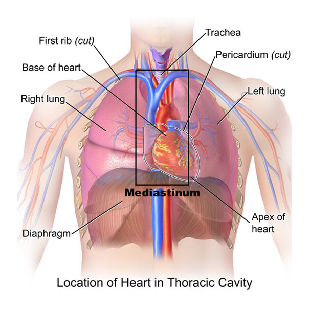
The heart has four chambers — two atria and two ventricles — lined by the endocardium, surrounded by the muscular myocardium, and enclosed in the pericardium sac. Four valves (mitral, aortic, tricuspid, pulmonary) control blood flow. Myxomas typically arise from the interatrial septum and dangle into the left atrium; fibroelastomas grow on valve surfaces.
The lungs each divide into lobes (three on the right, two on the left), fed by branching airways (trachea, bronchi, bronchioles) that end in gas-exchanging alveoli and are covered by the pleura. Blood arrives via the pulmonary arteries and returns oxygenated through the pulmonary veins to the left atrium — the same chamber where myxomas form. This shared circulation explains why heart and lung tumors interact and spread readily within the chest.
5. How the Disease Affects the Body
Tumors damage the body through several mechanisms, which explains their wide range of symptoms.
- Obstruction: A left atrial myxoma can swing into the mitral valve opening, intermittently blocking blood flow — mimicking mitral stenosis and causing breathlessness, fainting or collapse. A central lung tumor can obstruct a bronchus, collapsing part of the lung and trapping infection behind it.
- Embolism: Fragile surface fragments or clots from a heart tumor can break off and travel to the brain (stroke), limbs or organs. Papillary fibroelastomas are a recognized source of stroke.
- Arrhythmia: Tumors within or against the heart muscle can irritate its electrical system, producing palpitations, atrial fibrillation, heart block or, rarely, sudden cardiac death.
- Invasion and compression: Malignant tumors invade neighboring tissue. Lung cancers may compress the airway, esophagus, superior vena cava or nerves, and can spread to the pericardium, causing effusion and tamponade.
- Systemic and paraneoplastic effects: Tumors release chemicals and provoke immune responses causing fever, weight loss, anemia, clotting problems and hormone-like syndromes (common with SCLC).
- Metastatic spread: Malignant tumors seed distant sites — bone, liver, brain, adrenal glands — causing further organ-specific harm.
The overall effect is a combination of local mechanical damage and whole-body illness.
6. Types and Classification
Benign cardiac tumors:
- Myxoma — most common; usually left atrium
- Papillary fibroelastoma — valve surfaces
- Rhabdomyoma — infants, often with tuberous sclerosis
- Fibroma, lipoma, hemangioma — less common
Malignant cardiac tumors:
- Primary sarcomas — angiosarcoma, undifferentiated sarcoma, leiomyosarcoma
- Primary cardiac lymphoma — rare
- Metastatic tumors — far more common than primary cancers
Lung tumors — malignant:
- NSCLC — adenocarcinoma, squamous cell carcinoma, large cell carcinoma
- SCLC — aggressive, strongly smoking-related
- Carcinoid tumors — slower-growing neuroendocrine
- Metastases from breast, colon, kidney, melanoma and others
Lung tumors — benign: hamartoma, papilloma. Lung cancers are further staged by the TNM system.
7. Causes of the Disease
The causes differ sharply between benign and malignant, and between heart and lung.
- Cardiac myxoma — usually sporadic with no clear cause; a minority are part of the inherited Carney complex.
- Rhabdomyoma — strongly associated with tuberous sclerosis, a genetic condition.
- Cardiac sarcoma — cause largely unknown; arises from DNA mutations in cardiac connective tissue.
- Lung cancer — the leading cause is tobacco smoking, responsible for the large majority of cases. Other established causes include radon gas, asbestos, air pollution, occupational carcinogens (arsenic, diesel exhaust), and prior chest radiation.
- Metastatic tumors — caused by an existing cancer elsewhere spreading to the heart or lungs.
For most tumors, cancer develops when accumulated genetic damage lets cells grow uncontrollably and evade normal death signals.
8. How the Disease Develops
Tumors begin when a cell acquires genetic mutations that disrupt the normal balance between cell growth and cell death. Over time, additional mutations accumulate, giving abnormal cells growth advantages, resistance to self-destruction, and eventually the ability to invade and spread.
Benign heart tumors such as myxoma grow slowly from the endocardial lining, gradually enlarging until they interfere with valve function or shed fragments into the circulation. They do not invade or metastasize, but their location makes even a benign tumor dangerous.
Lung cancer typically develops over years. In smokers, repeated carcinogen exposure drives a sequence from normal cells to dysplastic cells to carcinoma in situ to invasive cancer. Once invasive, tumor cells penetrate blood vessels and lymphatics and travel to lymph nodes and distant organs; small cell lung cancer disseminates especially rapidly.
Cardiac sarcomas grow aggressively within the heart wall and metastasize early. Metastatic tumors develop when cells from a primary cancer elsewhere lodge and grow in heart or lung tissue. The pace — indolent for benign tumors, rapid for aggressive cancers — largely determines how and when the disease is found.
9. Risk Factors
Modifiable (mainly for lung cancer):
- Smoking (by far the strongest) and secondhand smoke
- Radon exposure in homes
- Occupational carcinogens — asbestos, arsenic, diesel exhaust, silica
- Air pollution
- Prior chest radiotherapy
Non-modifiable:
- Increasing age
- Family history of cancer or specific syndromes
- Genetic conditions — Carney complex (myxoma), tuberous sclerosis (rhabdomyoma)
- Personal history of another cancer (metastasis risk)
- Pre-existing lung disease such as COPD or pulmonary fibrosis
Benign cardiac tumors have few identifiable risk factors beyond genetic syndromes.
10. Genetic and Family-History Factors
While most tumors are sporadic, genetics matter in specific settings. About 1 in 10 cardiac myxomas occur as part of Carney complex, an inherited disorder (linked to the PRKAR1A gene) featuring multiple/recurrent myxomas, skin pigmentation and endocrine tumors — worth suspecting in young patients or those with several myxomas. Cardiac rhabdomyomas are closely tied to tuberous sclerosis, an inherited condition.
For lung cancer, having a first-degree relative with the disease modestly increases risk, and inherited mutations (for example in EGFR pathways) can influence tumor behavior and treatment. Genetic/molecular testing of the tumor itself — EGFR, ALK, ROS1, KRAS, PD-L1 — is now central to choosing targeted therapy and immunotherapy. Families with hereditary cancer syndromes may benefit from genetic counseling.
11. Who Is Most at Risk?
- Long-term and current smokers — highest risk group for lung cancer
- Older adults, particularly those over 50–60
- People with occupational or radon exposure
- Patients with an existing cancer elsewhere (heart/lung metastases)
- Young to middle-aged adults, more often women — atrial myxoma
- Infants and children — rhabdomyoma and fibroma
- People with Carney complex or tuberous sclerosis
- Those with a strong family history of cancer or with chronic lung disease
12. Prevalence and Epidemiology
Primary cardiac tumors are rare, found in well under 1% of autopsy series; roughly three-quarters are benign, and myxoma accounts for a large share of these. Metastatic tumors of the heart are many times more common than primary ones, seen in a notable minority of patients with widespread cancer.
Lung cancer, by contrast, is among the most common cancers globally and the leading cause of cancer death in both men and women. NSCLC makes up the majority of cases; SCLC a smaller but aggressive fraction. Incidence tracks closely with smoking rates and rises steeply with age. These figures are broad, approximate, and vary considerably by country, sex, and smoking prevalence.
13. Signs and Symptoms
Symptoms depend on tumor type, size and location, and many small tumors cause no symptoms at all (found incidentally).
Cardiac tumors classically produce three patterns:
- Obstructive — breathlessness, fatigue, fainting (syncope), positional symptoms, signs mimicking valve disease
- Embolic — stroke or “mini-stroke” (TIA), limb ischemia, from tumor fragments breaking off
- Constitutional/systemic — fever, weight loss, joint pains, anemia; plus palpitations or arrhythmia
Lung tumors commonly cause:
- Persistent cough or a change in a chronic cough
- Coughing up blood (hemoptysis)
- Chest pain, often worse on deep breathing
- Shortness of breath and wheeze
- Recurrent chest infections in the same area
- Hoarseness and unexplained weight loss, fatigue, loss of appetite
Advanced cancers may show symptoms from spread — bone pain, neurological signs (brain), or face and neck swelling (superior vena cava obstruction). Because early tumors are often silent, symptoms should never be ignored, especially in smokers or anyone with a known cancer.
14. Early-Stage Symptoms
Early tumors are frequently asymptomatic and discovered incidentally on a scan done for another reason. When early symptoms do appear they are often subtle and easy to dismiss:
- A new or persistent cough, or a mild change in an existing cough
- Occasional breathlessness on exertion
- Mild fatigue or reduced exercise tolerance
- Small amounts of blood-streaked sputum
- Brief palpitations or lightheadedness (cardiac tumors)
- Vague chest discomfort
Because these overlap with everyday minor illnesses, early diagnosis often depends on being alert to symptoms that persist beyond two to three weeks.
15. Advanced-Stage Symptoms
- Severe breathlessness at rest or with minimal effort
- Coughing up larger amounts of blood
- Persistent chest, shoulder or bone pain
- Marked weight loss, muscle wasting and weakness (cachexia)
- Fainting or heart-failure symptoms — swelling of legs, orthopnea (from obstructing cardiac tumors)
- Facial/neck swelling and distended veins (superior vena cava obstruction)
- Hoarseness or difficulty swallowing
- Neurological symptoms — headaches, confusion, seizures (brain metastases)
- Pericardial effusion / tamponade — a cardiac emergency with low blood pressure and collapse
Advanced symptoms reflect either large local tumor bulk or distant spread, and warrant urgent specialist assessment.
16. Symptoms in Women, Men and Older Adults
Women: Lung cancer in women is more likely to be adenocarcinoma and can occur in never-smokers; symptoms may be atypical and diagnosis sometimes delayed. Atrial myxoma is more common in women, often presenting in the 30–60 age range.
Men: Historically higher lung cancer rates linked to smoking patterns; squamous cell types are relatively more frequent. Presentation is often with cough, hemoptysis and weight loss.
Older adults: Symptoms may be blunted or attributed to other chronic conditions (COPD, heart failure), leading to later diagnosis. Fatigue, confusion, and functional decline may dominate. Coexisting illnesses also shape how aggressively tumors can be treated. Across all groups, persistent or unexplained symptoms deserve investigation regardless of age or sex.
17. Emergency Warning Signs
Seek emergency care immediately for:
- Sudden collapse or fainting (possible obstructing cardiac tumor)
- Coughing up large amounts of blood
- Signs of stroke — face droop, arm weakness, speech difficulty (tumor embolism)
- Severe breathlessness or chest pain
- Rapid facial/neck swelling with distended veins
- Low blood pressure, dizziness and collapse (possible cardiac tamponade)
18. When to Seek Medical Help
See a doctor promptly if you have a cough lasting more than three weeks, any coughing up of blood, unexplained breathlessness, chest pain, weight loss or fatigue, or new palpitations or fainting. Smokers, ex-smokers, people with occupational exposures, and anyone with a known cancer should have a low threshold for review. Early assessment greatly improves the chance of curative treatment.
19. Disease Stages, Grades and Severity
Benign cardiac tumors are not “staged” like cancers; severity depends on size, mobility and location and the risk of obstruction or embolism.
Lung cancer is staged using the TNM system:
- T — size and extent of the primary tumor
- N — spread to lymph nodes
- M — distant metastasis
These combine into Stages I–IV (I = small and localized; IV = distant spread). SCLC is often described more simply as limited-stage or extensive-stage. Tumors are also graded by how abnormal cells look (differentiation), which reflects aggressiveness. Cardiac sarcomas are graded and staged as soft-tissue sarcomas. Accurate staging drives treatment choice and prognosis.
20. Disease Progression
Benign heart tumors enlarge slowly; the danger is mechanical (obstruction, embolism) rather than spread, and complete removal usually halts progression. Untreated lung cancer progresses from a localized mass to lymph node involvement and then distant metastasis, with small cell cancer advancing especially fast. Cardiac sarcomas progress aggressively, invading local structures and metastasizing early. Progression rate depends on tumor biology, grade, and how early treatment begins — which is why timely diagnosis is critical.
21. Possible Complications
- Blood-flow obstruction and heart-failure symptoms (cardiac tumors)
- Stroke or systemic embolism from tumor fragments/clots
- Arrhythmias and, rarely, sudden cardiac death
- Pericardial effusion and tamponade
- Airway obstruction, lung collapse and post-obstructive pneumonia
- Massive hemoptysis (bleeding into the airway)
- Superior vena cava obstruction
- Metastatic complications — bone fractures, brain dysfunction, liver failure
- Paraneoplastic syndromes (hormonal, neurological, clotting)
- Treatment-related complications — surgical, chemotherapy or radiation side effects
22. Related and Associated Medical Conditions
These tumors frequently coexist with or resemble other conditions. Carney complex and tuberous sclerosis predispose to cardiac tumors. Lung cancer strongly overlaps with COPD and, given shared risk factors, coronary artery disease. Cardiac tumors can mimic valvular heart disease or infective endocarditis. Patients may also have atrial fibrillation, heart failure, venous thromboembolism and anemia, and paraneoplastic syndromes can affect the nervous, endocrine and clotting systems.
23. Screening and Early Detection
There is no routine screening for cardiac tumors — they are usually found incidentally on echocardiography or scans done for other reasons, or when symptoms prompt imaging.
For lung cancer, screening is well established for high-risk people: annual low-dose CT (LDCT) scanning is recommended for current or former heavy smokers within certain age ranges (broadly the 50s to 70s), following guidelines such as those from the USPSTF and NHS Targeted Lung Health Checks. LDCT can detect tumors while still small and curable. Eligible individuals should discuss screening with their doctor. Anyone with symptoms should be investigated promptly rather than waiting for screening.
24. How the Disease Is Diagnosed
Diagnosis combines a careful history, examination, imaging and — for suspected cancers — tissue sampling.
For cardiac tumors, the cornerstone is echocardiography (ultrasound of the heart), which shows size, location, attachment and mobility; a transesophageal echocardiogram (TEE) gives even clearer views. Cardiac MRI and CT further characterize the tumor and help distinguish benign from malignant and tumor from clot. Because myxomas are often removed on imaging alone, preoperative biopsy is frequently avoided (to prevent embolism); diagnosis is confirmed by examining the tissue after surgery.
For lung tumors, a chest X-ray may first reveal a mass, followed by a CT scan to define size, location and lymph nodes, and a PET-CT to assess metabolic activity and spread. A biopsy is essential to confirm cancer and its exact type — obtained by bronchoscopy, endobronchial ultrasound (EBUS), CT-guided needle biopsy or surgical sampling. The tissue is tested for molecular markers (EGFR, ALK, PD-L1 and others) that guide targeted and immune therapies. Staging is completed with further imaging (often brain MRI) before a multidisciplinary team plans treatment. Accurate diagnosis and staging are the foundation of every decision.
25. Physical Examination and Medical History
The doctor reviews smoking history, occupational exposures, prior cancers, family history, and the pattern and duration of symptoms. On examination they look for signs such as abnormal heart sounds or a “tumor plop” (with a myxoma), murmurs, irregular pulse, and signs of heart failure. For lung tumors they assess breathing, reduced air entry, dullness on percussion, wheeze, lymph node enlargement, hoarseness, clubbing of the fingers, and signs of spread (weight loss, bone tenderness, neurological changes, facial swelling). Examination guides which tests are ordered next.
26. Diagnostic Tests and Imaging
- Echocardiography (TTE and TEE) — first-line for cardiac tumors
- Cardiac MRI and CT — tissue characterization, benign vs malignant
- Chest X-ray — initial lung imaging
- CT chest — defines lung mass and nodes
- PET-CT — metabolic activity and staging for spread
- Bronchoscopy and EBUS — visualize airways and biopsy/sample nodes
- CT-guided needle biopsy — peripheral lung lesions
- Brain MRI and bone scans — assess metastasis
- ECG — arrhythmias and conduction effects
Imaging is chosen to both diagnose and stage the tumor.
27. Blood Tests, Biomarkers and Genetic Testing
No single blood test diagnoses these tumors, but labs support the picture. Full blood count may show anemia or a raised white count; inflammatory markers (ESR, CRP) are often elevated with myxoma. Liver, kidney and calcium tests help detect metastasis or paraneoplastic effects. Tumor molecular testing is now essential in lung cancer — the biopsy is analyzed for EGFR, ALK, ROS1, BRAF, KRAS mutations and PD-L1 to select targeted therapy and immunotherapy. Liquid biopsy (circulating tumor DNA) is increasingly used, and genetic counseling is considered when Carney complex or tuberous sclerosis is suspected.
28. Understanding Test Results
Test results are interpreted together, not in isolation. An echo report describing a mobile mass attached to the atrial septum strongly suggests myxoma. A CT/PET showing a lung mass with enlarged, metabolically active nodes suggests cancer with spread. The biopsy and pathology report give the definitive tumor type and grade, while molecular results determine eligibility for specific drugs. TNM stage summarizes how far cancer has spread and shapes whether treatment is curative or focused on control. Your specialist will explain what each result means for your particular situation; ask for plain-language explanations of any unfamiliar terms.
29. Differential Diagnosis
Several conditions can mimic heart or lung tumors and must be excluded:
- Blood clot (thrombus) in a heart chamber — can look like a tumor on imaging
- Vegetations from infective endocarditis — resemble valve tumors
- Valvular heart disease — myxoma can mimic mitral stenosis
- Benign lung nodules — infections (TB, fungal), scar tissue, hamartoma
- Pneumonia or lung abscess
- Sarcoidosis and other inflammatory masses
- Metastasis vs primary tumor — determining origin
- Mediastinal masses (lymphoma, thymoma)
Imaging plus biopsy usually resolves the distinction.
30. Specialist and Multidisciplinary Evaluation
These tumors are managed by a multidisciplinary team. For cardiac tumors this includes a cardiologist and cardiac surgeon, supported by imaging specialists. For lung and malignant tumors, care centers on a tumor board bringing together a pulmonologist, thoracic surgeon, medical oncologist, radiation oncologist, radiologist, pathologist and specialist nurses. Together they review imaging, pathology and staging to agree the best plan. You can find experienced heart and thoracic teams through our doctors directory and network of hospitals. Multidisciplinary evaluation consistently improves outcomes.
31. Treatment Goals
- Remove or control the tumor and relieve obstruction or symptoms
- Prevent complications — embolism, arrhythmia, bleeding, spread
- Cure, where the tumor is benign or early-stage cancer
- Prolong survival and control disease in advanced cancer
- Preserve heart and lung function and quality of life
- Support the whole patient — symptom relief, nutrition, emotional wellbeing
Goals are tailored: cure is realistic for a myxoma or early lung cancer, whereas advanced cancer may focus on control and comfort.
32. When Is Treatment Required?
Benign cardiac tumors such as myxoma warrant prompt surgical removal once diagnosed, because of the ongoing risk of obstruction, embolism or sudden death — even when symptoms are mild. Papillary fibroelastomas are usually removed if mobile or symptomatic. Lung cancer requires treatment as soon as it is confirmed and staged; the type and intensity depend on the stage and the patient’s fitness. Some very small, indolent nodules or slow-growing tumors in frail patients may be monitored rather than immediately treated. Metastatic disease is treated to control symptoms and extend life.
33. Active Monitoring and Watchful Waiting
Not every growth needs immediate intervention. Small, low-risk lung nodules discovered incidentally are commonly followed with serial CT scans over months to years to see whether they grow — many never do. Certain benign, stable, asymptomatic cardiac tumors or small fibroelastomas may be watched with periodic echocardiograms. Watchful waiting is also appropriate for frail patients where treatment risks outweigh benefits. Monitoring must be structured, with clear scan intervals and a plan to act if the tumor changes. Any growth, new symptoms, or concerning features prompt a switch to active treatment.
34. Medications
Medications play a central role in malignant tumors and a supportive role otherwise.
- Chemotherapy — kills or slows cancer cells; used in many lung cancers and cardiac sarcomas
- Targeted therapy — drugs directed at specific mutations (EGFR, ALK, ROS1 inhibitors) in suitable NSCLC
- Immunotherapy — checkpoint inhibitors (e.g., PD-1/PD-L1 agents) that harness the immune system
- Radiotherapy-sensitizing and supportive drugs
- Anticoagulants — to reduce clot/embolism risk in selected cases
- Anti-arrhythmic and heart-failure medications — manage cardiac effects
- Symptom control — pain relief, anti-nausea, appetite support
Benign heart tumors are treated by surgery, not drugs, though medications may manage associated symptoms.
35. Minimally Invasive Treatments
Minimally invasive approaches reduce trauma and speed recovery. For lung tumors, video-assisted thoracoscopic surgery (VATS) and robotic-assisted thoracic surgery allow lobectomy or wedge resection through small incisions, with less pain and shorter stays than open surgery. Bronchoscopic techniques can relieve airway obstruction. For cardiac tumors, minimally invasive and endoscopic cardiac surgery through smaller incisions can remove some myxomas and valve tumors in suitable patients. Stereotactic ablative radiotherapy (SABR/SBRT) offers a non-surgical, precise option for small early lung cancers in patients unfit for surgery. Learn more about minimally invasive cardiac surgery and VATS.
36. Catheter-Based and Endovascular Treatments
Catheter-based options are more limited for these tumors than for coronary or valve disease, but have a role. Interventional bronchoscopy can place airway stents, perform laser or cryotherapy debulking, and control bleeding to relieve obstruction. Bronchial artery embolization — a catheter procedure — can stop massive hemoptysis. Percutaneous ablation (radiofrequency or microwave) may treat small lung tumors or metastases in non-surgical candidates. For pericardial fluid, catheter drainage (pericardiocentesis) relieves tamponade. General endovascular techniques are covered under endovascular stenting.
37. Surgical Treatment Options
Surgery is the definitive, often curative, treatment for many of these tumors.
Cardiac tumors: A benign myxoma is removed via open-heart surgery using cardiopulmonary bypass. The surgeon excises the tumor with a margin of the attachment site (usually part of the atrial septum) to minimize recurrence, then repairs the defect. Results are excellent and most patients are effectively cured. Papillary fibroelastomas are shaved off the valve, preserving valve function where possible. Cardiac sarcomas are far more challenging: resection is attempted when feasible, but complete removal is often difficult and outcomes are guarded.
Lung tumors: For localized lung cancer, surgery offers the best chance of cure. Options, matched to tumor size and location, include:
- Wedge resection / segmentectomy — removal of a small portion (early tumors, limited lung reserve)
- Lobectomy — removal of an entire lobe, the standard for many operable NSCLC
- Pneumonectomy — removal of a whole lung for large or central tumors
These can be performed open (thoracotomy) or minimally invasively (VATS/robotic), usually with lymph node sampling for staging, and are often combined with chemotherapy, radiotherapy or immunotherapy. Explore video-assisted thoracic surgery and endoscopic heart surgery. Suitability depends on tumor stage, lung function and overall fitness.
38. Advanced and Emerging Treatments
The field is advancing rapidly, especially for lung cancer.
- Next-generation targeted therapies for a growing list of mutations
- Immunotherapy combinations and use earlier in treatment
- Robotic surgery with greater precision and shorter recovery
- Stereotactic radiotherapy (SBRT) for early tumors and oligometastatic disease
- Liquid biopsy for monitoring and early relapse detection
- Antibody-drug conjugates and novel immune agents
- Heart transplantation — considered in rare, selected cases of unresectable primary cardiac tumors at specialized centers
Many of these are available through clinical trials or leading international centers.
39. Treatment Options Compared
- Surgery — best chance of cure for benign heart tumors and early lung cancer; requires fitness for an operation.
- Minimally invasive/VATS/robotic — similar cure rates to open surgery for suitable tumors, with faster recovery.
- Radiotherapy (including SBRT) — an alternative for early tumors in unfit patients and part of combined treatment for advanced disease.
- Chemotherapy — controls and shrinks cancers, used before/after surgery or as main treatment in advanced disease.
- Targeted therapy/immunotherapy — effective for tumors with the right molecular profile; often better tolerated than chemotherapy.
- Watchful waiting — appropriate for tiny, stable or low-risk lesions.
The best choice balances tumor type, stage, molecular profile and patient fitness.
40. How Doctors Choose the Right Treatment
Treatment is individualized by the multidisciplinary team based on:
- Tumor type (benign vs malignant) and exact histology
- Stage (TNM) and whether disease is localized or spread
- Molecular/genetic markers (for targeted and immune therapy)
- Location and size, and impact on heart/lung function
- Patient fitness, lung reserve and other illnesses
- Age and personal preferences
- Symptoms and urgency
For a myxoma the answer is usually straightforward — surgery. For lung cancer, the team weighs many factors and often combines treatments.
41. Benefits and Risks of Treatment
Benefits: potential cure (benign tumors, early cancer), symptom relief, longer survival, prevention of embolism, obstruction and bleeding, and improved quality of life.
Risks: all treatments carry some risk. Surgery risks bleeding, infection, arrhythmia, air leak (lung), reduced lung capacity, and anesthetic complications. Chemotherapy causes nausea, fatigue, low blood counts and infection risk. Radiotherapy may cause skin and lung inflammation. Immunotherapy can trigger immune-related side effects. Targeted drugs have their own profiles. Your team explains the specific balance for your situation; for most benign cardiac tumors the benefits of surgery greatly outweigh the small risks.
42. What Happens If the Disease Is Left Untreated?
Leaving these tumors untreated is dangerous. An untreated myxoma can suddenly obstruct blood flow, cause a stroke through embolism, or trigger fatal collapse. Untreated lung cancer progresses relentlessly — spreading to lymph nodes and distant organs, causing worsening breathlessness, bleeding, pain and, ultimately, a poor outcome, with survival far shorter than with treatment. Cardiac sarcomas left alone are rapidly fatal. Early treatment transforms the outlook for benign tumors and early cancers, which is why prompt action after diagnosis is so important.
43. Treatment Success and Expected Outcomes
Outcomes vary widely by tumor. Benign cardiac tumors, especially myxoma, have excellent outcomes — most patients are cured by surgery, with low recurrence when removed completely. Papillary fibroelastoma removal is likewise highly successful. Early-stage lung cancer treated with surgery offers a good chance of cure, with success falling as stage advances. Targeted therapy and immunotherapy have meaningfully improved survival for many with advanced NSCLC. Cardiac sarcomas and extensive SCLC have more guarded outcomes. Results also depend heavily on the experience of the treating center. These are general expectations, not guarantees for any individual.
44. Prognosis and Long-Term Outlook
Prognosis spans the full range from cure to serious life-limiting disease. For a benign cardiac myxoma, the long-term outlook after complete surgical removal is very good, with most patients returning to normal life; a small proportion — particularly with Carney complex — may develop recurrences, so periodic echocardiographic follow-up is advised.
For lung cancer, prognosis is driven mainly by stage at diagnosis. Early-stage, surgically removed NSCLC carries a substantially better outlook than locally advanced or metastatic disease. Histology, molecular profile, treatment response and general health all influence survival. Progress in targeted therapies and immunotherapy has extended survival and, for some, turned advanced lung cancer into a longer-term controllable condition. Small cell lung cancer and primary cardiac sarcoma have poorer prognoses. Because outcomes differ so much, discuss your specific prognosis with your team. Overall, earlier diagnosis and treatment at an experienced center consistently improve the outlook.
45. Recovery and Rehabilitation
Recovery depends on the treatment. After cardiac tumor surgery, most patients spend a few days in hospital, then recover over several weeks; cardiac rehabilitation supports a return to activity. After lung surgery, recovery is faster with VATS/robotic approaches than open surgery; breathing exercises and pulmonary rehabilitation rebuild lung capacity. Patients on chemotherapy, radiotherapy or immunotherapy recover in cycles, managing side effects with their team. Good nutrition, gradual reactivation, smoking cessation, and emotional support all speed recovery. Rehabilitation is tailored to each person’s operation and fitness.
46. Follow-Up Tests and Long-Term Monitoring
- Myxoma: periodic echocardiograms to detect recurrence, especially in younger patients or Carney complex
- Lung cancer: scheduled CT scans, clinical review and sometimes PET/blood markers to detect recurrence
- Lung function tests after resection
- Molecular/liquid-biopsy monitoring in advanced cancer to detect resistance
- Ongoing screening of the remaining lung in treated smokers
Follow-up is most intensive in the first two to three years, then less frequent if stable.
47. Managing Recurrence or Disease Progression
If a tumor returns or progresses, the team reassesses with imaging and, where needed, repeat biopsy. Recurrent myxoma may be surgically removed again. For progressing lung cancer, options include switching systemic therapy (a different chemotherapy, next-line targeted drug for a new resistance mutation, or immunotherapy), radiotherapy to problem sites, or further surgery in selected cases. Palliative care is integrated early to control symptoms and maintain quality of life. Clinical trials may offer additional options. A clear, individualized plan is made with the multidisciplinary team.
48. Living with the Disease
Living with a heart or lung tumor — or its treatment — involves practical and emotional adjustment. Many patients treated for a benign tumor return fully to normal life. Those with cancer may live with ongoing treatment and monitoring. Helpful steps include stopping smoking, staying as active as able, eating well, keeping up with appointments and medications, pacing activity around fatigue, and leaning on family, support groups and professional help. Managing other conditions (heart, lung, diabetes) and planning around treatment cycles help maintain independence and wellbeing.
49. Diet and Nutrition Guidelines
- Eat a balanced diet rich in vegetables, fruits, whole grains and lean protein
- Ensure adequate protein and calories to combat cancer-related weight loss and support healing
- Small, frequent meals help when appetite is poor or during treatment
- Stay hydrated, particularly during chemotherapy
- Limit processed meats, excess alcohol and heavily processed foods
- Ask about a dietitian referral for weight loss, swallowing difficulty, or treatment side effects
Good nutrition supports treatment tolerance, recovery and immune function.
50. Exercise and Physical-Activity Guidelines
Regular, appropriate activity benefits nearly all patients. Aim for gentle aerobic activity such as walking, building up gradually as tolerated, plus light strengthening. Breathing exercises and pulmonary rehabilitation are especially valuable after lung surgery or with reduced lung function. After heart surgery, follow a cardiac rehabilitation program. Listen to your body, rest when fatigued (common during chemotherapy/radiotherapy), and avoid strenuous exertion until cleared. Always confirm your exercise plan with your care team, who can tailor it to your surgery, fitness and treatment stage.
51. Medications, Activities and Habits to Avoid
- Smoking — the single most important habit to stop; it worsens outcomes and healing
- Excess alcohol
- Unsupervised herbal supplements — some interact with chemotherapy or immunotherapy
- Over-the-counter drugs without checking — especially anticoagulant interactions
- Strenuous activity too soon after surgery
- Exposure to infections during low-immunity phases of chemotherapy (practice good hygiene, avoid sick contacts)
- Occupational carcinogen exposure where possible
Always review new medications and supplements with your oncology or cardiac team.
52. Preventing the Disease or Reducing Its Risks
Benign cardiac tumors are largely not preventable, though people with Carney complex benefit from surveillance. Lung cancer, however, is highly preventable:
- Do not smoke; if you smoke, stop — the most effective single measure
- Avoid secondhand smoke
- Test and mitigate radon in the home
- Reduce occupational exposure to asbestos and other carcinogens (use protection)
- Minimize air-pollution exposure where possible
- Attend LDCT screening if you are in a high-risk group
- Maintain a healthy diet and activity level
Stopping smoking reduces risk substantially over time, at any age.
53. Pregnancy and the Disease
Tumors of the heart and lung are uncommon in pregnancy but require specialist care when they occur. Cardiac myxoma in pregnancy can cause obstruction or embolism and may need surgery, timed carefully with obstetric and cardiac teams. Lung cancer in pregnancy is rare and managed by a multidisciplinary team balancing maternal treatment against fetal safety, with decisions on timing of therapy and delivery. Women with Carney complex or tuberous sclerosis benefit from pre-pregnancy counseling. Any pregnant woman with concerning cardiac or respiratory symptoms should be assessed promptly by specialists.
54. Disease in Children and Young Adults
Cardiac tumors in children differ from adults: rhabdomyoma is the most common, often linked to tuberous sclerosis, and many regress spontaneously without surgery unless they obstruct blood flow. Fibromas may need removal. Myxoma is less common in children but occurs, sometimes as part of Carney complex. Primary lung cancer is very rare in young people, though other chest tumors and metastases (e.g., from bone or kidney cancers) can occur. Care is delivered by pediatric cardiology, cardiac surgery and oncology teams; explore congenital heart procedures for related pediatric care.
55. Disease in Older Adults
Older adults face specific considerations. Lung cancer is most common in this age group, but coexisting COPD, heart disease and frailty can limit aggressive surgery and shape treatment toward radiotherapy, gentler systemic therapy or supportive care. Diagnosis may be delayed because symptoms overlap with existing conditions. Cardiac tumors in older patients are still treatable, but surgical risk is weighed against overall health. A comprehensive geriatric assessment helps tailor treatment intensity, and shared decision-making that respects the patient’s priorities and quality of life is essential.
56. Emotional Health and Patient Support
A diagnosis of a heart or lung tumor — particularly cancer — can bring fear, anxiety and low mood. These feelings are normal and support is available. Counseling, cancer-support groups, and mental-health services help patients and families cope. Specialist nurses guide you through treatment. Peer support from others who have faced the same diagnosis can be reassuring. Do not hesitate to raise emotional concerns with your team — psychological wellbeing is part of comprehensive care and can influence how well you cope with and complete treatment.
57. Preparing for Your Specialist Appointment
- Write down your symptoms, when they began and how they’ve changed
- List all medications, supplements and allergies
- Note your smoking history and occupational exposures
- Gather previous test results, scans and reports
- Record your family and past medical history
- Prepare your questions in advance (see next section)
- Bring a family member or friend for support and to help remember information
- Consider taking notes or asking permission to record
Good preparation makes appointments more productive.
58. Questions to Ask Your Doctor
- What type of tumor do I have, and is it benign or malignant?
- Where exactly is it, and has it spread?
- What stage and grade is it, and what does that mean for me?
- What are all my treatment options, including surgery?
- Is a minimally invasive (VATS/robotic) approach possible for me?
- What are the benefits, risks and side effects of each option?
- Will I need chemotherapy, radiotherapy, targeted therapy or immunotherapy?
- Has my tumor been tested for molecular markers?
- What is my expected recovery and long-term outlook?
- How experienced is this center with my type of tumor, and should I get a second opinion?
59. Cost of Diagnosis and Treatment
Costs vary enormously by tumor type, treatment intensity, country and hospital. The ranges below are broad approximations for guidance only.
| Region | Cardiac tumor surgery (approx.) | Lung cancer surgery / treatment (approx.) |
|---|---|---|
| USA | $50,000 – $150,000+ | $40,000 – $120,000+ (more with ongoing systemic therapy) |
| UK / Western Europe | £20,000 – £60,000 | £20,000 – £70,000 |
| India | $6,000 – $15,000 | $5,000 – $15,000 |
| Turkey | $8,000 – $20,000 | $7,000 – $18,000 |
| Thailand / Singapore | $10,000 – $30,000 | $9,000 – $28,000 |
Medical-tourism destinations such as India, Turkey and Thailand often cost roughly 50–90% less than the US or UK for comparable surgery. Ongoing drug therapy (targeted/immunotherapy) adds significant, variable cost everywhere. Always request a detailed, itemized quote.
60. Factors Affecting Treatment Cost
- Tumor type and stage — benign removal vs prolonged cancer treatment
- Type of surgery — open vs minimally invasive/robotic
- Need for chemotherapy, radiotherapy, targeted or immune drugs (often the largest cost)
- Length of hospital stay and ICU time
- Country and hospital (accreditation, reputation)
- Surgeon and team experience
- Diagnostic workup — PET-CT, MRI, molecular testing, biopsies
- Complications and follow-up care
- For international patients — travel, accommodation, interpreter and coordination fees
61. Choosing the Right Specialist
Look for a board-certified cardiac surgeon (for heart tumors) or thoracic surgeon and oncology team (for lung tumors) with specific, high-volume experience treating your tumor type. Ask about their case numbers, outcomes and complication rates, whether they offer minimally invasive/robotic options, and whether care is delivered through a multidisciplinary tumor board. Good communication and willingness to answer questions matter. Browse our vetted doctors directory to compare experienced heart and thoracic specialists.
62. Choosing the Right Hospital or Treatment Centre
Choose a center with:
- International accreditation (e.g., JCI) for quality and safety
- High volume of cardiac/thoracic surgery and cancer care
- Full multidisciplinary services — surgery, oncology, radiotherapy, imaging, pathology
- Advanced technology — robotic surgery, PET-CT, molecular labs, modern radiotherapy
- Strong published outcomes and infection-control records
- Dedicated international patient services for overseas patients
Explore accredited options through our hospitals and destinations directories.
63. Getting a Second Medical Opinion
A second opinion is wise before major treatment, especially for a cancer diagnosis or complex surgery. Another specialist may confirm the diagnosis, re-review pathology and imaging, suggest alternative options (such as minimally invasive surgery or a clinical trial), or reassure you the recommended plan is sound. Reputable doctors welcome second opinions. Many international centers offer remote second opinions using your scans and reports. Learn how to arrange one via our contact page.
64. Treatment Abroad and Medical-Travel Considerations
Many patients travel abroad for high-quality, more affordable cardiac and thoracic cancer care. When considering treatment overseas:
- Choose a JCI-accredited hospital with proven outcomes
- Confirm the surgeon’s and oncologist’s experience with your tumor
- Clarify total costs, inclusions and follow-up arrangements
- Plan for fitness to fly and adequate recovery before travel home
- Arrange continuity of care and records transfer with home doctors
Popular destinations include India, Turkey, Thailand and Singapore. Explore options via our destinations and hospitals pages.
65. Frequently Asked Questions
Are heart tumors usually cancer? No. Most primary heart tumors are benign, with myxoma the commonest. Malignant primary heart tumors (sarcomas) are rare, though cancer that spreads to the heart from elsewhere is more common.
Can a benign heart tumor still be dangerous? Yes. Even benign tumors like myxoma can block blood flow or shed fragments causing stroke, so they are usually removed promptly.
Is lung cancer always caused by smoking? Smoking is the leading cause, but lung cancer can also affect non-smokers due to radon, asbestos, air pollution, genetics or other factors.
Can these tumors be treated with minimally invasive surgery? Often yes — VATS and robotic surgery treat many lung tumors, and minimally invasive/endoscopic techniques can remove some heart tumors.
Will I need chemotherapy after surgery? It depends on the tumor type and stage. Benign heart tumors need no chemotherapy; some lung cancers require additional chemotherapy, radiotherapy or immunotherapy.
Can a heart myxoma come back after removal? Recurrence is uncommon after complete removal, but is more likely with Carney complex, so follow-up echocardiograms are advised.
How is lung cancer detected early? Through low-dose CT screening in high-risk people and by promptly investigating persistent symptoms such as a lasting cough or coughing up blood.
Can I get treated abroad affordably? Yes — accredited hospitals in India, Turkey and Thailand offer quality care at substantially lower cost than the US or UK.
66. Patient Stories and Treatment Experiences
The following are representative, anonymized examples for illustration only.
Meera, India (age 45): After months of unexplained breathlessness and one fainting spell, an echocardiogram revealed a large left-atrial myxoma. She had open-heart surgery to remove it and recovered fully within weeks, returning to work symptom-free.
James, United Kingdom (age 62): A former smoker, James had a small early-stage lung cancer found on a low-dose CT screening scan. He underwent VATS lobectomy, went home in a few days, and needed no chemotherapy — a reminder of the value of early detection.
Ahmed, UAE (treated in Turkey): Diagnosed with advanced NSCLC carrying an EGFR mutation, Ahmed traveled to an accredited center for targeted therapy after molecular testing. His disease has been controlled for an extended period with good quality of life.
67. Latest Research and Clinical Trials
Research is transforming outcomes, particularly for lung cancer. Key directions include expanding targeted therapies for newly identified mutations, combination and earlier-stage immunotherapy, antibody-drug conjugates, and liquid-biopsy monitoring to detect relapse and drug resistance sooner. Surgical advances include robotic and lung-sparing resections, and stereotactic radiotherapy for early or limited-spread tumors. For rare cardiac tumors, research explores better imaging characterization and multimodal treatment of sarcomas. Reputable clinical trials are run through cancer centers and cooperative groups worldwide; ask your team whether a trial suits you. (This is a general summary, not a reference to specific published studies.)
68. Related Diseases and Conditions
- Lung Disease
- Coronary Artery Disease
- Heart Failure
- Infective Endocarditis
- Disorders of Heart Rhythm
- Valvular Heart Disease
69. Related Treatments and Procedures
- Video-Assisted Thoracic Surgery (VATS)
- Minimally Invasive Cardiac Surgery
- Endoscopic Heart Surgery
- Endovascular Stenting
- Other Procedures
70. Medical Glossary
- Myxoma — the most common benign primary heart tumor, usually in the left atrium
- Papillary fibroelastoma — a small benign tumor on heart valves
- Rhabdomyoma — a benign heart-muscle tumor, common in children
- Sarcoma — a malignant tumor of connective tissue (rare in the heart)
- NSCLC — non-small cell lung cancer
- SCLC — small cell lung cancer
- Metastasis — spread of cancer to another part of the body
- Embolism — blockage of a vessel by a fragment (clot or tumor) that traveled through the blood
- Echocardiography — ultrasound imaging of the heart
- VATS — video-assisted thoracoscopic surgery, a minimally invasive lung operation
- Lobectomy — surgical removal of a lobe of the lung
- TNM staging — system describing tumor size, node and metastasis status
- Targeted therapy — drugs aimed at specific tumor mutations
- Immunotherapy — treatment that stimulates the immune system to fight cancer
- Pericardial effusion — fluid around the heart, which can cause tamponade
71. Medical Review, Editorial Policy and Last Updated Date
Last updated: 11 July 2026.
This article was prepared by the BestHeartSurgery.com editorial team and reviewed for accuracy against established guidance from bodies such as the ACC/AHA, ESC, WHO, NHS, GINA and major oncology and thoracic-surgery societies. Our editorial policy emphasizes accurate, current, patient-friendly information, with periodic review as clinical knowledge evolves.
Disclaimer: This content is provided for general educational purposes only and is not a substitute for professional medical advice, diagnosis or treatment. Always consult a qualified healthcare provider about your specific condition. Never disregard or delay seeking medical advice because of something you have read here.
72. Clinical Guidelines and Medical References
This information aligns with general guidance from reputable organizations, including:
- American College of Cardiology (ACC) and American Heart Association (AHA)
- European Society of Cardiology (ESC)
- Society of Thoracic Surgeons (STS)
- World Health Organization (WHO) cancer resources
- National Institute for Health and Care Excellence (NICE) and NHS
- National Comprehensive Cancer Network (NCCN) oncology guidelines
- International Association for the Study of Lung Cancer (IASLC) — TNM staging
Please consult these bodies and your care team for detailed, current clinical recommendations.
73. Book an Appointment or Request a Second Opinion
If you or a loved one has been diagnosed with a heart or lung tumor, expert care can make a decisive difference. Our network connects you with accredited hospitals, experienced cardiac and thoracic surgeons, and oncology teams worldwide.
- Book an appointment: /contact/
- Request a second opinion or ask a question: Contact us
- Explore top hospitals, doctors and destinations for cardiac and thoracic tumor care.
Take the next step toward expert, affordable treatment and greater peace of mind.