1. Disease Overview
Restrictive cardiomyopathy (RCM) is an uncommon but serious disease of the heart muscle in which the walls of the lower pumping chambers (ventricles) become abnormally stiff and non-compliant. The heart squeezes reasonably well, so the ejection fraction is usually preserved or near-normal, but the stiff ventricles cannot relax and fill properly between beats. This is a problem of diastolic filling rather than pumping.
Because the chambers cannot expand to accept returning blood, pressure backs up into the atria, lungs and body veins. The atria enlarge while the ventricles stay normal-sized or small, and patients develop the congestive symptoms of heart failure — breathlessness, fatigue, and swelling — despite a heart that “looks” like it is contracting normally.
RCM is the least common of the three classic cardiomyopathies (the others being dilated and hypertrophic). It may be idiopathic or result from a systemic disease that infiltrates or scars the myocardium, such as amyloidosis, sarcoidosis, hemochromatosis or endomyocardial fibrosis. A central challenge is distinguishing RCM from constrictive pericarditis, which mimics it but is often surgically curable. Management focuses on treating the underlying cause, relieving congestion, controlling rhythm, and — in advanced disease — considering heart transplantation.
2. Key Facts at a Glance
| Fact | Detail |
|---|---|
| Also known as | RCM, restrictive heart muscle disease, infiltrative cardiomyopathy |
| Body system affected | Cardiovascular (heart muscle / myocardium), often with systemic disease |
| Common in | Adults (varies by cause); endomyocardial fibrosis mainly in tropical regions; children in idiopathic and familial forms |
| Severity range | Mild diastolic dysfunction to advanced, refractory heart failure |
| Key treatments | Treat underlying cause, diuretics, rate/rhythm control, anticoagulation, device therapy, heart transplant |
| Outlook | Highly variable — depends on cause; often guarded when idiopathic or advanced |
3. Alternative Names and Medical Terminology
- RCM — the standard abbreviation.
- Restrictive heart muscle disease.
- Infiltrative cardiomyopathy — when caused by deposition of abnormal material (amyloid, iron).
- Endomyocardial fibrosis (EMF) and Löffler endocarditis (eosinophilic endomyocardial disease) — specific endomyocardial subtypes.
- Cardiac amyloidosis — a common infiltrative cause, subdivided into AL (light-chain) and ATTR (transthyretin) types.
- Related descriptive terms include diastolic dysfunction and heart failure with preserved ejection fraction (HFpEF), which overlap with RCM physiology.
4. Relevant Heart, Lung or Vascular Anatomy
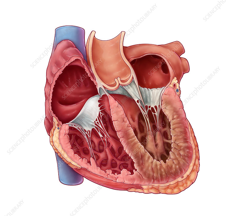
The heart has four chambers: two upper atria and two lower ventricles. The muscular wall (myocardium) must contract to eject blood and, just as importantly, relax to allow filling during diastole. The inner lining is the endocardium; the outer sac is the pericardium.
In RCM the abnormality lies in the ventricular myocardium and/or endocardium, which stiffens from infiltration, fibrosis or scarring. As filling pressures rise, the atria dilate markedly, and backpressure transmits through the pulmonary veins into the lungs (breathlessness) and through the venae cavae into the liver, abdomen and legs (congestion and swelling). RCM primarily impairs the filling phase while sparing chamber size and contraction — the key to understanding its symptoms and treatment.
5. How the Disease Affects the Body
In a healthy heart, the ventricles relax easily to draw in blood at low pressure. In RCM, the stiffened walls resist stretching, so even a small amount of returning blood raises chamber pressure sharply — a hallmark of impaired diastolic compliance. Because contraction is preserved, output at rest may be reasonable, but the heart cannot fill enough to increase output during exertion.
The consequences ripple outward. Elevated left-sided pressures push fluid back into the lungs, causing breathlessness. Elevated right-sided pressures cause jugular venous distension, liver congestion, ascites, and leg swelling. The dilated, overworked atria become prone to atrial fibrillation and blood clots, raising stroke risk.
Depending on the cause, other organs are affected too — amyloidosis involves kidneys, nerves and soft tissues; sarcoidosis the lungs and lymph nodes; hemochromatosis the liver, pancreas and joints. Over time, cardiac output falls and — in advanced cases — the heart can no longer meet the body’s needs, leading to low-output heart failure. The stiff heart also disturbs conduction, causing arrhythmias and heart block.
6. Types and Classification
RCM is usually classified by cause and by which layer of the heart is involved:
- Infiltrative — abnormal substances deposit between muscle fibres: amyloidosis (AL and ATTR), sarcoidosis.
- Storage diseases — accumulation within cells: hemochromatosis (iron), Fabry disease, glycogen storage disorders.
- Endomyocardial — disease of the inner lining and adjacent muscle: endomyocardial fibrosis, Löffler (hypereosinophilic) endocarditis, carcinoid heart disease, radiation-induced fibrosis.
- Non-infiltrative / idiopathic — no identifiable deposit; includes idiopathic RCM and some familial (genetic) forms, and diabetic/scleroderma-associated disease.
Physiologically, all forms share restrictive filling with preserved or near-normal ejection fraction and biatrial enlargement.
7. Causes of the Disease
Common and important causes include:
- Cardiac amyloidosis — one of the most frequent causes, from AL (plasma-cell) or ATTR (transthyretin) protein deposition.
- Sarcoidosis — granulomatous inflammation infiltrating the myocardium.
- Hemochromatosis — iron overload, hereditary or from repeated transfusions.
- Endomyocardial fibrosis — a major cause in tropical Africa, Asia and South America.
- Löffler endocarditis — from prolonged eosinophil elevation.
- Idiopathic RCM — no cause found.
- Other causes: radiation therapy to the chest, scleroderma, carcinoid syndrome, and certain genetic mutations.
8. How the Disease Develops
The pathway to RCM depends on its cause, but the end result is a stiff, poorly compliant ventricle. In infiltrative disease, abnormal proteins or cells accumulate between muscle fibres, preventing the walls from stretching. In amyloidosis, misfolded proteins deposit and stiffen the myocardium over months to years. In hemochromatosis, excess iron stored in heart cells damages them and impairs relaxation.
In endomyocardial forms, inflammation — often driven by eosinophils in Löffler disease — damages the inner lining, which then scars and thickens, sometimes trapping ventricular inflow or the valve apparatus. In idiopathic and familial RCM, progressive interstitial fibrosis stiffens the muscle without a visible deposit.
As stiffness increases, filling pressures rise, the atria enlarge, and congestion develops. The disease is typically progressive, though its speed varies — from indolent over years to rapid decline in aggressive AL amyloidosis. Early on the heart compensates by raising filling pressure; over time this fails, output drops, arrhythmias appear, and end-stage heart failure ensues.
9. Risk Factors
- Underlying systemic diseases — amyloidosis, sarcoidosis, hemochromatosis, hypereosinophilic syndromes, scleroderma, carcinoid.
- Genetic predisposition — familial cardiomyopathy or amyloidosis mutations, hereditary hemochromatosis.
- Older age — ATTR amyloidosis in particular rises with age.
- Prior chest radiation — for lymphoma, breast or lung cancer.
- Repeated blood transfusions — leading to iron overload.
- Geographic/environmental exposure — tropical residence for endomyocardial fibrosis.
- Chronic eosinophilia — from parasitic infection, allergy or myeloproliferative disease.
10. Genetic and Family-History Factors
A meaningful proportion of RCM is inherited. Hereditary ATTR amyloidosis results from TTR gene mutations and can run in families, sometimes with neuropathy. Hereditary hemochromatosis is linked to HFE mutations causing iron overload. Idiopathic and familial RCM can arise from sarcomere gene mutations (troponin, myosin) — some shared with hypertrophic cardiomyopathy.
A detailed three-generation family history is important, and genetic counselling and testing are recommended when a hereditary cause is suspected. Identifying a genetic form allows screening of at-risk relatives, who may benefit from earlier monitoring or, in ATTR, disease-modifying therapy.
11. Who Is Most at Risk?
- Older adults, especially men, for wild-type ATTR cardiac amyloidosis.
- People with plasma-cell disorders (multiple myeloma, MGUS) for AL amyloidosis.
- Those with a family history of amyloidosis, hemochromatosis or cardiomyopathy.
- Patients with chronic eosinophilia or hypereosinophilic syndrome.
- People living in tropical regions for endomyocardial fibrosis.
- Cancer survivors treated with chest radiation.
- Patients requiring frequent transfusions (e.g., thalassemia).
12. Prevalence and Epidemiology
RCM is the rarest of the major cardiomyopathies, accounting for only a small share of cardiomyopathy cases in Western countries. Its frequency varies dramatically by cause and geography. Endomyocardial fibrosis is comparatively common in equatorial regions and is a significant cause of heart failure there, whereas it is rare in temperate countries. Cardiac amyloidosis, once thought uncommon, is now recognized far more often thanks to improved imaging and awareness — ATTR amyloidosis is increasingly diagnosed in older adults with what was previously labelled HFpEF. These figures are approximate and variable; RCM as a whole remains uncommon, but individual causes cluster in specific populations.
13. Signs and Symptoms
The symptoms of RCM are those of congestive heart failure and often develop gradually. Because the right side is frequently affected, signs of systemic congestion are prominent — sometimes more so than lung symptoms. Common features include:
- Breathlessness (dyspnoea) on exertion, and later at rest.
- Difficulty breathing lying flat (orthopnoea) or waking at night short of breath.
- Fatigue and reduced exercise tolerance from limited cardiac output.
- Swelling of the legs, ankles and abdomen (ascites), with weight gain.
- Loss of appetite, nausea or abdominal fullness from liver and gut congestion.
- Palpitations or an irregular pulse — atrial fibrillation is common.
- Cough (often at night) and light-headedness or fainting (syncope).
Because the ejection fraction often looks normal on a scan, symptoms can be wrongly attributed to other conditions. Prominent right-sided congestion with a “normal” pumping heart is a clue that should prompt evaluation for RCM.
14. Early-Stage Symptoms
In early RCM, symptoms are often subtle and easy to overlook. Patients may notice reduced stamina, becoming breathless with activities they used to manage easily, or tiredness out of proportion to effort. Mild ankle swelling at the end of the day, occasional palpitations, or a sense of abdominal fullness after meals can appear. Some people are diagnosed incidentally when an echocardiogram done for another reason shows enlarged atria or abnormal filling patterns. Because these signs are non-specific, early disease is frequently mistaken for deconditioning, ageing or other illnesses.
15. Advanced-Stage Symptoms
As RCM progresses, congestion becomes marked and cardiac output falls. Features of advanced disease include:
- Severe breathlessness at rest and marked orthopnoea.
- Massive fluid retention — tense abdominal ascites, pronounced leg and sometimes body-wide swelling.
- Profound fatigue and weakness from low output.
- Cardiac cachexia — unintended weight and muscle loss.
- Recurrent atrial fibrillation and other arrhythmias, with palpitations and syncope.
- Signs of liver congestion — jaundice, right-sided abdominal discomfort.
- Low blood pressure, cold extremities, and kidney impairment in end-stage disease.
At this point, patients often require repeated hospital admissions, and evaluation for transplant or advanced therapies becomes appropriate.
16. Symptoms in Women, Men and Older Adults
The core symptoms are similar across sexes, but presentation differs by cause. Wild-type ATTR amyloidosis predominantly affects older men, often preceded by carpal tunnel syndrome or lumbar spinal stenosis years earlier. AL amyloidosis affects both sexes and may present with additional signs such as bruising around the eyes, an enlarged tongue, or kidney disease. In older adults, breathlessness and swelling may be blamed on age, other heart conditions or lung disease, delaying recognition. Women may report more fatigue and atypical symptoms. Regardless of demographic, unexplained heart failure with a preserved ejection fraction should raise suspicion of RCM.
17. Emergency Warning Signs
Seek emergency care for:
- Severe or sudden breathlessness or an inability to lie flat.
- Chest pain, especially with sweating or faintness.
- Fainting (syncope) or near-collapse.
- A very fast, very slow or wildly irregular heartbeat with dizziness.
- Signs of stroke — face drooping, arm weakness, speech difficulty.
- Coughing up pink, frothy sputum (possible pulmonary oedema).
18. When to Seek Medical Help
Arrange a prompt medical review if you have persistent or worsening breathlessness, new or increasing swelling of the legs or abdomen, unexplained fatigue limiting daily activity, palpitations, or rapid weight gain from fluid. Anyone with a known systemic disease that can involve the heart — amyloidosis, sarcoidosis, hemochromatosis or hypereosinophilia — should report new cardiac symptoms without delay. Because RCM can masquerade as other conditions, don’t hesitate to ask your doctor about referral to a cardiologist if symptoms are unexplained.
19. Disease Stages, Grades and Severity
RCM does not have a single formal staging system; severity is assessed using several complementary tools:
- NYHA functional class (I–IV) — grades how much symptoms limit activity.
- ACC/AHA heart-failure stages (A–D) — from at-risk to advanced refractory failure.
- Echocardiographic diastolic-function grading — the degree of filling abnormality and estimated filling pressures.
- Biomarker levels (BNP/NT-proBNP, troponin) — reflect strain and injury and, in amyloidosis, feed into cause-specific staging systems that estimate prognosis.
- Cause-specific markers — e.g., light-chain levels in AL amyloidosis.
Together these help gauge how advanced the disease is and guide treatment intensity.
20. Disease Progression
The natural course of RCM is variable and cause-dependent. Some patients stay relatively stable for years with well-controlled congestion, while others deteriorate steadily. AL amyloidosis can progress rapidly; ATTR amyloidosis advances more slowly; endomyocardial fibrosis may stabilize after its inflammatory phase but leaves fixed scarring; idiopathic RCM often progresses gradually toward refractory failure. As disease advances, atrial fibrillation, conduction disease, thromboembolism and low output become more common, and the risk of hospitalization and death rises.
21. Possible Complications
- Progressive heart failure — the central complication, eventually refractory to medication.
- Atrial fibrillation and other arrhythmias — from stretched, diseased atria.
- Conduction block — especially in sarcoidosis and amyloidosis, sometimes needing a pacemaker.
- Blood clots and stroke — from stagnant blood in dilated atria or ventricular scar.
- Sudden cardiac death — from dangerous arrhythmias.
- Liver and kidney dysfunction and pulmonary hypertension — from chronic congestion.
- Valve involvement — particularly in endomyocardial fibrosis, causing regurgitation.
22. Related and Associated Medical Conditions
RCM is closely linked to the systemic disease that causes it, so associated conditions include multiple myeloma or MGUS (AL amyloidosis), neuropathy and carpal tunnel syndrome (ATTR), pulmonary and lymph-node sarcoidosis, liver disease, diabetes and arthritis (hemochromatosis), and hypereosinophilic syndrome (Löffler). It overlaps physiologically with HFpEF and must be distinguished from constrictive pericarditis and hypertrophic cardiomyopathy. Atrial fibrillation, chronic kidney disease and pulmonary hypertension commonly coexist.
23. Screening and Early Detection
There is no population-wide screening for RCM, but targeted screening is valuable in high-risk groups. Relatives of patients with hereditary ATTR, familial RCM or hemochromatosis may undergo genetic testing and periodic cardiac evaluation. People with known systemic diseases affecting the heart should have baseline echocardiography and biomarkers, with follow-up if symptoms develop. Increasingly, older patients labelled “HFpEF” or with unexplained wall thickening plus red-flag features are screened for cardiac amyloidosis using bone scintigraphy and blood tests, substantially improving early detection.
24. How the Disease Is Diagnosed
Diagnosing RCM is a stepwise process combining the clinical picture with imaging, laboratory tests and sometimes tissue sampling. The first step is recognizing heart failure with a preserved ejection fraction and prominent congestion, then confirming a restrictive filling pattern and searching for a cause. The typical pathway is:
- History and examination — congestive signs, systemic clues (neuropathy, bruising, liver disease).
- ECG — often shows low voltages (classically in amyloidosis) or conduction abnormalities.
- Echocardiography — the key initial test: normal-sized ventricles, thickened walls, markedly enlarged atria, and a restrictive diastolic filling pattern with preserved ejection fraction.
- Cardiac MRI — characterizes the muscle and detects infiltration, fibrosis or iron.
- Blood tests and biomarkers — including light chains, iron studies and BNP/troponin.
- Nuclear scintigraphy — for ATTR amyloidosis.
- Cardiac catheterization — to measure pressures and, crucially, to distinguish RCM from constrictive pericarditis.
- Endomyocardial biopsy — reserved for cases where the cause remains unclear, to identify amyloid, iron, granulomas or eosinophilic disease.
Establishing the specific cause is essential, because treatment and prognosis differ greatly between treatable hemochromatosis and aggressive AL amyloidosis.
25. Physical Examination and Medical History
The history focuses on congestive symptoms and clues to a systemic cause — family history, neuropathy, carpal tunnel surgery, prior radiation, transfusions or tropical residence. On examination, doctors look for right-sided congestion: raised jugular venous pressure (rising on inspiration — Kussmaul’s sign), an enlarged tender liver, ascites and leg swelling. Heart sounds may include a third or fourth heart sound, an irregular pulse suggests atrial fibrillation, and blood pressure may be low. Extra-cardiac findings — an enlarged tongue, periorbital bruising, skin or joint changes — can point directly to the cause.
26. Diagnostic Tests and Imaging
- Electrocardiogram (ECG) — may show low voltage, atrial abnormality, arrhythmias or heart block.
- Echocardiography — the cornerstone: assesses wall thickness, chamber sizes, diastolic filling, atrial size and valve function.
- Cardiac MRI (CMR) — tissue characterization; late gadolinium enhancement patterns and T1/T2 mapping help identify amyloid, fibrosis and iron overload.
- Nuclear imaging — bone-avid scintigraphy (e.g., PYP/DPD scans) strongly supports ATTR cardiac amyloidosis; FDG-PET helps assess cardiac sarcoidosis.
- Cardiac CT — evaluates the pericardium (thickness/calcification) to separate constriction from restriction.
- Cardiac catheterization — measures intracardiac pressures; hemodynamic tracings help distinguish RCM from constrictive pericarditis.
- Endomyocardial biopsy — provides a definitive tissue diagnosis when required.
27. Blood Tests, Biomarkers and Genetic Testing
Laboratory testing helps identify the cause and gauge severity:
- BNP / NT-proBNP — elevated with cardiac strain; often disproportionately high in amyloidosis.
- Troponin — may be persistently raised in infiltrative disease.
- Serum and urine free light chains, and immunofixation — screen for AL amyloidosis / plasma-cell disorders.
- Iron studies (ferritin, transferrin saturation) — for hemochromatosis.
- Eosinophil count — for Löffler/hypereosinophilic disease.
- Serum ACE and calcium — may support sarcoidosis.
- Genetic testing — for hereditary ATTR, familial RCM and hemochromatosis, with counselling.
28. Understanding Test Results
Interpreting results together is more informative than any single test. A preserved ejection fraction with enlarged atria and a restrictive filling pattern suggests RCM physiology. Low ECG voltages despite thick walls on echo is a classic amyloidosis clue. A positive bone scan with negative light chains effectively confirms ATTR amyloidosis without biopsy in the right context. High ferritin and transferrin saturation point to iron overload. Importantly, normal or near-normal pumping function does not mean the heart is healthy — in RCM the problem is filling, not squeezing. Your specialist will explain how your specific results fit together and what they mean for treatment.
29. Differential Diagnosis
The key distinction is between RCM and constrictive pericarditis, which share symptoms yet constriction is often surgically curable by removing the thickened pericardium. Pericardial imaging (CT/MRI), hemodynamic patterns at catheterization, and respiratory variation help separate them. Other conditions to exclude include hypertrophic cardiomyopathy, hypertensive heart disease, HFpEF from other causes, valve disease, and pericardial effusion/tamponade. Because getting this right changes treatment completely, evaluation at an experienced centre is valuable.
30. Specialist and Multidisciplinary Evaluation
RCM is best managed by a multidisciplinary team led by a cardiologist with heart-failure or cardiomyopathy expertise. Depending on the cause, it may include a haematologist (AL amyloidosis), neurologist (ATTR neuropathy), hepatologist (hemochromatosis), rheumatologist or pulmonologist (sarcoidosis, scleroderma), cardiac imaging specialists, electrophysiologists, and transplant/advanced-heart-failure teams, with genetic counsellors for inherited forms. This ensures both the heart and the systemic disease are treated. Find experienced teams via our doctors and hospitals directories.
31. Treatment Goals
The goals of treatment in RCM are to:
- Treat the underlying cause wherever possible (e.g., chemotherapy for AL amyloidosis, iron removal for hemochromatosis, stabilizers for ATTR).
- Relieve congestion and control fluid overload to ease symptoms.
- Maintain heart rate and rhythm and prevent arrhythmia complications.
- Reduce the risk of stroke through anticoagulation when indicated.
- Preserve quality of life and functional capacity.
- Identify candidates for advanced therapy or transplant at the right time.
Because contraction is usually preserved, treatment centres on improving filling conditions and managing the systemic disease rather than boosting pumping strength.
32. When Is Treatment Required?
Treatment generally begins as soon as RCM is diagnosed, even if symptoms are mild, because addressing the underlying cause early can slow progression. Cause-directed therapy — such as ATTR stabilizers or hemochromatosis phlebotomy — is often most effective before extensive, irreversible damage occurs. Congestive symptoms (breathlessness, swelling) warrant prompt diuretic therapy. Atrial fibrillation, conduction block or high thromboembolic risk each triggers specific interventions. In advanced, refractory disease, timely referral for transplant evaluation is essential rather than delayed.
33. Active Monitoring and Watchful Waiting
Even when symptoms are minimal, patients need structured monitoring rather than passive observation, because RCM tends to progress. This involves regular review, echocardiography, biomarker tracking (BNP/NT-proBNP), and rhythm monitoring. Symptom-free relatives with a genetic form may be followed with periodic screening. “Watchful waiting” here means vigilant surveillance for early congestion, arrhythmia or worsening filling pressures so treatment can be stepped up promptly — not simply waiting until the patient is seriously unwell.
34. Medications
Medication mainly aims to manage congestion, control rhythm, prevent clots and treat the underlying cause:
- Diuretics — the mainstay for fluid overload and breathlessness; used carefully, since stiff ventricles depend on adequate filling.
- Rate-control agents — cautiously, to allow filling time; some are avoided in amyloidosis.
- Anticoagulants — to prevent stroke, especially with atrial fibrillation or intracardiac clot.
- Antiarrhythmic drugs — to control atrial fibrillation and other rhythms.
- Cause-specific therapy — TTR stabilizers/silencers for ATTR, chemotherapy for AL amyloidosis, immunosuppression for sarcoidosis and Löffler disease.
Standard drugs used for weak-pump failure are less effective and sometimes poorly tolerated in RCM, so treatment is individualized. Never start or stop drugs without specialist advice.
35. Minimally Invasive Treatments
Because RCM is a muscle disease, few purely minimally invasive procedures treat the underlying problem, but several help manage complications. Therapeutic phlebotomy is the cornerstone of hemochromatosis treatment, reducing iron load in a simple outpatient procedure; iron chelation is used when phlebotomy is unsuitable. Paracentesis can drain tense ascites for relief. Catheter procedures for arrhythmia and pacing are covered next. See our treatments directory for supportive options.
36. Catheter-Based and Endovascular Treatments
Catheter-based interventions in RCM focus on rhythm and conduction management rather than the muscle itself:
- Pacemaker implantation — for symptomatic bradycardia or heart block, common in sarcoidosis and amyloidosis.
- Implantable cardioverter-defibrillator (ICD) — considered in selected patients at risk of dangerous arrhythmias.
- Catheter ablation — may be used for atrial fibrillation or flutter in appropriate patients.
- Diagnostic catheterization — to measure pressures and separate restriction from constriction.
These are performed percutaneously (through a vein) and generally require only a short recovery. Explore related procedures for more detail.
37. Surgical Treatment Options
Surgery has a limited but important role in RCM, and its value depends heavily on the cause.
-
Heart transplantation is the definitive treatment for advanced, refractory RCM in suitable candidates, particularly younger patients with idiopathic or familial disease and no significant extra-cardiac involvement. In selected AL amyloidosis patients it may be combined with treatment of the plasma-cell disorder; in some hereditary ATTR cases, combined heart–liver transplant has historically been considered, though modern drugs have reduced this need.
-
Endomyocardial fibrosis surgery — endocardectomy (removing the thickened fibrotic inner lining) with valve repair or replacement when the mitral or tricuspid apparatus is involved, relieving inflow obstruction in selected patients.
-
Device and supportive surgery — pacemakers or defibrillators, and rarely mechanical circulatory support as a bridge to transplant, though the small stiff ventricles make durable pump support challenging.
Crucially, if constrictive pericarditis is the true diagnosis, surgical pericardiectomy can be curative — underscoring why accurate diagnosis matters. Surgical decisions are made by an experienced surgery team after multidisciplinary review.
38. Advanced and Emerging Treatments
The most transformative advances are in cardiac amyloidosis. TTR stabilizers (preventing transthyretin misfolding) and gene-silencing therapies (reducing TTR production) have improved ATTR outcomes, and newer antibody therapies to clear amyloid and gene-editing strategies are under investigation. For AL amyloidosis, modern anti-plasma-cell regimens have improved cardiac survival. Research also continues into anti-fibrotic drugs, refined mechanical support for small stiff ventricles, and better risk stratification. Emerging treatments should be discussed with a specialist centre, often through trials.
39. Treatment Options Compared
- Cause-directed therapy (ATTR stabilizers, AL chemotherapy, phlebotomy, immunosuppression) — targets the root problem; most effective early; benefit depends on cause.
- Diuretics and congestion control — reliably relieve symptoms but do not alter the disease; require careful dosing.
- Rhythm/device therapy (pacemaker, ICD, ablation, anticoagulation) — prevents arrhythmia and stroke complications; does not cure the muscle disease.
- Surgery for endomyocardial fibrosis — can improve selected advanced cases but is not curative and carries operative risk.
- Heart transplantation — the most definitive option for end-stage disease, but limited by donor availability, systemic disease and eligibility.
The best strategy usually combines several of these, tailored to the cause and stage.
40. How Doctors Choose the Right Treatment
Treatment choice depends on several factors:
- The underlying cause — the single biggest determinant.
- Disease stage and symptom severity (NYHA class, biomarkers).
- Presence of arrhythmia, conduction block or clot risk.
- Extent of extra-cardiac (systemic) involvement.
- Patient age, fitness and other medical conditions.
- Transplant eligibility and access.
- Patient preferences and goals of care.
A multidisciplinary team weighs these to build an individualized plan, and a second opinion can be valuable for complex decisions.
41. Benefits and Risks of Treatment
Treatments offer real benefits — symptom relief, slowed progression, fewer hospitalizations, reduced stroke and, with transplant, a chance at long-term survival — but each carries risks. Diuretics can over-dry a filling-dependent heart, dropping blood pressure or harming kidneys. Anticoagulants increase bleeding risk. Antiarrhythmic and rate-control drugs may be poorly tolerated, especially in amyloidosis. Chemotherapy or immunosuppression have their own side effects, and surgery and transplantation carry operative risks plus lifelong immunosuppression. The balance is individualized, and shared decision-making is essential.
42. What Happens If the Disease Is Left Untreated?
Untreated RCM tends to progress to worsening heart failure, with harder-to-control congestion causing relentless breathlessness, swelling and fatigue. Atrial fibrillation, conduction block and thromboembolic stroke become more likely, as does sudden cardiac death. Chronic congestion damages the liver and kidneys, and output eventually falls to end-stage, low-output failure. Untreated AL amyloidosis can be rapidly fatal. Because some causes (hemochromatosis, ATTR, sarcoidosis) respond well when caught early, delay can mean losing the window for disease-modifying therapy.
43. Treatment Success and Expected Outcomes
Outcomes vary widely and depend chiefly on the cause and how early treatment starts. Hemochromatosis treated with iron removal can improve substantially before permanent scarring. ATTR amyloidosis on modern stabilizers/silencers often shows slowed progression and better survival, cardiac sarcoidosis may respond to immunosuppression, and AL amyloidosis outcomes have improved markedly, though advanced cardiac involvement remains serious. Successful transplantation can restore years of good-quality life. Overall, treatment aims for stabilization, symptom control and slowed decline, with the best results when caught early.
44. Prognosis and Long-Term Outlook
The prognosis of RCM is highly variable and cause-dependent, so it cannot be summarized in a single figure. Idiopathic RCM generally carries a guarded outlook. AL amyloidosis with significant cardiac involvement has historically had the most serious prognosis, though modern therapy has improved it. ATTR amyloidosis progresses more slowly, and disease-modifying drugs have meaningfully extended survival. Hemochromatosis and some sarcoidosis can have a good outlook when treated early; endomyocardial fibrosis varies with severity and access to surgery.
Prognosis is worse with advanced heart failure, marked atrial enlargement, high biomarkers, significant arrhythmia or conduction disease, and extensive systemic involvement, and better when the cause is treatable and caught early. For many patients RCM becomes a chronic condition managed over years, with quality of life maintained through cause-directed therapy, congestion control and, when needed, device or transplant therapy. Individual prognosis should always be discussed with your own specialist.
45. Recovery and Rehabilitation
Because RCM is usually chronic rather than a one-off event, “recovery” often means stabilization and adaptation. After treatment or a procedure, patients follow a plan of gradual activity, fluid and salt management, and medication adherence. Cardiac rehabilitation — supervised gentle exercise with education and support — can improve stamina and confidence in suitable patients. After heart transplantation, a structured rehabilitation and immunosuppression programme guides recovery over months, supported by close follow-up and family involvement.
46. Follow-Up Tests and Long-Term Monitoring
Long-term monitoring generally includes regular clinical reviews, echocardiography, ECG/rhythm checks and biomarker measurements (BNP/NT-proBNP) to track disease activity and filling pressures. Cause-specific monitoring is added as needed — iron studies in hemochromatosis, light chains in AL amyloidosis, imaging in sarcoidosis. Patients with devices have periodic pacemaker/ICD checks, and those on anticoagulation are monitored accordingly. The frequency is tailored to disease stage — more often when unstable, less often when well controlled.
47. Managing Recurrence or Disease Progression
RCM tends to be progressive, so management focuses on detecting and responding to worsening early. Rising biomarkers, increasing congestion, new arrhythmia or declining exercise tolerance prompt treatment intensification — adjusting diuretics, optimizing cause-directed therapy, addressing rhythm, or escalating toward transplant evaluation. After transplant, monitoring guards against rejection and, for some genetic causes, graft recurrence. Staying alert to daily weight, swelling and breathlessness and reporting changes promptly helps keep progression in check.
48. Living with the Disease
Living well with RCM is a partnership between patient and care team. Practical steps include daily weight checks, watching for swelling and breathlessness, taking medications reliably, keeping follow-up appointments, and managing the underlying disease. Many patients continue meaningful work and activity, adjusting pace to symptoms. Salt and fluid awareness, vaccinations, and a support network all help. Learning the early warning signs of decompensation lets patients seek help promptly and avoid admissions.
49. Diet and Nutrition Guidelines
- Limit dietary salt to reduce fluid retention and ease congestion.
- Follow individualized fluid advice — moderate intake if advised, but avoid extremes, since the stiff heart depends on adequate filling.
- Eat a balanced, heart-healthy diet rich in vegetables, fruit, whole grains and lean protein.
- In hemochromatosis — avoid iron and vitamin-C supplements and excess alcohol; limit high-iron foods as advised.
- Watch for malnutrition in advanced disease (cardiac cachexia) and seek dietitian support.
Always tailor nutrition to your specific cause with professional guidance.
50. Exercise and Physical-Activity Guidelines
Regular, moderate activity is encouraged within limits set by your cardiologist. Gentle aerobic exercise such as walking helps maintain fitness and wellbeing, and supervised cardiac rehabilitation is ideal for building capacity safely. Patients should avoid sudden heavy exertion, competitive or high-intensity sport, and straining, as the stiff heart cannot rapidly increase output. Anyone with significant arrhythmia, syncope or advanced disease needs individualized restrictions. Listen to your body, pace activities, and stop for chest pain, severe breathlessness or dizziness.
51. Medications, Activities and Habits to Avoid
- Avoid excess salt and, where advised, excess fluids.
- Limit or avoid alcohol — especially in hemochromatosis and heart failure.
- Don’t smoke.
- Be cautious with drugs that impair filling or drop blood pressure — some calcium-channel blockers and certain other agents are avoided in amyloidosis; digoxin is used cautiously.
- Avoid NSAIDs (anti-inflammatory painkillers), which cause fluid retention.
- Avoid iron and high-dose vitamin C supplements in hemochromatosis unless prescribed.
- Avoid strenuous, competitive exertion and dehydration.
- Do not start or stop any medication without specialist advice.
52. Preventing the Disease or Reducing Its Risks
Many causes cannot be prevented, but some risks can be reduced. Early treatment of hereditary hemochromatosis prevents iron-related damage, and genetic screening of relatives in familial ATTR, hemochromatosis or RCM allows early monitoring. Prompt treatment of hypereosinophilic conditions may prevent Löffler endocarditis, and careful chest-radiation planning limits radiation fibrosis. General cardiovascular health — not smoking, staying active, treating associated conditions — supports heart function. For unpreventable causes, the emphasis is on early detection to limit damage.
53. Pregnancy and the Disease
RCM in pregnancy is uncommon but high-risk, because pregnancy sharply increases the heart’s workload and blood volume, which the stiff, filling-limited ventricles tolerate poorly. Women with RCM who are pregnant or planning pregnancy need pre-conception counselling and specialist care from a combined cardiology–obstetric team. Some inherited forms carry a risk of transmission to the child, making genetic counselling important. Medications must be reviewed for safety, and close monitoring is essential throughout pregnancy and delivery.
54. Disease in Children and Young Adults
RCM is rare in children, but when it occurs it is often idiopathic or familial and tends to be more aggressive than in adults, frequently presenting with heart failure and carrying a serious prognosis. Children may also develop RCM from storage diseases or, in some regions, endomyocardial fibrosis. Because progression can be rapid, affected children are often referred early for transplant evaluation. Genetic testing and family screening are especially important. Care is delivered by paediatric cardiology and cardiomyopathy specialists; see our congenital heart disease resources for related information.
55. Disease in Older Adults
In older adults, RCM is most often due to cardiac amyloidosis, particularly wild-type ATTR, increasingly recognized as a cause of HFpEF in the elderly. Symptoms may be wrongly attributed to ageing or other heart and lung conditions, delaying diagnosis. Older patients often have other conditions that complicate treatment and may make them ineligible for transplant, so management focuses on cause-directed therapy, congestion control, and quality of life, with careful gentle medication use given frailty and kidney function.
56. Emotional Health and Patient Support
A diagnosis of RCM — a rare, chronic and sometimes life-limiting condition — can bring anxiety, low mood and uncertainty for patients and families. Emotional wellbeing is part of good care. Counselling, psychological support, and cardiomyopathy or heart-failure support groups help people cope, share experiences and reduce isolation. Family involvement, social connection, and open conversations about prognosis and goals all help. Ask your team about mental-health resources — emotional health directly affects quality of life.
57. Preparing for Your Specialist Appointment
To make the most of your appointment:
- Write down your symptoms — what they are, when they started, and how they affect daily life.
- List all medications and supplements, including doses.
- Note your medical and family history, especially heart disease, amyloidosis, hemochromatosis or unexplained heart failure in relatives.
- Bring previous test results and imaging if available.
- Prepare your questions in advance (see next section).
- Bring a family member or friend to help remember information.
- Record recent weights and symptom changes if you track them.
58. Questions to Ask Your Doctor
- What is the underlying cause of my restrictive cardiomyopathy?
- Is it inherited, and should my relatives be screened?
- Could this actually be constrictive pericarditis, which might be curable?
- What is my prognosis, given my specific cause and stage?
- Which treatments are available for my situation, and what are their benefits and risks?
- Are there cause-directed or disease-modifying therapies I should consider?
- Do I need a pacemaker, defibrillator or anticoagulation?
- Am I a candidate for a clinical trial or heart transplantation?
- What lifestyle, diet and activity changes should I make?
- What warning signs should prompt urgent care?
59. Cost of Diagnosis and Treatment
Costs vary widely by country, hospital and the complexity of care. The figures below are approximate and for general guidance only.
| Item / Region | United States / UK (approx.) | India / Turkey / Thailand (approx.) |
|---|---|---|
| Echocardiogram | $500–$2,500 | $50–$300 |
| Cardiac MRI | $1,500–$5,000 | $200–$700 |
| Nuclear/amyloid scintigraphy | $2,000–$6,000 | $300–$1,000 |
| Endomyocardial biopsy | $5,000–$15,000+ | $1,000–$3,000 |
| Pacemaker / ICD implant | $20,000–$60,000+ | $5,000–$15,000 |
| Heart transplant (with care) | $1,000,000+ | $70,000–$150,000 |
Medical-tourism destinations such as India, Turkey, Thailand and Singapore often cost roughly 50–90% less than the US or UK for comparable care. Explore destinations and hospitals for options.
60. Factors Affecting Treatment Cost
- The underlying cause — expensive amyloidosis drugs or chemotherapy add substantially.
- Diagnostic intensity — MRI, nuclear imaging, biopsy and genetic testing.
- Type of intervention — medication vs. devices vs. transplant.
- Hospital type and accreditation (e.g., JCI-accredited centres).
- Length of hospital stay and intensive-care needs.
- Country and city of treatment.
- Long-term medication and follow-up, including post-transplant immunosuppression.
- Travel, accommodation and insurance for international patients.
61. Choosing the Right Specialist
Look for a cardiologist experienced in cardiomyopathy and heart failure, ideally at a centre with a dedicated cardiomyopathy or amyloidosis programme. Because RCM has diverse causes, access to a multidisciplinary team (haematology, neurology, imaging, electrophysiology, transplant) is a major advantage. Consider the specialist’s experience with your specific cause, their use of advanced imaging, their outcomes and volume, and their willingness to explain options clearly. Our doctors directory can help you identify experienced specialists.
62. Choosing the Right Hospital or Treatment Centre
Choose a hospital with:
- Recognized accreditation (e.g., JCI) and strong quality standards.
- A dedicated heart-failure/cardiomyopathy or amyloidosis service.
- Advanced cardiac imaging (MRI, nuclear scintigraphy) and pathology for biopsy.
- Electrophysiology and device capability, and, if needed, a transplant programme.
- High procedural volume and published outcomes.
- Good international-patient support for those travelling.
Browse our hospitals and destinations directories to compare centres.
63. Getting a Second Medical Opinion
A second opinion is especially valuable in RCM because it is rare, easily confused with constrictive pericarditis, and getting the cause exactly right changes treatment and prognosis. If your diagnosis is uncertain, the recommended treatment is major (transplant, long-term chemotherapy), or you simply want reassurance, review at an experienced cardiomyopathy centre is encouraged. You can request a second opinion through our platform.
64. Treatment Abroad and Medical-Travel Considerations
Many patients travel abroad for high-quality cardiac care at lower cost. Destinations such as India, Turkey, Thailand and Singapore offer JCI-accredited hospitals, experienced specialists and international-patient services, often at a fraction of Western prices. When planning, consider the centre’s experience with RCM and its causes, continuity of follow-up at home, medication availability, language support, travel fitness, and clear cost estimates, and coordinate with both home and destination teams. Our destinations and hospitals directories can help you plan.
65. Frequently Asked Questions
Is RCM the same as heart failure? Not exactly. RCM is a heart-muscle disease that causes heart failure — specifically with a preserved ejection fraction, because filling, not squeezing, is impaired.
Why is my ejection fraction normal if my heart is failing? Pumping strength is usually preserved; the problem is that the stiff ventricles cannot fill properly. A normal ejection fraction does not mean the heart is healthy.
How is RCM different from constrictive pericarditis? They cause similar symptoms, but constriction is a disease of the outer sac and is often surgically curable, whereas RCM is a muscle disease. Distinguishing them is a key step.
Is RCM curable? The muscle disease itself usually isn’t cured, but some causes (hemochromatosis, certain sarcoidosis) respond very well, and transplantation can be curative in end-stage disease.
Is it inherited? Some forms are — hereditary ATTR amyloidosis, hemochromatosis and familial RCM. Genetic counselling and family screening are recommended when a hereditary cause is found.
Can I exercise with RCM? Usually yes, in moderation and as guided by your cardiologist. Gentle activity and supervised rehabilitation are encouraged; heavy or competitive exertion is discouraged.
What is the most common cause? In Western countries, cardiac amyloidosis and idiopathic disease; in tropical regions, endomyocardial fibrosis.
Will I need a heart transplant? Only a minority do. Transplant is considered for advanced, refractory disease in suitable candidates.
66. Patient Stories and Treatment Experiences
The following stories are illustrative and anonymized, representing typical experiences rather than specific individuals.
Rajan, India — In his early sixties, Rajan had increasing breathlessness and leg swelling despite a “normal” heart scan. A bone scan and blood tests confirmed ATTR cardiac amyloidosis. On a stabilizer drug and diuretics, his symptoms stabilized and he continues his daily walks.
Amara, Nigeria — A young woman with progressive fatigue and abdominal swelling from endomyocardial fibrosis, Amara underwent endocardectomy with valve repair; her congestion improved markedly and she returned to work.
David, United Kingdom — Diagnosed with idiopathic RCM in his forties, David progressed despite medication. After thorough transplant evaluation, he underwent a successful heart transplant and regained a full, active life.
67. Latest Research and Clinical Trials
Research in RCM is advancing rapidly, especially for cardiac amyloidosis. New TTR stabilizers and gene-silencing therapies have changed the ATTR outlook, and antibody treatments that clear amyloid and gene-editing approaches are being studied. In AL amyloidosis, anti-plasma-cell regimens continue to improve. Researchers are also exploring better non-invasive diagnosis, anti-fibrotic drugs, and mechanical support for small stiff ventricles. Patients interested in clinical trials should ask their specialist or a cardiomyopathy centre; participation is voluntary and supervised. (No specific studies are cited here; discuss current trials with your team.)
68. Related Diseases and Conditions
- Dilated Cardiomyopathy
- Ischemic Cardiomyopathy
- Heart Failure
- Disorders of Heart Rhythm
- Mitral Valve Disease
- Tricuspid Valve Disease
69. Related Treatments and Procedures
- Electrophysiological Procedures — for arrhythmia and device therapy.
- Mitral Valve Procedures — when valves are involved, as in endomyocardial fibrosis.
- Minimally Invasive Cardiac Surgery — selected surgical options.
- Other Procedures — including transplant-related and specialized care.
70. Medical Glossary
- Restrictive cardiomyopathy (RCM) — heart-muscle disease with stiff ventricles and impaired filling.
- Diastole — the relaxation/filling phase of the heartbeat.
- Ejection fraction — the percentage of blood pumped out per beat; usually preserved in RCM.
- Myocardium — the heart muscle.
- Endocardium — the inner lining of the heart.
- Amyloidosis — deposition of abnormal proteins that stiffen tissues, including the heart.
- ATTR / AL amyloidosis — transthyretin-type and light-chain-type amyloidosis.
- Hemochromatosis — iron overload that can damage the heart.
- Sarcoidosis — granulomatous inflammatory disease that can infiltrate the myocardium.
- Endomyocardial fibrosis — scarring of the inner heart layer, common in the tropics.
- Constrictive pericarditis — stiffening of the outer heart sac; mimics RCM but often curable.
- Atrial fibrillation — a common irregular heart rhythm.
- BNP / NT-proBNP — blood markers of cardiac strain.
- Endomyocardial biopsy — sampling heart tissue for diagnosis.
- Diastolic dysfunction — impaired filling of the ventricles.
71. Medical Review, Editorial Policy and Last Updated Date
Last updated: 11 July 2026.
This article was prepared by the BestHeartSurgery.com editorial team and reviewed for accuracy in line with guidance from bodies including the ACC/AHA, ESC, NHS and WHO. Our policy is to present evidence-based, clearly explained information, use ranges and qualifiers rather than invented statistics, and update content as practice evolves.
Disclaimer: This content is for general education only and is not a substitute for professional medical advice, diagnosis or treatment. Always consult a qualified healthcare provider about your situation.
72. Clinical Guidelines and Medical References
Readers seeking authoritative information may consult general guidance from reputable bodies, including:
- American College of Cardiology (ACC) and American Heart Association (AHA) — cardiomyopathy and heart-failure guidelines.
- European Society of Cardiology (ESC) — cardiomyopathy and heart-failure guidelines, including cardiac amyloidosis position statements.
- National Health Service (NHS) and National Institute for Health and Care Excellence (NICE) — patient and clinical resources.
- World Health Organization (WHO) — cardiovascular disease information.
- Amyloidosis and cardiomyopathy patient-support and specialist organizations.
Consult these alongside your own cardiologist; specific studies are not cited here to avoid misattribution.
73. Book an Appointment or Request a Second Opinion
If you or a loved one has restrictive cardiomyopathy, expert evaluation can make a real difference — especially in confirming the cause and ruling out treatable mimics. Take the next step today.
Our platform connects you with experienced cardiologists, leading hospitals, and trusted medical-tourism destinations worldwide, so you can access the right care with confidence.