1. Disease Overview
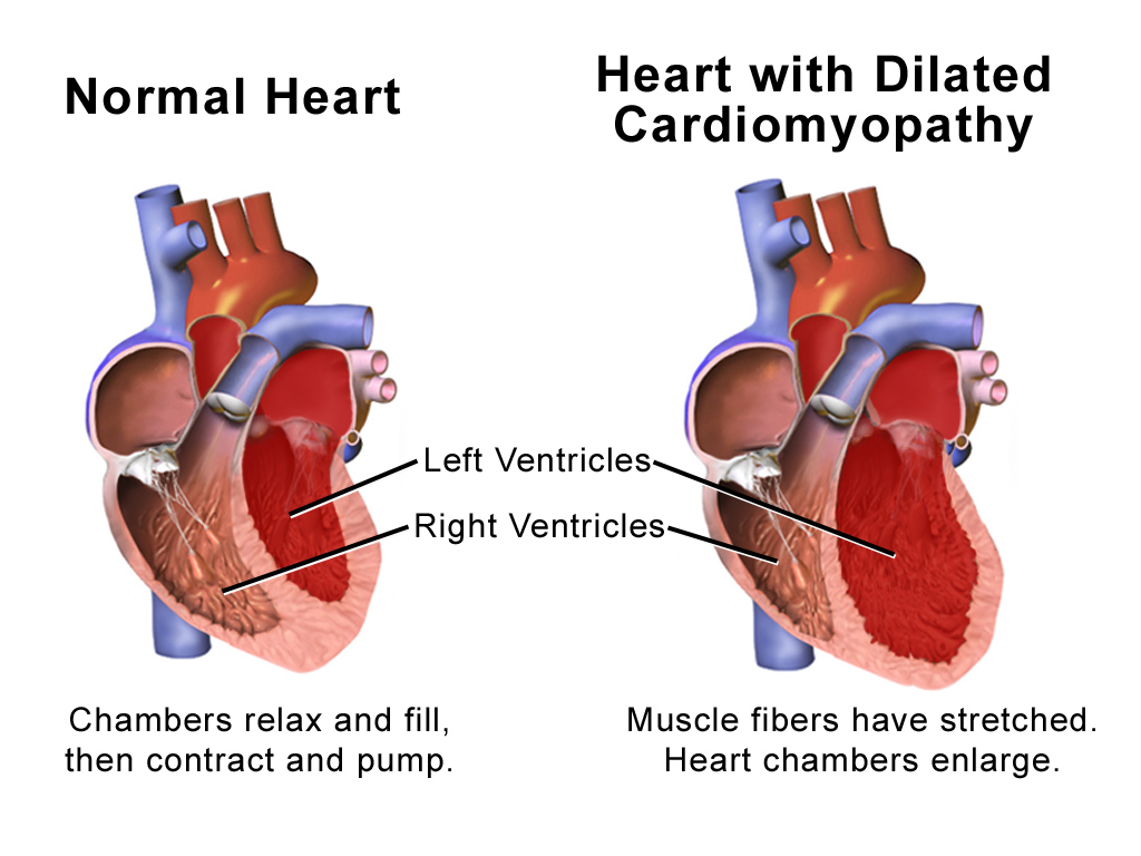
Dilated cardiomyopathy (DCM) is a disease of the heart muscle in which the main pumping chamber — the left ventricle — becomes enlarged (dilated), stretched and thin. As the muscle wall weakens, it can no longer squeeze forcefully, so the heart pumps out a smaller fraction of blood with each beat. Doctors describe this as a reduced ejection fraction, and it is the most common cause of chronic heart failure in younger and middle-aged adults.
DCM often develops gradually. Some people feel well for years while the heart slowly enlarges; others notice breathlessness, fatigue and ankle swelling as the first sign. The condition can be inherited (genetic), triggered by a viral infection or myocarditis, caused by heavy alcohol use, appear around pregnancy (peripartum), or have no identifiable cause, in which case it is called idiopathic DCM.
Although DCM is a serious, lifelong condition, the outlook has improved dramatically. Modern guideline-directed medical therapy, device therapy (pacemakers, defibrillators and resynchronisation), and — for the most severe cases — mechanical heart pumps and heart transplantation allow many patients to live full, active lives. Early diagnosis and consistent treatment are the keys to a good outcome, which is why this guide explains the disease, its treatment and how to access expert cardiac care worldwide.
2. Key Facts at a Glance
| Fact | Detail |
|---|---|
| Also known as | DCM, congestive cardiomyopathy, dilated heart muscle disease |
| Body system affected | Cardiovascular system — mainly the left ventricle |
| Common in | Adults aged 20–60; more often in men; can affect children |
| Severity range | Mild (symptom-free) to advanced end-stage heart failure |
| Key treatments | Heart-failure medications, ICD/CRT devices, LVAD, transplant |
| Outlook | Variable; many stabilise or improve with modern therapy |
3. Alternative Names and Medical Terminology
DCM is referred to by several names in medical records and literature:
- DCM — the standard abbreviation.
- Congestive cardiomyopathy — older term reflecting fluid congestion.
- Idiopathic dilated cardiomyopathy — when no cause is found.
- Familial dilated cardiomyopathy — inherited forms.
- Non-ischaemic cardiomyopathy — distinguishes it from heart-muscle disease caused by blocked coronary arteries.
- Cause-specific labels include alcoholic cardiomyopathy, peripartum cardiomyopathy and viral (myocarditic) cardiomyopathy.
4. Relevant Heart, Lung or Vascular Anatomy
The heart has four chambers: two upper atria and two lower ventricles. The left ventricle is the main pumping chamber, ejecting oxygen-rich blood into the aorta and around the body. Heart valves — the mitral and aortic on the left side — ensure blood flows in one direction.
In DCM, the left ventricle enlarges and its walls thin. The mitral valve ring stretches, so the valve may leak (functional mitral regurgitation). The right ventricle and atria can dilate too. Because blood backs up behind the failing left ventricle, pressure rises in the pulmonary veins and lungs, causing breathlessness.
5. How the Disease Affects the Body
DCM impairs the heart’s core job: pumping enough blood to meet the body’s needs. As the left ventricle dilates and weakens, the ejection fraction falls and less oxygenated blood reaches the organs and muscles. This produces fatigue, breathlessness and exercise intolerance.
The body responds with compensatory mechanisms that are helpful at first but harmful long-term. The sympathetic nervous system and the renin–angiotensin–aldosterone system are activated, raising heart rate, tightening blood vessels and causing the body to retain salt and water. This retained fluid accumulates in the lungs (causing breathlessness and cough) and in the legs, ankles and abdomen (causing swelling, or oedema). Over time these hormonal signals drive further adverse remodelling — the heart keeps enlarging in a vicious cycle.
A stretched left ventricle also disturbs the heart’s electrical system, predisposing to arrhythmias such as atrial fibrillation and dangerous ventricular rhythms. The dilated, sluggish chambers allow blood to pool and clot, raising the risk of stroke and other embolisms. A leaking mitral valve adds further volume load. Understanding this cascade explains why treatment targets not just symptoms but the underlying neurohormonal overactivity that fuels the disease.
6. Types and Classification
DCM is classified mainly by cause and inheritance:
- Idiopathic DCM — no identifiable cause (a diagnosis of exclusion).
- Familial/genetic DCM — caused by inherited gene mutations.
- Ischaemic-appearing vs non-ischaemic — DCM is a non-ischaemic cardiomyopathy, distinct from heart-muscle damage caused by coronary artery blockages.
- Acquired forms — viral/myocarditic, alcoholic, drug- or toxin-induced (e.g. chemotherapy), peripartum, and tachycardia-induced.
Severity is graded functionally using the NYHA (New York Heart Association) classes I–IV and by ejection fraction.
7. Causes of the Disease
DCM has many possible causes:
- Genetic mutations — the single largest identifiable group.
- Viral myocarditis — infection and inflammation of the heart muscle.
- Excessive alcohol intake over years (alcoholic cardiomyopathy).
- Pregnancy — peripartum cardiomyopathy, appearing late in pregnancy or after delivery.
- Toxins and drugs — chemotherapy agents (e.g. anthracyclines), cocaine, some medications.
- Nutritional deficiencies (thiamine, selenium) and thyroid disorders.
- Sustained rapid heart rhythms (tachycardia-induced).
- Autoimmune and infiltrative diseases. In many patients no cause is found — idiopathic DCM.
8. How the Disease Develops
DCM usually develops through an initial injury or genetic vulnerability in the heart muscle. A viral infection, toxin, sustained fast rhythm or an inherited weakness in the proteins that give heart cells their structure sets the process in motion. Individual muscle cells become dysfunctional or die, and the heart tries to compensate by enlarging its chamber and recruiting neurohormonal systems.
At first this compensation maintains blood flow, and the person may feel normal. But the same hormonal signals that boost output also promote fibrosis (scarring), cell death and progressive chamber enlargement — a process called adverse remodelling. As the ventricle grows rounder and thinner, it becomes mechanically less efficient (by the law of Laplace, a larger chamber needs more wall tension to generate the same pressure). The mitral valve begins to leak, adding volume overload.
This self-reinforcing cycle means DCM tends to progress unless interrupted. The encouraging news is that modern therapies can slow, halt and sometimes reverse remodelling, allowing the ventricle to shrink and the ejection fraction to recover — especially when treatment starts early and the trigger (such as alcohol or a fast rhythm) is removed.
9. Risk Factors
Non-modifiable:
- Family history of DCM or unexplained heart failure/sudden death.
- Male sex and age 20–60 (though any age can be affected).
- Certain genetic mutations and ethnic backgrounds.
Modifiable / acquired:
- Heavy alcohol consumption.
- Uncontrolled high blood pressure.
- Viral infections and untreated myocarditis.
- Exposure to cardiotoxic drugs (some chemotherapies, cocaine).
- Poorly controlled diabetes, thyroid disease and nutritional deficiencies.
10. Genetic and Family-History Factors
Up to a third or more of DCM cases are familial, inherited in most families in an autosomal dominant pattern (a 50% chance of passing the gene to each child). Dozens of genes are implicated; mutations in TTN (titin) are the most common, alongside LMNA, MYH7 and others. Some genes, notably LMNA and FLNC, carry a higher risk of dangerous arrhythmias and sudden death, which influences early defibrillator decisions.
Because relatives may carry the gene before symptoms appear, guidelines recommend cardiac screening (echocardiogram and ECG) of first-degree relatives and referral for genetic counselling and testing when a familial cause is suspected.
11. Who Is Most at Risk?
The people at highest risk of DCM include:
- First-degree relatives of someone with familial DCM.
- Adults aged 20–60, particularly men.
- People with a history of viral myocarditis.
- Heavy long-term drinkers.
- Pregnant women and new mothers (peripartum form), especially with multiple pregnancies or older maternal age.
- Cancer survivors treated with cardiotoxic chemotherapy.
- People with untreated thyroid disease or sustained arrhythmias.
12. Prevalence and Epidemiology
DCM is one of the most common cardiomyopathies and a leading reason for heart transplantation worldwide. Population studies suggest it affects roughly 1 in 250 to 1 in 500 people, though many mild cases go undiagnosed, so the true figure is likely higher. It occurs across all countries and ethnic groups, is somewhat more common in men, and is a major cause of heart failure in adults under 50. In children, DCM is the most frequent form of cardiomyopathy leading to transplant. These figures are approximate and vary by region and diagnostic criteria.
13. Signs and Symptoms
Symptoms of DCM arise mainly from heart failure and reduced blood flow. Some people have no symptoms early on, while others present with marked breathlessness. Common features include:
- Shortness of breath — on exertion, then at rest and when lying flat (orthopnoea).
- Waking at night gasping for breath (paroxysmal nocturnal dyspnoea).
- Fatigue and weakness, reduced exercise tolerance.
- Swelling (oedema) of the ankles, legs and abdomen.
- Palpitations or an awareness of irregular heartbeats.
- Dizziness, light-headedness or fainting (syncope).
- Persistent cough or wheeze, sometimes with frothy sputum.
- Weight gain from fluid retention.
- Reduced appetite and abdominal bloating from liver and gut congestion.
Because symptoms often come on gradually and resemble everyday tiredness or a chest infection, DCM is sometimes advanced by the time it is recognised. Any unexplained breathlessness, swelling or fainting deserves prompt medical assessment.
14. Early-Stage Symptoms
In its early stages DCM may cause few or subtle symptoms. People often notice:
- Getting breathless sooner during exercise or climbing stairs.
- Unusual tiredness and reduced stamina.
- Mild ankle swelling by the end of the day.
- Occasional palpitations. Some are diagnosed by chance on a scan or ECG done for another reason, before any symptoms appear.
15. Advanced-Stage Symptoms
As DCM progresses, symptoms become more limiting:
- Breathlessness at rest or with minimal activity.
- Inability to lie flat; needing several pillows to sleep.
- Marked swelling of legs, abdomen (ascites) and sometimes the whole body.
- Severe fatigue and muscle wasting (cardiac cachexia).
- Fainting or dangerous arrhythmias.
- Cold, pale extremities and confusion from poor circulation. These indicate advanced heart failure requiring specialist evaluation for devices, mechanical support or transplant.
16. Symptoms in Women, Men and Older Adults
- Women are more likely to develop the peripartum form around pregnancy and may report fatigue and breathlessness that is easily mistaken for normal late-pregnancy tiredness.
- Men are affected more often overall and may present at a younger age, sometimes with alcohol-related or genetic DCM.
- Older adults often have coexisting conditions (hypertension, coronary disease, kidney impairment) that blur the picture; they may present with confusion, falls or general decline rather than classic breathlessness.
17. Emergency Warning Signs
Call emergency services or go to hospital immediately for:
- Severe or sudden breathlessness, or coughing up pink, frothy sputum.
- Chest pain or pressure.
- Fainting or collapse.
- A very fast, irregular or pounding heartbeat that does not settle.
- Blue lips, cold clammy skin or confusion. These may signal acute heart failure, a dangerous arrhythmia or a blood clot.
18. When to Seek Medical Help
See a doctor promptly if you have persistent breathlessness, unexplained fatigue, ankle or abdominal swelling, recurrent palpitations or fainting — especially if you have a family history of cardiomyopathy, heart failure or sudden death. Early assessment allows treatment to begin before the heart weakens further.
19. Disease Stages, Grades and Severity
Severity in DCM is described using complementary systems:
- NYHA functional classes I–IV — from no symptoms (I) to symptoms at rest (IV).
- ACC/AHA heart-failure stages A–D — from at-risk (A) to advanced, refractory heart failure (D).
- Ejection fraction — the percentage of blood pumped per beat; DCM typically shows a reduced EF (below 40%), with severe disease often under 30%.
- Additional measures include ventricular size on echocardiography and biomarker levels. Together these guide treatment intensity and prognosis.
20. Disease Progression
DCM follows a variable course. Some patients stabilise or recover substantially once the cause is treated and medications take effect; the ventricle can shrink and the ejection fraction improve over months. Others experience a slow decline despite therapy, and a minority deteriorate rapidly. Progression is driven by ongoing adverse remodelling, worsening mitral leak, arrhythmias and repeated episodes of fluid overload (decompensation). Consistent medication, monitoring and prompt treatment of setbacks greatly influence which path a patient follows.
21. Possible Complications
DCM can lead to several serious complications:
- Heart failure — progressive breathlessness and fluid overload.
- Arrhythmias — atrial fibrillation and life-threatening ventricular rhythms.
- Sudden cardiac death from ventricular arrhythmia.
- Functional mitral (and tricuspid) regurgitation from stretched valves.
- Blood clots forming in the sluggish chambers, causing stroke or pulmonary embolism.
- Liver and kidney impairment from chronic congestion and low output.
22. Related and Associated Medical Conditions
DCM frequently coexists with or overlaps other conditions:
- Atrial fibrillation and other rhythm disorders.
- Hypertension and coronary artery disease (which must be excluded).
- Chronic kidney disease (cardiorenal syndrome).
- Diabetes and metabolic syndrome.
- Thyroid disorders.
- Sleep apnoea, which worsens heart strain.
- Other inherited cardiomyopathies within the same family.
23. Screening and Early Detection
Because DCM is often familial, screening of first-degree relatives is recommended when someone is diagnosed with genetic or unexplained DCM. Screening usually involves an ECG and echocardiogram, sometimes repeated every few years, plus genetic testing where a mutation is known. Athletes and people with a family history of sudden death may also be screened. Detecting DCM before symptoms appear allows earlier, more effective treatment.
24. How the Disease Is Diagnosed
Diagnosing DCM combines the clinical picture with imaging and tests to confirm an enlarged, weakly contracting left ventricle and to identify the cause. The pathway usually begins when a patient presents with heart-failure symptoms or an abnormal ECG, or when a relative is screened.
The cornerstone test is the echocardiogram (heart ultrasound), which measures chamber size, wall thickness, ejection fraction and valve function, revealing the characteristic dilated, poorly contracting ventricle. An ECG detects rhythm problems and conduction delays. Blood tests (including BNP/NT-proBNP) support the diagnosis and gauge severity.
Crucially, doctors must exclude coronary artery disease as the cause, because blocked arteries produce a similar picture (ischaemic cardiomyopathy) but need different treatment. This is done with coronary angiography or CT coronary angiography. Cardiac MRI provides detailed images of muscle structure and scarring and can point to specific causes such as prior myocarditis or infiltration. Additional tests — thyroid function, genetic testing, and occasionally heart-muscle biopsy — help pin down the underlying trigger. A confident diagnosis therefore rests on demonstrating a dilated, weakened ventricle and systematically ruling out reversible or specific causes.
25. Physical Examination and Medical History
The doctor reviews symptoms, alcohol intake, recent infections, pregnancy, medication and cancer treatment, and a detailed family history of heart disease or sudden death. On examination, findings may include a displaced, diffuse heart apex, an added heart sound (third heart sound / gallop), a mitral regurgitation murmur, raised jugular venous pressure, lung crackles, an enlarged liver and swollen ankles. Blood pressure and pulse, including any irregularity, are assessed.
26. Diagnostic Tests and Imaging
Key investigations include:
- Echocardiogram — the primary test; measures ventricular size and ejection fraction.
- Electrocardiogram (ECG) — rhythm, conduction delays (e.g. left bundle branch block).
- Chest X-ray — enlarged heart and lung congestion.
- Cardiac MRI — tissue characterisation, scarring, and clues to cause.
- Coronary angiography / CT coronary angiography — to exclude coronary artery disease.
- Holter or event monitoring — to capture arrhythmias.
- Exercise or cardiopulmonary testing — to assess functional capacity.
27. Blood Tests, Biomarkers and Genetic Testing
Blood work supports diagnosis, finds treatable causes and tracks severity:
- BNP / NT-proBNP — natriuretic peptides raised in heart failure.
- Full blood count, kidney and liver function, electrolytes.
- Thyroid function tests.
- Iron studies, glucose/HbA1c.
- Troponin — if myocarditis or injury is suspected.
- Genetic testing — a panel of cardiomyopathy genes, with genetic counselling, when a familial cause is likely; results can guide family screening and defibrillator decisions.
28. Understanding Test Results
Results are interpreted together. A reduced ejection fraction with an enlarged ventricle on echo confirms DCM; the lower the EF, generally the more severe the disease. Raised BNP/NT-proBNP indicates heart strain and fluid overload, and falling levels suggest treatment is working. Normal coronary arteries rule out an ischaemic cause. MRI scarring patterns or a positive genetic test point to a specific type. Your cardiologist will explain what your particular numbers mean and how they shape your treatment plan.
29. Differential Diagnosis
Several conditions can mimic DCM and must be distinguished:
- Ischaemic cardiomyopathy — heart weakness from coronary artery disease.
- Hypertensive heart disease.
- Valvular heart disease causing secondary dilation.
- Restrictive or hypertrophic cardiomyopathy.
- Myocarditis (which may resolve).
- Tachycardia-induced cardiomyopathy (reversible if rhythm controlled).
- Infiltrative diseases such as amyloidosis or sarcoidosis.
30. Specialist and Multidisciplinary Evaluation
Optimal DCM care involves a team: a cardiologist (often a heart-failure specialist), cardiac imaging experts, electrophysiologists for rhythm and devices, cardiac surgeons and transplant/LVAD specialists for advanced cases, plus heart-failure nurses, pharmacists, dietitians and genetic counsellors. This multidisciplinary approach ensures medication is optimised, devices are offered at the right time, and advanced options are considered promptly. You can find experienced doctors and specialist hospitals through this site.
31. Treatment Goals
The aims of DCM treatment are to:
- Relieve symptoms — breathlessness, swelling and fatigue.
- Slow, halt or reverse the heart’s adverse remodelling.
- Prevent complications — arrhythmias, sudden death and stroke.
- Reduce hospital admissions for heart failure.
- Improve quality of life and survival, and treat any reversible cause. Treatment is tailored to each patient’s cause, symptoms and heart function.
32. When Is Treatment Required?
Treatment generally begins as soon as DCM is diagnosed, even in people with few symptoms, because early therapy protects the heart and can reverse remodelling. Guideline-directed medical therapy is started and gradually built up. More intensive options — devices, mechanical support or transplant — are introduced when symptoms persist despite medication, the ejection fraction stays very low, dangerous arrhythmias appear, or heart failure becomes advanced.
33. Active Monitoring and Watchful Waiting
Pure “watchful waiting” is uncommon in DCM, because even symptom-free patients benefit from medication. However, once therapy is established, patients with mild, stable disease are actively monitored with periodic reviews, echocardiograms, ECGs and blood tests to detect progression early. Relatives found to carry a gene mutation but with normal hearts are followed with regular screening rather than immediate treatment.
34. Medications
Medications are the foundation of DCM care. Four pillars of guideline-directed medical therapy for heart failure with reduced ejection fraction are used together and up-titrated:
- ACE inhibitors / ARBs or ARNI (sacubitril–valsartan) — relax blood vessels and block harmful hormones.
- Beta-blockers (e.g. bisoprolol, carvedilol) — slow the heart and protect against remodelling and arrhythmia.
- Mineralocorticoid receptor antagonists (MRAs) — spironolactone or eplerenone.
- SGLT2 inhibitors (dapagliflozin, empagliflozin) — improve outcomes even without diabetes.
Additional drugs include diuretics to remove excess fluid, anticoagulants for atrial fibrillation or clots, anti-arrhythmics, and ivabradine or digoxin in selected cases. Doses are increased slowly to the highest tolerated level, as this improves survival.
35. Minimally Invasive Treatments
Beyond tablets, several less-invasive options help selected patients:
- Cardiac resynchronisation therapy (CRT) — a specialised pacemaker that re-coordinates the ventricles in people with conduction delay.
- Implantable cardioverter-defibrillators (ICDs) — implanted under the skin to stop lethal arrhythmias.
- Percutaneous mitral valve repair (e.g. edge-to-edge clip) — reduces a leaking mitral valve without open surgery. See minimally invasive cardiac procedures for related techniques.
36. Catheter-Based and Endovascular Treatments
Catheter-based procedures are performed through small punctures in blood vessels rather than open surgery:
- Catheter ablation — to treat atrial fibrillation or ventricular arrhythmias, and to cure tachycardia-induced DCM by restoring normal rhythm.
- Transcatheter mitral valve repair/replacement — for functional mitral regurgitation.
- Lead placement for CRT and ICD devices via veins. These electrophysiological procedures offer shorter recovery than open surgery.
37. Surgical Treatment Options
Surgery is reserved for advanced DCM or specific problems that medication and catheter therapy cannot solve. Options include:
- Mitral valve surgery — repair or replacement when severe functional mitral regurgitation adds to the heart’s burden; see mitral valve procedures.
- Left ventricular assist device (LVAD) — a surgically implanted mechanical pump that takes over the work of the failing left ventricle. It can be a bridge to transplant or a permanent (destination) therapy for those not eligible for transplant.
- Heart transplantation — the definitive treatment for end-stage DCM that no longer responds to any other therapy. A donor heart replaces the failing one, and modern immunosuppression allows many recipients to return to active lives.
- Surgical ventricular restoration and other adjunctive procedures in selected centres.
The choice depends on how advanced the disease is, the patient’s age and other health problems, and the availability of donor organs and mechanical support. Because LVAD implantation and transplantation are highly specialised, they are performed at experienced cardiac surgery centres with dedicated advanced heart-failure teams. Many patients travel to accredited international hospitals for these procedures, which combine complex surgery with lifelong follow-up.
38. Advanced and Emerging Treatments
Research is expanding the options for DCM:
- Newer mechanical pumps that are smaller, more durable and less prone to clotting.
- Gene-specific and gene-editing therapies for inherited forms.
- Cell-based and regenerative therapies aiming to repair heart muscle.
- Refined resynchronisation techniques such as conduction-system pacing.
- Anti-inflammatory and immunomodulatory therapy for myocarditis-related DCM. Many are available through clinical trials at specialist centres.
39. Treatment Options Compared
- Medications — first-line for everyone; low risk, must be lifelong and up-titrated.
- Devices (ICD/CRT) — reduce sudden death and improve symptoms in selected patients; require a minor implant procedure.
- Catheter procedures — treat arrhythmias and valve leak with quick recovery.
- Surgery/LVAD — for advanced disease; more invasive but life-saving.
- Transplant — best long-term outcome for end-stage disease but limited by donor availability and lifelong immunosuppression. Most patients move stepwise from medication toward advanced therapies only if the disease progresses.
40. How Doctors Choose the Right Treatment
Treatment selection weighs several factors: the underlying cause, ejection fraction and ventricular size, NYHA class and symptom burden, presence of arrhythmias or conduction delay, valve function, kidney and liver health, age and other illnesses, genetic findings, and the patient’s preferences and goals. Reversible causes (alcohol, fast rhythms, thyroid problems) are corrected first. Decisions are usually made by a multidisciplinary heart team to match therapy to the individual.
41. Benefits and Risks of Treatment
- Medications — proven to prolong life and reduce admissions; possible side effects include low blood pressure, dizziness, kidney changes and electrolyte shifts, managed by careful monitoring.
- Devices — reduce sudden death and improve symptoms; small risks of infection, lead problems or inappropriate shocks.
- Surgery/LVAD — can be life-saving but carry risks of bleeding, infection, stroke and clotting.
- Transplant — offers the best quality of life for end-stage disease but requires lifelong immunosuppression with its own risks. Benefits generally outweigh risks when therapies are matched appropriately.
42. What Happens If the Disease Is Left Untreated?
Untreated DCM tends to progress. The ventricle enlarges further, the ejection fraction falls, and heart failure worsens with escalating breathlessness and swelling. The risks of dangerous arrhythmias, sudden cardiac death, stroke and blood clots rise, and repeated episodes of fluid overload lead to recurrent hospital admissions and damage to the kidneys and liver. Without treatment, advanced DCM carries a poor prognosis — which is why early, sustained therapy is so important.
43. Treatment Success and Expected Outcomes
Outcomes have improved markedly with modern therapy. Many patients on optimised medication experience significant recovery of ejection fraction and symptoms, sometimes near-normalising heart function — particularly when a reversible cause is removed. Devices reduce sudden death, and LVADs and transplantation offer good survival and quality of life for advanced disease. Outcomes vary with cause, severity, how early treatment starts and adherence, so your cardiologist can give a realistic estimate for your situation. Overall, most patients can expect meaningful improvement in symptoms and prognosis with comprehensive care.
44. Prognosis and Long-Term Outlook
The outlook for DCM is highly variable and, on average, far better than in past decades. A substantial proportion of patients stabilise or improve on guideline-directed therapy, and some recover normal heart function, especially those with recent-onset disease, a treatable cause (such as alcohol or tachycardia), or a good early response to medication.
Factors linked to a poorer prognosis include a very low ejection fraction, advanced symptoms (NYHA III–IV), significant scarring on MRI, serious arrhythmias, certain high-risk genetic mutations (such as LMNA and FLNC), and coexisting kidney disease. Even so, many people with these features do well thanks to ICDs, resynchronisation, LVADs and transplantation.
Long-term, DCM is a chronic condition requiring ongoing treatment and monitoring, but with consistent care, healthy lifestyle measures and prompt attention to setbacks, most patients live active lives for many years. Family members should be screened, since early detection in relatives leads to better outcomes. The single most powerful influence on prognosis is early diagnosis followed by adherence to comprehensive, guideline-based treatment.
45. Recovery and Rehabilitation
Recovery in DCM is usually a gradual process of stabilising the heart and rebuilding fitness. Cardiac rehabilitation — a supervised programme of monitored exercise, education and lifestyle support — improves symptoms, confidence and quality of life. After device implantation, recovery takes days to weeks; after LVAD surgery or transplant, it involves a hospital stay followed by months of rehabilitation and close follow-up. Taking medications reliably, managing fluid and salt, and attending reviews are central to a durable recovery.
46. Follow-Up Tests and Long-Term Monitoring
DCM requires lifelong monitoring: regular clinic visits, echocardiograms to track ejection fraction and chamber size, ECGs and Holter monitoring for rhythm, and blood tests including kidney function and BNP/NT-proBNP. Device patients have their pacemaker/ICD checked in person or remotely. Monitoring detects progression early and allows medications and therapies to be adjusted before problems escalate.
47. Managing Recurrence or Disease Progression
If DCM worsens despite treatment, the team reassesses and escalates care: optimising and adding medications, considering CRT/ICD if not already in place, treating a leaking mitral valve, and evaluating for LVAD or transplant. Recurrence of a reversible cause — for example, a return to heavy drinking — must be addressed. Prompt treatment of decompensation episodes (fluid overload) helps prevent long-term decline.
48. Living with the Disease
Living well with DCM means combining medical treatment with daily self-care: taking medicines as prescribed, weighing yourself regularly to spot fluid retention, limiting salt and fluids as advised, staying active within limits, avoiding alcohol and tobacco, and attending reviews. Learning to recognise warning signs and having an action plan reduces emergencies. Many people continue to work, travel and enjoy family life with good disease control and support.
49. Diet and Nutrition Guidelines
- Limit salt (sodium) to reduce fluid retention and breathlessness.
- Follow any fluid restriction advised by your team.
- Eat a heart-healthy diet rich in vegetables, fruit, whole grains, fish and lean protein.
- Avoid or strictly limit alcohol, which can directly damage the heart muscle.
- Weigh yourself daily and report rapid weight gain (a sign of fluid build-up).
- Correct deficiencies (e.g. thiamine) and manage diabetes and weight with dietitian support.
50. Exercise and Physical-Activity Guidelines
Regular, moderate activity is beneficial for most people with stable DCM and is best introduced through cardiac rehabilitation. Suitable activities include walking, cycling and light aerobic exercise, guided by symptoms. Patients should avoid sudden intense exertion and, for some genetic or arrhythmia-prone forms, may be advised to avoid competitive or high-intensity sport. Always agree an activity plan with your cardiologist, and stop and seek help for chest pain, severe breathlessness or fainting.
51. Medications, Activities and Habits to Avoid
- Avoid alcohol and recreational drugs (especially cocaine).
- Stop smoking.
- Avoid NSAID painkillers (e.g. ibuprofen), which cause fluid retention, unless approved by your doctor.
- Be cautious with excess salt and fluids.
- Avoid strenuous or competitive sport if advised, particularly with high-risk genotypes.
- Do not stop heart-failure medications abruptly, and check new medicines and supplements with your team.
52. Preventing the Disease or Reducing Its Risks
Not all DCM is preventable, but risks can be reduced by:
- Moderating or avoiding alcohol.
- Controlling blood pressure, diabetes and thyroid disease.
- Treating infections and myocarditis promptly.
- Avoiding cardiotoxic drugs where possible and monitoring the heart during chemotherapy.
- Screening relatives of people with familial DCM so treatment can start early.
- Maintaining a healthy weight, diet and activity level.
53. Pregnancy and the Disease
Pregnancy is an important consideration in DCM. Peripartum cardiomyopathy develops in late pregnancy or after delivery and needs specialist care. Women with existing DCM face higher risks during pregnancy, and some heart-failure medications are unsafe in pregnancy. Any woman with DCM who is pregnant or planning pregnancy should have pre-conception counselling and be managed by a joint cardiology–obstetric team.
54. Disease in Children and Young Adults
DCM is the most common cardiomyopathy in children and a leading reason for paediatric heart transplantation. Causes include genetic mutations, viral myocarditis and metabolic disorders. Young adults may have inherited forms that first appear in their teens or twenties. Care is provided by paediatric or young-adult heart-failure specialists, with family screening and genetic counselling, and treatment principles similar to adults, adapted by age and size.
55. Disease in Older Adults
In older adults, DCM often coexists with hypertension, coronary disease, kidney impairment and other illnesses, making diagnosis and treatment more complex. Medication doses may need careful adjustment, and treatment decisions balance benefits against frailty and quality of life. Advanced therapies such as transplant are generally reserved for younger, fitter patients, but devices and medical therapy benefit many older people.
56. Emotional Health and Patient Support
A diagnosis of DCM can bring anxiety, low mood and worry about the future, for patients and families alike. Emotional support is an essential part of care. Talking to your team, joining cardiomyopathy or heart-failure support groups, and accessing counselling or psychological services can help. Involving family, learning about the condition, and connecting with others who share the experience all improve coping and wellbeing.
57. Preparing for Your Specialist Appointment
To make the most of your appointment:
- List your symptoms, when they started and what makes them better or worse.
- Note your family history of heart disease and sudden death.
- Bring a list of all medications and supplements.
- Record your alcohol intake, recent infections or pregnancy.
- Bring previous test results and scans.
- Write down your questions and bring someone with you to help remember the discussion.
58. Questions to Ask Your Doctor
- What is causing my dilated cardiomyopathy?
- What is my ejection fraction and what does it mean?
- Could my condition be genetic, and should my family be screened?
- Which medications do I need and what are their side effects?
- Do I need a device such as an ICD or CRT?
- Might I need an LVAD or transplant in future?
- What lifestyle changes should I make?
- How will you monitor my heart over time?
- What are the warning signs I should watch for?
- Is it safe for me to exercise, travel or (for women) become pregnant?
59. Cost of Diagnosis and Treatment
Costs vary widely by country, hospital and the complexity of treatment. The figures below are approximate USD ranges for guidance only.
| Item | US / UK / Western Europe | India / Turkey / Thailand |
|---|---|---|
| Diagnosis (echo, ECG, MRI, bloods) | $2,000 – $8,000 | $400 – $2,000 |
| ICD/CRT device implant | $30,000 – $80,000 | $8,000 – $25,000 |
| Mitral valve surgery | $50,000 – $150,000 | $6,000 – $15,000 |
| LVAD implantation | $150,000 – $400,000+ | $60,000 – $130,000 |
| Heart transplant | $800,000 – $1,500,000+ | $80,000 – $180,000 |
Medical tourism to destinations such as India, Turkey, Thailand and Singapore can cost roughly 50–90% less than the US or UK for comparable, accredited care. Explore destinations and hospitals for options.
60. Factors Affecting Treatment Cost
- Type and complexity of treatment (medication vs device vs transplant).
- Country and hospital chosen, and its accreditation and reputation.
- Length of hospital stay and intensive-care needs.
- Device and implant brand and model.
- Surgeon and specialist fees.
- Complications, follow-up and lifelong medication (e.g. immunosuppressants after transplant).
- Travel, accommodation and interpreter costs for international patients.
61. Choosing the Right Specialist
Look for a cardiologist with heart-failure and cardiomyopathy expertise, ideally at a centre offering the full range of therapies including devices, LVAD and transplant. Consider board certification, experience with your specific type of DCM, procedure volumes and outcomes, access to a multidisciplinary team, and clear communication. This site can help you find qualified doctors.
62. Choosing the Right Hospital or Treatment Centre
Choose a hospital with:
- Recognised accreditation (e.g. JCI for international centres).
- A dedicated advanced heart-failure and transplant/LVAD programme.
- High procedure volumes and published outcomes.
- Full cardiac imaging, electrophysiology and surgery on site.
- Strong follow-up and rehabilitation services.
- Experience with international patients if travelling. Browse accredited hospitals and destinations.
63. Getting a Second Medical Opinion
A second opinion is valuable in DCM, especially before major decisions such as device implantation, LVAD or transplant, or when the cause is unclear. Another specialist may confirm the diagnosis, suggest additional treatable causes, or offer therapies not available locally. Reputable centres welcome second opinions. You can request a second opinion through this site.
64. Treatment Abroad and Medical-Travel Considerations
Many patients travel abroad for high-quality, affordable DCM care. When planning medical travel, consider hospital accreditation and outcomes, the surgeon’s experience, clear cost estimates, and arrangements for follow-up and complications after returning home. Factor in travel time, fitness to fly, visas, language support and care for accompanying family. Coordinate closely with your home cardiologist so long-term therapy continues seamlessly. Explore destinations and treatments for guidance.
65. Frequently Asked Questions
Is dilated cardiomyopathy curable? There is usually no outright cure, but many patients recover substantial heart function with treatment, and some near-normalise, especially when a reversible cause is removed.
Is DCM inherited? A significant proportion is genetic. If you have familial DCM, first-degree relatives should be offered screening and genetic counselling.
Can I exercise with DCM? Yes — moderate activity, ideally through cardiac rehabilitation, is beneficial. High-intensity or competitive sport may be restricted in some cases.
Will I need a heart transplant? Only a minority reach end-stage disease requiring transplant or LVAD. Most are managed well with medications and devices.
Can drinking alcohol cause DCM? Yes — heavy long-term drinking can cause alcoholic cardiomyopathy, which may improve if alcohol is stopped.
Is DCM the same as a heart attack? No. A heart attack is caused by blocked coronary arteries; DCM is a disease of the heart muscle itself, though coronary disease must be excluded.
Can women with DCM have children? Pregnancy carries higher risks and needs specialist pre-conception counselling and joint cardiology–obstetric care.
How is DCM monitored? With regular clinic visits, echocardiograms, ECGs, blood tests and device checks.
66. Patient Stories and Treatment Experiences
The following are representative, anonymised illustrations, not real individuals.
Rahul, India — Diagnosed with idiopathic DCM in his thirties after months of breathlessness, Rahul started guideline-directed medication and joined cardiac rehab. Within a year his ejection fraction had improved substantially and he returned to work and light sport.
Elena, Spain — After a viral illness, Elena developed heart failure and was found to have DCM. An ICD and optimised medication stabilised her condition, and she now manages well with regular monitoring.
James, UK — With advanced familial DCM, James received an LVAD as a bridge to transplant, travelled to an accredited international centre, and successfully underwent a heart transplant, returning to an active life.
67. Latest Research and Clinical Trials
Research is advancing rapidly. Active areas include gene-specific therapies and gene editing for inherited DCM, regenerative and cell-based approaches to repair heart muscle, smaller and safer mechanical pumps, refined conduction-system pacing, and improved medications building on the four-pillar heart-failure regimen. Better genetic testing and risk prediction are helping tailor defibrillator decisions. Patients interested in clinical trials should ask their specialist centre; major cardiology bodies regularly update guidance as new evidence emerges.
68. Related Diseases and Conditions
- Ischemic Cardiomyopathy
- Restrictive Cardiomyopathy
- Heart Failure
- Mitral Valve Disease
- Disorders of Heart Rhythm
- Infective Endocarditis
69. Related Treatments and Procedures
- Minimally Invasive Cardiac Surgery
- Mitral Valve Procedures
- Electrophysiological Procedures
- Hybrid Cardiac Procedures
- Other Procedures
70. Medical Glossary
- Cardiomyopathy — disease of the heart muscle.
- Dilated — enlarged and stretched.
- Ejection fraction (EF) — percentage of blood pumped out with each beat.
- Left ventricle — the heart’s main pumping chamber.
- Heart failure — the heart cannot pump enough blood for the body’s needs.
- Remodelling — structural change in the heart over time.
- Mitral regurgitation — a leaking mitral valve.
- Arrhythmia — abnormal heart rhythm.
- ICD — implantable cardioverter-defibrillator, stops lethal rhythms.
- CRT — cardiac resynchronisation therapy, coordinates the ventricles.
- LVAD — left ventricular assist device, a mechanical heart pump.
- BNP/NT-proBNP — blood markers of heart strain.
- Peripartum — around the time of pregnancy and childbirth.
- Idiopathic — of unknown cause.
- Guideline-directed medical therapy — the proven combination of heart-failure medications.
71. Medical Review, Editorial Policy and Last Updated Date
Last updated: 11 July 2026.
This article is reviewed by qualified medical professionals and written to align with current cardiology guidance from bodies such as the ACC/AHA, ESC, NHS and WHO. Our editorial policy emphasises accuracy, clarity and balance, with regular updates as evidence evolves.
Disclaimer: This content is for education only and is not a substitute for professional medical advice, diagnosis or treatment. Always consult a qualified healthcare provider about your individual condition.
72. Clinical Guidelines and Medical References
This article draws on general, guideline-based knowledge from leading cardiology organisations, including:
- American College of Cardiology / American Heart Association (ACC/AHA) heart-failure and cardiomyopathy guidelines.
- European Society of Cardiology (ESC) guidelines on heart failure and cardiomyopathies.
- National Health Service (NHS) and World Health Organization (WHO) patient information.
- Standard cardiology textbooks and peer-reviewed literature. These sources are cited in general terms; consult your specialist for detailed, current references.
73. Book an Appointment or Request a Second Opinion
If you or a loved one has dilated cardiomyopathy, expert help is available. Our partner network connects you with experienced heart-failure specialists and accredited hospitals worldwide.
- Book an appointment: /contact/
- Request a second opinion or ask a question: Contact us
- Explore top hospitals, doctors and destinations for dilated cardiomyopathy treatment.
Take the first step toward expert cardiac care today.