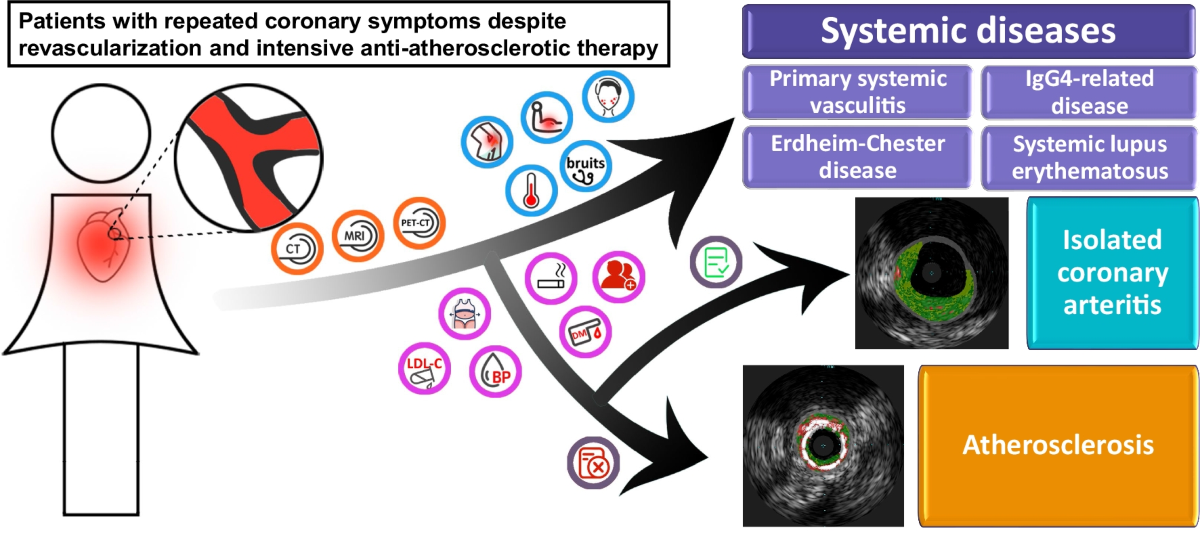
1. Disease Overview
Isolated coronary artery disease (CAD) is the narrowing or blockage of the coronary arteries—the vessels that supply oxygen-rich blood to the heart muscle—caused by atherosclerosis (plaque buildup). The word “isolated” means the disease affects primarily the coronary arteries, without significant involvement of the heart valves, aorta, or congenital defects—in practice, often single-vessel or focal disease treatable by targeting the coronary arteries alone.
CAD ranges from mild narrowing that causes no symptoms to severe blockage that triggers a heart attack. It is the most common heart disease worldwide, yet the outlook is generally excellent: with modern medications, lifestyle change, angioplasty, and bypass surgery, most patients relieve symptoms, lower their heart-attack risk, and return to normal life.
2. Key Facts at a Glance
| Aspect | Details |
|---|---|
| Also known as | Coronary heart disease, atherosclerotic heart disease, ischemic heart disease |
| Body system affected | Cardiovascular system (coronary arteries and heart muscle) |
| Common in | Adults over 45, especially with smoking, diabetes, high cholesterol, high blood pressure, family history, or obesity |
| Severity range | Silent (no symptoms) → stable angina → unstable angina → heart attack |
| Key treatments | Lifestyle change, medications (aspirin, statins, beta-blockers), angioplasty with stenting, coronary artery bypass grafting (CABG) |
| Outlook | Excellent with early treatment and risk-factor control; most patients return to normal activities |
3. Alternative Names and Medical Terminology
- Coronary heart disease (CHD) — often used interchangeably with CAD
- Ischemic heart disease (IHD) — emphasizes reduced blood flow (ischemia)
- Atherosclerotic cardiovascular disease (ASCVD) — broader term including CAD and stroke
- Myocardial ischemia — inadequate blood supply to heart muscle
- Angina pectoris — chest pain caused by CAD (stable or unstable)
- Acute coronary syndrome (ACS) — emergency presentation including heart attack and unstable angina
4. Relevant Heart, Lung or Vascular Anatomy
The coronary arteries arise from the base of the aorta:
- Left coronary artery — divides into the left anterior descending (LAD), supplying the front and much of the left ventricle, and the left circumflex (LCx), supplying the side and back
- Right coronary artery — supplies the right ventricle and the inferior left ventricle
These arteries run along the heart’s surface and branch into the muscle. The left ventricle, the main pumping chamber, has the highest oxygen demand and is most vulnerable to CAD; when a coronary artery narrows, the muscle it feeds receives too little blood, especially during exercise.
5. How the Disease Affects the Body
In CAD, cholesterol, calcium, cellular debris, and inflammatory cells accumulate in the artery wall to form atherosclerotic plaques. Over years these plaques narrow the lumen and limit blood flow, stiffen the artery so it cannot widen when demand rises, and may rupture suddenly, triggering a clot that abruptly blocks the vessel—a heart attack.
When the heart muscle is starved of oxygen it cannot pump efficiently, especially during exertion; this shortfall produces angina. A complete blockage causes heart muscle to die (myocardial infarction), replacing working tissue with scar that cannot contract, which can lead to heart failure, dangerous arrhythmias, or sudden cardiac death. Because the process is gradual, significant disease can exist for years before symptoms appear.
6. Types and Classification
CAD is classified several ways. By clinical presentation:
- Asymptomatic (silent ischemia) — significant narrowing without symptoms
- Stable angina — predictable chest discomfort with exertion, relieved by rest
- Unstable angina — new, worsening, or rest angina; high heart-attack risk
- Acute myocardial infarction — complete blockage with muscle damage
By anatomical extent: single-vessel, two-vessel, and three-vessel or left-main disease (most severe). By plaque type: stable versus vulnerable (thin-capped, rupture-prone).
Isolated CAD means coronary disease without significant valvular or congenital disease—typically single-territory or focal disease.
7. Causes of the Disease
The root cause of CAD is atherosclerosis, a chronic inflammatory process: the artery lining is injured (by high blood pressure, smoking, diabetes, or high cholesterol), LDL (“bad”) cholesterol enters the wall, macrophages ingest it to become “foam cells,” and foam cells, smooth muscle, and calcium build up into plaque that enlarges over decades.
The main drivers are high LDL and low HDL cholesterol, high blood pressure, smoking, diabetes, chronic inflammation, and genetic predisposition.
8. How the Disease Develops
CAD develops slowly, usually beginning in early adulthood: fatty streaks (teens–20s, potentially reversible), plaque formation (30s–40s, with a fibrous cap), progressive narrowing (40s–50s, reaching 50–70% and possibly causing exertional angina), vulnerable plaque (50s–60s, with large fatty cores and thin caps), and finally an acute event when a plaque ruptures and triggers a clot.
Importantly, moderate plaques (30–50%) are often more likely to rupture than severely narrowed ones with thick, stable caps. This is why many heart attacks strike “moderately” diseased arteries, and why controlling risk factors matters even when a blockage looks minor.
9. Risk Factors
Modifiable:
- Smoking / tobacco — doubles to triples risk
- High blood pressure and high LDL / low HDL cholesterol
- Diabetes or prediabetes — strongly accelerates atherosclerosis
- Obesity (especially abdominal) and physical inactivity
- Unhealthy diet, excess alcohol, chronic stress, and sleep apnea
Non-modifiable:
- Age — risk rises after 45 in men, 55 in women
- Male sex — women’s risk catches up after menopause
- Family history of early CAD and genetic conditions such as familial hypercholesterolemia
- Ethnicity — higher rates in South Asian, African, and Hispanic populations
10. Genetic and Family-History Factors
- Family history — a first-degree relative with early CAD roughly doubles your risk, even after accounting for other factors.
- Familial hypercholesterolemia (FH) — very high LDL (often >190 mg/dL) from birth, leading to early CAD if untreated.
- Polygenic risk — many common gene variants each add a small amount of risk, together shaping susceptibility.
- Genetics also act indirectly through inherited high blood pressure, diabetes, and clotting tendencies.
Genetic testing can identify FH, allowing early treatment and cascade screening of relatives. Importantly, most CAD risk remains modifiable through lifestyle and medication, even with a genetic predisposition.
11. Who Is Most at Risk?
Highest-risk groups include:
- Adults over 45–50 with multiple risk factors and smokers
- People with diabetes — risk comparable to already having CAD
- Metabolic syndrome and chronic kidney disease (which accelerates vascular calcification)
- South Asian ethnicity — higher baseline risk, often at younger ages
- Autoimmune/inflammatory disease (rheumatoid arthritis, lupus, psoriasis)
- Prior heart attack or stroke and strong family history
Risk rises sharply when several factors combine.
12. Prevalence and Epidemiology
- Extremely common — on the order of 100+ million people worldwide, and a leading cause of death.
- Age and sex — most common over 60; men typically develop CAD 7–10 years earlier than women, whose risk climbs after menopause.
- Geography and ethnicity — historically higher in industrialized nations but rising elsewhere; notably high and earlier-onset in South Asian populations.
Many people have undiagnosed “silent” CAD, particularly those with diabetes—hence the value of screening high-risk groups.
13. Signs and Symptoms
Symptoms depend on the severity of narrowing and how quickly it developed. The most common include:
- Chest discomfort or angina — pressure, squeezing, or tightness in the center or left chest, with exertion (stable) or at rest (unstable)
- Shortness of breath — often with exertion, sometimes without chest pain
- Unusual fatigue with activity, or palpitations, lightheadedness, or sweating
- Pain radiating to the left arm, shoulder, neck, jaw, or upper back
- Nausea, indigestion, or heartburn — easily mistaken for a stomach problem
Some patients—especially those with diabetes—have silent ischemia with no symptoms despite significant narrowing, so any new, exertional, or unexplained chest discomfort deserves evaluation.
14. Early-Stage Symptoms
Early CAD often causes no symptoms, or mild ones that are easy to dismiss:
- Mild chest “tightness” or “heaviness” only with strenuous effort
- Reduced exercise tolerance — can’t walk as far or climb stairs as before
- Fatigue with previously easy tasks, or mild breathlessness blamed on being “out of shape”
- Occasional indigestion-like sensations or intermittent arm/shoulder discomfort with activity
Many people quietly adapt by cutting back on activity, masking the change. Screening in high-risk individuals often detects early CAD before symptoms appear.
15. Advanced-Stage Symptoms
With more severe disease (typically >70% narrowing):
- Typical angina with minimal exertion—short walks, climbing stairs, even dressing or eating
- Frequent attacks, sometimes several times a week
- Angina at rest or nocturnal angina waking you from sleep (unstable angina)
- A worsening pattern of increasing frequency, severity, or duration
- Significant breathlessness, and ankle swelling or arrhythmias if heart failure or muscle damage develops
Advanced CAD signals a high heart-attack risk and needs prompt evaluation.
16. Symptoms in Women, Men and Older Adults
Women more often have atypical symptoms—nausea, indigestion, fatigue, or breathlessness rather than classic chest pressure; discomfort may be in the back, shoulders, or jaw and may occur at rest.
Men more commonly have the classic pattern—central chest pressure radiating to the left arm, clearly linked to exertion.
Older adults (especially over 75) may have no chest pain at all, instead presenting with confusion, weakness, or breathlessness often blamed on “just getting older.” People with diabetes at any age frequently have atypical or absent symptoms.
17. Emergency Warning Signs
Call emergency services immediately (911, 112, or your local number) for:
- Chest pressure, squeezing, or pain lasting more than a few minutes, or that comes and goes
- Chest pain spreading to the arm(s), shoulder, neck, or jaw
- Sudden severe shortness of breath
- Cold sweat, nausea, or vomiting with chest discomfort
- Fainting, severe weakness, or a sense of impending doom
Do not drive yourself. It is safer to be evaluated and told it is not a heart attack than to wait and miss one.
18. When to Seek Medical Help
Seek same-day urgent care for new or worsening chest discomfort, stable angina that becomes more frequent or prolonged, breathlessness at rest or with minimal effort, fainting, or new palpitations.
Schedule a routine evaluation for risk-factor assessment (especially with family history), a new diagnosis of diabetes, hypertension, or high cholesterol, or before starting vigorous exercise. Waiting to see whether heart symptoms pass is dangerous—every minute counts.
19. Disease Stages, Grades and Severity
CAD is graded by both anatomy and symptoms.
Anatomical (by angiography):
| Stage | Narrowing | Presentation |
|---|---|---|
| Mild | <50% | Usually asymptomatic |
| Moderate | 50–70% | Symptoms with heavy exertion |
| Severe | >70% | Typical angina with moderate exertion |
| Critical | >90% | Symptoms with minimal activity or rest |
Clinical: asymptomatic → stable angina (graded CCS Class I–IV) → unstable angina (a medical emergency) → acute MI. By vessels: single-, two-, and three-vessel or left-main disease (the last usually requiring surgery). Isolated CAD is most often single-vessel or limited disease.
20. Disease Progression
Untreated, CAD usually progresses gradually. Risk factors first injure the artery lining and lay down fatty streaks; plaques then enlarge, and narrowing of 50–70% may cause reduced exercise tolerance or mild angina, though many remain asymptomatic. As narrowing passes 70%, stable angina develops, while vulnerable plaques may rupture unpredictably. Over years, chronic ischemia can weaken the muscle into heart failure, and scar tissue can trigger arrhythmias.
Progression is not linear or predictable—some patients stay stable for years while others suddenly reach a heart attack. Aggressive risk-factor control can slow, halt, or partly reverse it.
21. Possible Complications
Acute (emergencies): heart attack (muscle death from a blocked artery), cardiac arrest, dangerous arrhythmias (ventricular tachycardia/fibrillation), acute heart failure or cardiogenic shock, and papillary muscle rupture causing sudden severe valve leakage.
Chronic: heart failure from a weakened pump (ischemic cardiomyopathy), arrhythmias including atrial fibrillation, left ventricular aneurysm (a bulging area that can harbor clots), ischemic mitral regurgitation, and sudden cardiac death. Procedure-related: bleeding, infection, stent thrombosis, and restenosis after angioplasty.
22. Related and Associated Medical Conditions
CAD shares risk factors with, and often coexists with, other conditions:
- Peripheral and carotid artery disease and abdominal aortic aneurysm — atherosclerosis elsewhere
- Chronic kidney disease — shared, mutually accelerating risk
- Ischemic cardiomyopathy / left ventricular dysfunction — the heart weakened by CAD
- Hypertension, diabetes, dyslipidemia, metabolic syndrome, and obesity — the driving risk factors
- Atrial fibrillation, ischemic mitral regurgitation, and depression/anxiety — frequent companions
23. Screening and Early Detection
All adults should have periodic checks of blood pressure, cholesterol, and blood sugar. Targeted cardiac screening is considered for men over 45 and women over 55 with multiple risk factors, people with diabetes, those with a strong family history (especially FH), and people with inflammatory diseases.
Screening tools include:
- Coronary artery calcium (CAC) score — a CT that detects calcified plaque and refines risk
- Exercise stress test or stress echocardiography — to detect exercise-induced ischemia
- CT angiography (CTA) — non-invasive imaging of the arteries
The goal is to find high-risk people before symptoms occur, so intensive prevention can head off a heart attack.
24. How the Disease Is Diagnosed
Diagnosis proceeds in steps, combining symptoms, risk factors, and testing.
Initial evaluation — a detailed history, exam, and resting ECG (often normal in stable CAD, but may reveal prior damage or arrhythmia).
Functional tests to detect ischemia — the exercise stress test, stress echocardiogram, nuclear perfusion imaging, or stress cardiac MRI.
Anatomic tests to visualize the arteries — coronary CT angiography (CTA), excellent for non-invasively ruling out disease, and invasive coronary angiography, the gold standard, which shows the exact site and severity and allows immediate treatment.
Blood tests include troponin, a lipid panel, and markers of inflammation, blood sugar, and kidney function. A firm diagnosis rests on showing both a narrowed artery and evidence that it limits blood flow or damages muscle.
25. Physical Examination and Medical History
The history is often the most revealing part. Your doctor will ask about the character of chest discomfort (location, quality, duration, triggers, and relief), its relationship to exertion, associated symptoms, functional capacity, risk factors, and medications.
The exam may show high blood pressure, a rapid pulse, extra heart sounds (S3/S4), a murmur, lung crackles or ankle swelling if heart failure is present, or sweating and pallor during an acute event—but in stable CAD it is frequently normal between episodes, so testing confirms the diagnosis.
26. Diagnostic Tests and Imaging
- ECG/EKG — records electrical activity; may show old damage, ischemia, or arrhythmia, but is often normal at rest.
- Exercise stress test — reveals ischemia during exertion, but limited sensitivity means a normal test does not fully exclude CAD.
- Stress echocardiography — shows wall-motion abnormalities in ischemic areas; more accurate than a plain stress test.
- Nuclear perfusion imaging — maps reduced blood flow and old scar accurately.
- Coronary CT angiography — non-invasive; excellent for ruling out significant disease.
- Invasive coronary angiography — the gold standard; contrast is injected directly into the arteries, and angioplasty can be performed in the same sitting. Cardiac MRI adds detail on scar and muscle viability.
27. Blood Tests, Biomarkers and Genetic Testing
- Cardiac troponin (I or T) — the key test in a suspected heart attack; released when heart muscle is damaged, measured in a series.
- Lipid panel — total, LDL (“bad”), HDL (“good”) cholesterol, and triglycerides; guides statin therapy, with LDL the main target.
- Other labs — high-sensitivity CRP (inflammation), HbA1c and glucose (diabetes), complete blood count, kidney function (before contrast), and BNP/NT-proBNP if heart failure is suspected.
- Genetic testing in selected cases — for familial hypercholesterolemia or pharmacogenetics.
28. Understanding Test Results
Lipid panel:
| Test | Optimal | Borderline | High |
|---|---|---|---|
| Total cholesterol | <200 | 200–239 | ≥240 mg/dL |
| LDL cholesterol | <100 | 100–159 | ≥160 mg/dL |
| HDL cholesterol | ≥60 (protective) | 40–59 | <40 mg/dL (risk) |
| Triglycerides | <150 | 150–199 | ≥200 mg/dL |
Stress test — “positive” means ischemia was demonstrated; “negative” means none at peak exertion; “inconclusive” often reflects inadequate effort.
Angiography — <50% is usually managed medically; 50–70% may need functional testing; >70% typically warrants revascularization; left-main >50% usually needs surgery.
Troponin — elevated indicates heart-muscle damage; normal serial values rule out a heart attack. Calcium score — 0 is very low risk, 1–100 low, 101–400 moderate, >400 high. Discuss your specific numbers with your cardiologist.
29. Differential Diagnosis
Many conditions mimic CAD chest pain and must be ruled out:
- Gastrointestinal — GERD/heartburn, esophageal spasm, peptic ulcer, gallbladder disease
- Musculoskeletal — costochondritis, chest-wall muscle strain
- Other cardiac/vascular — pericarditis, myocarditis, aortic dissection (a tearing pain and emergency), pulmonary embolism, pneumothorax
- Psychological — panic attacks, anxiety, hyperventilation
Testing distinguishes true coronary disease from the alternatives.
30. Specialist and Multidisciplinary Evaluation
CAD is best managed by a team: the primary care physician (screening and risk-factor control), the cardiologist (diagnosis, medications, testing), the interventional cardiologist (angioplasty/stenting), and the cardiac surgeon (bypass), supported by rehab specialists, dietitians, and mental-health professionals.
For complex cases a Heart Team (cardiologist, interventionalist, and surgeon) agrees the best approach. Explore our doctors.
31. Treatment Goals
The goals of treatment are to relieve symptoms (angina and breathlessness), prevent heart attacks by stabilizing plaque and preventing clots, improve survival and prevent complications such as heart failure and arrhythmias, slow or reverse progression, and restore quality of life. The approach is individualized: asymptomatic patients focus on prevention, stable angina on symptom relief, and acute syndromes on emergency stabilization.
32. When Is Treatment Required?
Every patient with CAD needs treatment, though urgency varies.
- Emergency — acute coronary syndrome, rest pain, or cardiac arrest needs immediate hospital care.
- Definite treatment — symptomatic disease, narrowing >70% in a major artery, left-main or multivessel disease with reduced heart function, prior heart attack, or CAD with diabetes.
- Recommended — mild-to-moderate or asymptomatic disease, with aggressive risk-factor modification.
- Active monitoring may suit very mild (<50%) narrowing in low-risk, asymptomatic patients.
Early treatment is most effective.
33. Active Monitoring and Watchful Waiting
For selected patients with mild disease, close monitoring rather than a procedure may be appropriate—typically asymptomatic, low-risk patients with <50% narrowing. It involves regular follow-up (usually every 6–12 months), repeat stress testing if symptoms change, aggressive risk-factor control, and education about warning signs. Monitoring is not appropriate for limiting angina, narrowing >70%, left-main or multivessel disease, declining heart function, or CAD with diabetes—most patients need active treatment.
34. Medications
Several drug classes are used, and almost all CAD patients should be on aspirin plus a statin unless contraindicated:
- Antiplatelets — aspirin prevents clots; a P2Y12 inhibitor (clopidogrel, prasugrel, ticagrelor) is added after a stent or heart attack, usually for at least 12 months.
- Cholesterol-lowering — statins lower LDL and stabilize plaque; ezetimibe and PCSK9 inhibitors add further lowering for high-risk patients.
- Blood-pressure and anti-anginal — ACE inhibitors/ARBs protect the heart, while beta-blockers and calcium-channel blockers reduce workload and relieve angina.
- Symptom relief — nitroglycerin for acute attacks, long-acting nitrates for prevention, ranolazine for refractory angina.
- Diabetes agents — metformin plus cardioprotective SGLT2 inhibitors or GLP-1 agonists.
35. Minimally Invasive Treatments
- Transradial catheterization — angiography and stenting through the wrist rather than the groin; less bleeding, faster recovery, and often same-day discharge.
- Enhanced external counterpulsation (EECP) — non-invasive leg cuffs that improve coronary flow and reduce angina, for patients unsuitable for stenting or surgery.
- Minimally invasive and robotic-assisted CABG — small-incision bypass avoiding a full sternotomy.
- Lifestyle-based programs — supervised cardiac rehabilitation, stress management, and diet.
See minimally invasive cardiac procedures.
36. Catheter-Based and Endovascular Treatments
Percutaneous coronary intervention (PCI), or angioplasty, is the main catheter-based treatment. A catheter is guided to the artery under X-ray, a balloon is inflated across the narrowing, and a stent—usually drug-eluting—is expanded to hold the vessel open.
Indications: significant narrowing (>70%), angina despite medication, acute coronary syndromes, and selected left-main disease. Benefits: rapid symptom relief, a small access site, a short stay (often overnight), and high success—especially well suited to the single-vessel or focal disease typical of isolated CAD.
Limitations: restenosis (reduced with drug-eluting stents), stent thrombosis, unsuitability for very diffuse disease, and occasional repeat procedures. Atherectomy is used for heavily calcified lesions. Learn more about angioplasty.
37. Surgical Treatment Options
Coronary artery bypass grafting (CABG) reroutes blood around blocked arteries using grafts. Under general anesthesia (usually with a heart-lung machine), the surgeon constructs bypasses using the internal mammary artery (best durability, used for the LAD), the saphenous vein (leg), and/or the radial artery (arm). The traditional approach uses a breastbone incision (sternotomy) with a 4–7 day stay; off-pump and minimally invasive variations exist for selected patients.
CABG is favored over stenting for left-main disease, three-vessel disease (especially with reduced heart function), diabetes with multivessel disease, and complex lesions unsuitable for stenting. Its advantages are excellent long-term graft patency (mammary grafts often last 15–20+ years), more complete revascularization, and better survival than medicine alone in severe disease. In isolated single-vessel disease, it is generally reserved for anatomy unsuitable for stenting. See coronary artery bypass grafting.
38. Advanced and Emerging Treatments
- Hybrid coronary revascularization — combines a minimally invasive mammary-to-LAD bypass with stenting of other vessels.
- Complex/high-risk PCI — rotational atherectomy for calcified plaque and chronic total occlusion (CTO) techniques for completely blocked arteries.
- Newer stent technology — bioresorbable scaffolds, polymer-free drug-eluting stents, and ultra-thin struts.
- Coronary sinus reducer — redistributes blood to ischemic areas for patients unsuitable for stenting or surgery.
- Regenerative therapies — investigational stem-cell and gene therapies to repair muscle or grow new vessels.
Most advanced therapies are offered at specialized centers, often within clinical trials.
39. Treatment Options Compared
| Treatment | Best for | Advantages | Disadvantages | Recovery |
|---|---|---|---|---|
| Medical therapy | Asymptomatic/mild disease, high surgical risk | No procedure, treats all arteries, stabilizes plaque | Doesn’t remove blockages; may need escalation | Lifestyle adjustment only |
| Angioplasty/stenting | 1–2 vessel or focal disease, acute syndromes | Rapid relief, less invasive, short stay | Possible restenosis; needs antiplatelets | ~1 week |
| CABG surgery | Left-main, 3-vessel, diabetic multivessel disease | Best long-term results for extensive disease, better survival | Invasive, longer recovery | 6–8 weeks |
| Hybrid procedure | Selected 2–3 vessel anatomy | Combines surgery and stenting; smaller incision | Two procedures; limited availability | 4–6 weeks |
The best choice depends on the number and location of blockages, symptom severity, heart function, diabetes status, and patient preference. For isolated single-vessel disease, medical therapy or a single stent is usually sufficient.
40. How Doctors Choose the Right Treatment
Selection is individualized around:
- Presentation — stable angina is treated medically first, then revascularized if symptoms persist; acute syndromes need emergency stenting.
- Anatomy — single-vessel disease is usually stented; three-vessel or left-main disease favors CABG.
- Heart function — a reduced ejection fraction (<40%) with extensive disease often favors surgery.
- Diabetes — diabetics with multivessel disease do better long-term with CABG.
- Patient factors — age, frailty, other illnesses, and preference.
Complex cases go to the Heart Team for a consensus recommendation guided by ACC/AHA and ESC guidelines.
41. Benefits and Risks of Treatment
Medical therapy — substantially reduces events and stabilizes plaque, but requires lifelong adherence and doesn’t remove blockages; side effects include statin muscle aches and aspirin-related bleeding.
Angioplasty/stenting — rapid relief with high success and short recovery. Risks include access-site bleeding, contrast kidney injury, stent thrombosis (~1–2%), restenosis (under 10%), and 12+ months of dual antiplatelet therapy.
CABG — the most complete revascularization and best durability, with a survival advantage in severe disease. Risks are higher upfront: death (~1–3% for elective surgery), stroke (1–2%), wound infection, temporary atrial fibrillation, and longer recovery. For well-selected patients, benefits outweigh risks.
42. What Happens If the Disease Is Left Untreated?
Untreated CAD tends to progress. Mild disease is often silent while plaques grow, while moderate-to-severe disease brings worsening angina, falling exercise tolerance, and eventually heart failure. Acute events—plaque rupture or dangerous arrhythmias—may occur, and in a meaningful proportion of people sudden cardiac death is the first sign.
Natural-history data show high mortality in untreated severe disease, and roughly half of heart-attack deaths occur before reaching hospital—so early intervention saves lives.
43. Treatment Success and Expected Outcomes
For well-selected patients, medical therapy meaningfully reduces events, stabilizes plaque, and controls symptoms in many patients. Angioplasty/stenting has procedural success above 95% in experienced centers, immediate symptom relief in over 90%, and restenosis under 10% with drug-eluting stents. CABG has procedural survival above 97% in elective cases and symptom relief in over 90%; the internal mammary graft stays open in the great majority at 10 years, while vein grafts are less durable. Outcomes are best with complete revascularization, preserved heart function, and an experienced team.
44. Prognosis and Long-Term Outlook
With modern treatment, prognosis is generally excellent, and isolated single-vessel disease carries the best outlook of all CAD patterns.
Prognosis depends on the extent of disease (single-vessel is far better than three-vessel), heart function, the completeness of revascularization, risk-factor control, and comorbidities. Lifelong risk-factor management, medication adherence, and follow-up are the keys to a good outcome.
45. Recovery and Rehabilitation
After angioplasty/stenting — usually an overnight (sometimes same-day) stay; avoid heavy lifting for about a week; sedentary work resumes in 3–5 days and manual work in about 2 weeks. Dual antiplatelet therapy is essential for at least 12 months.
After CABG — a 4–7 day stay, then 4–6 weeks of limited activity and 6–8 weeks to full recovery. Avoid lifting over ~10 lbs and driving for several weeks; chest-wall discomfort gradually eases.
Cardiac rehabilitation—supervised exercise, education, and support over about 12 weeks—speeds recovery and lowers mortality.
46. Follow-Up Tests and Long-Term Monitoring
An early office visit (2–4 weeks) follows any procedure; after CABG, stress testing and imaging at 6–12 weeks confirm graft function. Thereafter, all CAD patients need lifelong follow-up, usually every 6–12 months, including:
- Lipid panels to confirm cholesterol is at goal
- Blood pressure and, for diabetics, HbA1c monitoring
- Symptom review and periodic stress testing or echocardiography if symptoms change
Patients help by reporting new symptoms promptly and adhering to medication.
47. Managing Recurrence or Disease Progression
CAD can progress despite successful initial treatment. Warning signs include returning or new angina, breathlessness, or falling exercise tolerance. Causes include stent restenosis, graft failure, progression in untreated vessels, or incomplete revascularization.
Management starts with prompt evaluation, then optimizing medication and, where needed, repeat angioplasty or CABG. Prevention is central: strict adherence to statins and antiplatelets, tight control of blood pressure, LDL, and blood sugar, and smoking cessation. Most recurrences respond to treatment.
48. Living with the Disease
Most CAD patients live full, active lives. After recovery, work, travel, driving, and sexual activity are generally safe. Practical priorities are:
- Diet — a Mediterranean, DASH, or plant-based pattern with portion control
- Medications — taken exactly as prescribed; never stop antiplatelets without medical advice
- Know your numbers — blood pressure, cholesterol, blood sugar, and weight
- Follow-up — keep appointments and seek support for anxiety or low mood after a diagnosis
49. Diet and Nutrition Guidelines
Heart-healthy eating significantly improves outcomes. The best-supported patterns are the Mediterranean diet (vegetables, fruit, whole grains, legumes, nuts, olive oil, and fish, with limited red meat and sweets), the DASH diet (similar, designed to lower blood pressure and sodium), and plant-based diets. Practical targets:
- Limit saturated fat (<7% of calories), avoid trans fats, keep sodium under ~2,300 mg/day
- Emphasize 25–35 g fiber daily, omega-3s (two servings of fatty fish per week), vegetables, fruit, whole grains, legumes, nuts, and olive oil
- Limit processed and red meat, full-fat dairy, fried foods, sweets, sugary drinks, and excess alcohol
50. Exercise and Physical-Activity Guidelines
Regular exercise is a cornerstone of CAD management. Aim for about 150 minutes a week of moderate aerobic activity (walking, cycling, swimming) plus strength training twice a week, and reduce prolonged sitting.
After stenting, walking is encouraged immediately, building to a regular program over 2–4 weeks; after CABG, activity progresses over 6–8 weeks, with most people returning to their previous level by 3–4 months. Cardiac rehabilitation is highly recommended.
Safety: stay at a “can talk but not sing” intensity, and stop and seek help for chest discomfort, severe breathlessness, or dizziness. Avoid heavy straining, contact sports until the sternum heals, and exercise in extreme temperatures.
51. Medications, Activities and Habits to Avoid
Medications and supplements to use cautiously or avoid:
- NSAIDs (ibuprofen, naproxen) — may raise cardiovascular risk and interfere with aspirin; use acetaminophen instead
- Decongestants (pseudoephedrine, phenylephrine) — raise blood pressure and can trigger angina
- Herbal products — St. John’s wort interferes with cardiac drugs; ginkgo, garlic, and ginseng may increase bleeding; ephedra and bitter orange can dangerously raise blood pressure
Activities and habits to avoid: heavy lifting after stenting or CABG (per your surgeon’s timeline); smoking (it must be stopped—it doubles the risk of stent thrombosis or graft failure); excess alcohol; recreational stimulants such as cocaine and methamphetamine; and a sedentary lifestyle.
52. Preventing the Disease or Reducing Its Risks
Primary prevention (before CAD) and secondary prevention (after diagnosis) share the same foundations.
Lifestyle: stop smoking (the single most important step—risk falls markedly within 1–2 years), follow a heart-healthy diet, exercise 150 minutes/week, manage weight, and reduce stress.
Medical control of risk factors:
- Blood pressure below ~130/80 mmHg
- LDL cholesterol below 100 mg/dL (primary) or 70 mg/dL (secondary), statins first-line
- Diabetes to HbA1c below 7%
- Aspirin lifelong for established CAD (and selectively for high-risk primary prevention)
53. Pregnancy and the Disease
Most women with CAD are postmenopausal, so pregnancy is uncommon but possible in younger women, particularly those with FH or diabetes. Pre-existing CAD makes pregnancy high-risk, needing joint cardiology and maternal-fetal medicine care because rising blood volume stresses the heart.
Medication safety matters: beta-blockers, calcium-channel blockers, and low-dose aspirin are generally acceptable, whereas ACE inhibitors and ARBs are avoided (fetal harm) and statins are usually stopped. Ideally women plan pregnancy with pre-conception counseling and a coordinated delivery plan. Preeclampsia and gestational diabetes both raise future CAD risk, so postpartum follow-up is important.
54. Disease in Children and Young Adults
True CAD is rare under 30, but early atherosclerosis and specific conditions matter:
- Familial hypercholesterolemia — very high LDL from birth; needs statins from around age 8–10 and cascade family screening, or CAD may strike in the 20s–30s.
- Kawasaki disease — a childhood vasculitis that can cause coronary aneurysms and lifelong CAD risk.
- Other drivers — childhood obesity, type 1 or 2 diabetes, and inflammatory diseases.
Young-adult CAD (<40) most often stems from FH, diabetes, smoking, or strong family history, and is easily overlooked. Prevention includes universal lipid screening at ages 9–11 and 17–21, and obesity and smoking prevention.
55. Disease in Older Adults
CAD is increasingly common after 65–70. Older adults often present atypically—silent ischemia, vague fatigue, breathlessness, or confusion rather than chest pain. Comorbidities, polypharmacy, and frailty all shape care.
Treatment is individualized: medical therapy is often favored in the very elderly, but angioplasty offers excellent symptom relief for healthy older patients, and CABG remains reasonable despite higher operative risk. Age alone is not a contraindication—comorbidity and frailty drive the decision.
56. Emotional Health and Patient Support
Emotional wellbeing is integral to recovery. Anxiety, depression, denial, and fear are common after a cardiac diagnosis. Depression is especially important: it has a two-way link with heart disease and, after a heart attack, roughly doubles the risk of further events, so screening and treatment are recommended.
Helpful strategies include relaxation and mindfulness, a gradual return to activity, cardiac rehabilitation, and support from family and peer groups. Seek professional help when anxiety or low mood is significant—treating the whole person improves outcomes.
57. Preparing for Your Specialist Appointment
Preparation makes appointments far more productive.
Before: gather previous test results, a full medication and supplement list, and relevant family history. Keep a symptom diary—when symptoms occur, what triggers and relieves them, and how they limit you. Write your questions in priority order.
During: be specific (“chest tightness after two flights of stairs” rather than “chest pain”), and take notes.
After: review your notes, start medications as directed, and schedule tests promptly.
58. Questions to Ask Your Doctor
- What exactly is my diagnosis—which arteries are affected, and how severely?
- What are my specific risk factors, and which can I change?
- What are my treatment options, and which do you recommend for me, and why?
- What are the risks and benefits of each option, and what happens if I don’t treat it?
- What lifestyle changes do I need, and how critical are they?
- What medications will I take, what does each do, and what side effects should I watch for?
- If a procedure is advised, how many have you performed and what are your outcomes?
- How often will I need follow-up, and which symptoms should prompt emergency care?
- Do I need cardiac rehabilitation, and how do I enroll?
59. Cost of Diagnosis and Treatment
Costs vary widely by country, hospital, and case complexity. All figures below are approximate (US dollars).
| Service | United States | India | Turkey | Thailand | Singapore |
|---|---|---|---|---|---|
| Cardiology consultation | $200–500 | $50–150 | $80–200 | $100–250 | $200–400 |
| Stress test | $500–3,000 | $150–400 | $200–500 | $250–600 | $400–800 |
| Coronary CT angiography | $1,500–4,000 | $300–800 | $400–1,000 | $500–1,200 | $800–1,500 |
| Invasive angiography | $5,000–15,000 | $800–2,500 | $1,200–3,500 | $1,500–4,000 | $2,500–6,000 |
| Angioplasty + 1 drug-eluting stent | $15,000–35,000 | $3,000–7,000 | $4,500–9,000 | $5,000–10,000 | $8,000–15,000 |
| CABG (single bypass) | $40,000–100,000 | $6,000–15,000 | $8,000–18,000 | $10,000–22,000 | $15,000–30,000 |
| Cardiac rehab (12 weeks) | $2,000–5,000 | $400–1,200 | $600–1,500 | $800–2,000 | $1,200–3,000 |
| Annual medications (generic) | $500–2,000 | $100–500 | $150–600 | $200–700 | $300–900 |
Notes: each additional stent or graft adds cost; medical tourism can save 50–90% versus the US or UK; and hidden costs (travel, accommodation, post-op care, complications) should be budgeted. Cost should never be the only consideration—accreditation, surgeon experience, and outcomes matter most.
60. Factors Affecting Treatment Cost
- Location and hospital — country, private vs. public, and reputation
- Provider — the specialist’s experience and volume
- Procedure complexity — number of blockages, calcified or occluded lesions, and emergency vs. elective care
- Patient factors — comorbidities, age and frailty, and any complications, all of which lengthen stays
- Add-on costs — pre-procedure testing, brand-name medications, follow-up, rehabilitation, and travel
- Insurance — coverage, out-of-pocket caps, and whether complications abroad are covered
61. Choosing the Right Specialist
Look for:
- Board certification — in cardiology, interventional cardiology, or thoracic surgery
- Experience and volume — high-volume operators generally have better outcomes; ask surgeons about annual CABG numbers and mortality
- Communication — a specialist who listens, explains clearly, and welcomes second opinions
- Practical fit — location, availability, and insurance network
Browse our doctors.
62. Choosing the Right Hospital or Treatment Centre
Hospital quality strongly affects outcomes. Consider:
- Accreditation — Joint Commission International (JCI) is the gold standard for international hospitals, alongside local accreditation
- Volume and full-service care — high procedure volumes plus interventional cardiology, cardiac surgery, a dedicated cardiac ICU, 24/7 catheter-lab coverage, and on-site rehabilitation
- Outcomes — mortality, complication, and readmission rates at or below benchmarks
- International services — language support, travel logistics, and clear post-discharge follow-up
Explore accredited hospitals and destinations.
63. Getting a Second Medical Opinion
A second opinion is valuable—especially at the initial diagnosis of significant CAD, when major surgery or multivessel stenting is recommended, when doctors disagree, or simply when you want confirmation.
To make it useful, bring all your records, be open about the first recommendation, and ask focused questions. A good second opinion confirms or refines the plan and builds your confidence. Reputable doctors welcome them; most insurers cover second opinions for major procedures.
64. Treatment Abroad and Medical-Travel Considerations
Cardiac medical tourism is increasingly common. Popular destinations include India (excellent centers, 50–80% savings, English-speaking), Turkey and Thailand (modern facilities, 50–70% savings), and Singapore (world-class, higher cost).
Advantages: major cost savings even after travel, many JCI-accredited hospitals, high-volume surgeons, and shorter waits.
Risks: language barriers, continuity of care after returning home, handling complications, limited legal recourse, insurance gaps, and long flights after a procedure.
Choosing well: verify JCI accreditation and surgeon credentials, request outcomes data, confirm English-speaking patient services, and arrange local follow-up before departure. Watch for red flags—unclear pricing, unrealistic promises, or pressure tactics. A hybrid approach—procedure abroad, follow-up at home—works well. See destinations.
65. Frequently Asked Questions
Q: Can coronary artery disease be reversed? A: Established plaques can’t be fully eliminated, but aggressive lifestyle change and statins stabilize plaque, reduce inflammation, and can modestly shrink it—and treatment dramatically lowers heart-attack risk.
Q: How long do stents last? A: Modern drug-eluting stents are durable—about 90–95% remain open at one year and most last well over a decade, especially when risk factors are controlled. Most patients never need another procedure on that stent.
Q: How long does bypass surgery last? A: Internal mammary grafts to the LAD have excellent patency and often last a lifetime, while saphenous vein grafts are less durable (many needing attention by 10–15 years); radial grafts fall in between.
Q: Will I take blood thinners forever after a stent? A: After a drug-eluting stent you need dual antiplatelet therapy for at least 12 months, then lifelong aspirin. Never stop antiplatelets without your cardiologist’s approval—stent thrombosis is often fatal.
Q: Can I exercise after a stent or bypass? A: Yes—exercise is essential. After angioplasty most people resume normal activity within a week; after CABG, cardiac rehab starts within 1–2 weeks and normal exercise returns over 2–3 months.
Q: What is a “widowmaker” heart attack? A: A complete blockage of the LAD, which supplies a large part of the heart’s front wall and can be fatal. With prompt treatment survival is excellent—so call emergency services immediately.
Q: How will CAD affect flying and work? A: After uncomplicated stenting most people can fly in 1–2 weeks (longer after a heart attack or CABG) and return to sedentary work in 3–5 days (6–8 weeks after CABG), with manual work later.
66. Patient Stories and Treatment Experiences
These stories are representative composites and do not identify specific individuals.
Maria, 52, Mexico — Maria dismissed months of chest pressure as indigestion until tightness climbing stairs prompted a visit. Angiography revealed 80% narrowing of her LAD; she had angioplasty with a drug-eluting stent and went home the next day. Three months on she is back at work, walking daily, and attending cardiac rehab. “I wish I’d gone sooner.”
Rajesh, 61, India — With a strong family history, Rajesh sought help when chest discomfort limited his golf. Angiography showed three-vessel disease with reduced heart function, and his Heart Team advised CABG because of his diabetes. A year after surgery he is playing golf again and has lost 15 kg. “Don’t ignore symptoms, especially with family history.”
Sophie, 45, UK — Sophie had only unusual fatigue, but because her mother had early CAD she was evaluated. A high calcium score and CT angiography showed 60% narrowing, and she began aggressive medical therapy. She overhauled her diet, and a repeat scan showed plaque stabilization. “Women often have atypical symptoms—we caught this early.”
67. Latest Research and Clinical Trials
Active areas of CAD research include:
- Advanced imaging — improved coronary CT, plaque characterization to spot vulnerable plaques, non-invasive FFR-CT, and AI-assisted interpretation
- Interventional innovation — bioresorbable scaffolds, drug-coated balloons, chronic-total-occlusion techniques, and robotic-assisted PCI
- Medical therapy — PCSK9 inhibitors, twice-yearly inclisiran, bempedoic acid, anti-inflammatory approaches, and cardioprotective SGLT2 inhibitors and GLP-1 agonists
- Prevention and genetics — polygenic risk scores and gene therapies for familial hypercholesterolemia
- Regenerative medicine — investigational stem-cell and gene therapies to repair muscle or grow new vessels
Clinical-trial participation may offer access to emerging treatments.
68. Related Diseases and Conditions
Patients with isolated CAD often have, or are at risk for, related conditions:
- Complex Coronary Artery Disease — more extensive multi-vessel disease
- Recurrent Coronary Artery Disease — disease that returns after treatment
- Left Ventricular Aneurysm — a bulging area of muscle after a heart attack
- Ischemic Cardiomyopathy — heart failure caused by CAD
- Mitral Valve Disease — from ischemia affecting the papillary muscles
- Disorders of Heart Rhythm — arrhythmias common in CAD
69. Related Treatments and Procedures
Common treatments for CAD and related conditions:
- Angioplasty — catheter-based balloon dilation and stenting, the mainstay for focal isolated CAD
- Coronary Artery Bypass Grafting — surgical bypass of blocked arteries
- Minimally Invasive Cardiac LIS — less invasive surgical approaches
- Hybrid Cardiac Procedures — combining surgery and stenting
- Electrophysiological Procedures — for associated arrhythmias
70. Medical Glossary
Angina pectoris — chest discomfort from reduced blood flow to the heart muscle; stable angina occurs with exertion, unstable angina at rest.
Atherosclerosis — plaque buildup (cholesterol, calcium, debris, inflammatory cells) that narrows and stiffens arteries.
Coronary arteries — the vessels supplying the heart muscle: the left coronary artery (dividing into LAD and LCx) and the right coronary artery.
Ejection fraction — the percentage of blood pumped with each beat; normal is 55–70%, below 40% indicates a weakened heart.
Ischemia — inadequate blood and oxygen supply to tissue; myocardial ischemia refers to the heart muscle.
Myocardial infarction (heart attack) — death of heart muscle from a completely blocked coronary artery.
Plaque — deposits in the artery wall that narrow the vessel and may rupture to cause a clot.
Revascularization — restoring blood flow by angioplasty/stenting or bypass surgery; restenosis is re-narrowing afterward (reduced by drug-eluting stents).
Stenosis — narrowing of an artery or valve.
Stent — a tiny mesh tube that holds a narrowed artery open; drug-eluting stents release medication to limit scar tissue.
Sudden cardiac arrest — abrupt loss of heart function, usually from a dangerous arrhythmia; needs immediate CPR and defibrillation.
71. Medical Review, Editorial Policy and Last Updated Date
Last updated: 11 July 2026
Medical review policy: This content is reviewed by cardiac specialists to ensure accuracy and alignment with current guidelines from major cardiology societies (ACC/AHA, ESC) and standard textbooks.
Editorial policy: Content is written for patient education and reflects evidence-based medicine. No specific commercial products or hospitals are endorsed, and cost estimates are approximate and vary by location.
Disclaimer: This information is for educational purposes only and is not a substitute for professional medical advice, diagnosis, or treatment. Always consult your physician with questions about a medical condition, and never disregard or delay seeking professional advice because of something you read here. If you think you may have a medical emergency, call emergency services immediately.
72. Clinical Guidelines and Medical References
This content is consistent with current clinical practice guidelines and standard references, including:
Professional society guidelines:
- ACC/AHA guidelines for chronic and stable ischemic heart disease and for coronary artery bypass surgery
- ESC guidelines for acute coronary syndromes and for myocardial revascularization
- ACC/AHA and ESC/EAS guidelines on blood cholesterol and dyslipidemia management
Standard textbooks: Braunwald’s Heart Disease; Hurst’s The Heart. Landmark trials frequently cited here include COURAGE (medical therapy vs. PCI), SYNTAX (CABG vs. PCI in complex disease), and FREEDOM (CABG vs. PCI in diabetics). This content is updated regularly to reflect new evidence.
73. Book an Appointment or Request a Second Opinion
Take control of your heart health today. If you or a loved one has been diagnosed with coronary artery disease—or has symptoms that concern you—don’t wait. Early intervention saves lives.
We can help you:
- Connect with top cardiac specialists and JCI-accredited hospitals worldwide
- Explore treatment options—medical therapy, angioplasty/stenting, or bypass surgery
- Get a second opinion and arrange affordable treatment abroad, including record transfer and travel support
Get started:
- Book online: /contact/
- Contact us: /contact/ · Email: info@bestheartsurgery.com
With modern cardiac care, most patients return to full, active lives.
Disclaimer: This service provides information and connection to healthcare providers, not medical advice or diagnosis. Always consult qualified professionals for medical decisions.