Introduction
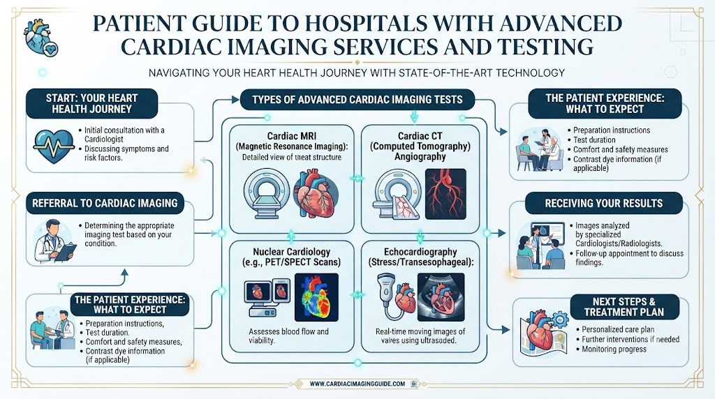
Evaluating cardiovascular health with precision requires sophisticated diagnostic capabilities that go beyond standard physical examinations. Modern cardiac imaging allows doctors to evaluate heart structure, blood flow, heart function, and coronary arteries with greater accuracy. Hospitals with comprehensive cardiac imaging programs often combine multiple imaging technologies with experienced cardiologists to support accurate diagnosis and treatment planning (BestHeartSurgery.com).This comprehensive educational guide explores what advanced cardiac imaging involves, the various diagnostic tests available, advanced technologies used in modern cardiology, and how patients can select the right hospital. For reliable educational resources, hospital comparisons, and patient guidance, readers can explore BESTHEARTSURGERY.
What Is Advanced Cardiac Imaging?
Advanced cardiac imaging refers to a specialized group of non-invasive and minimally invasive diagnostic procedures designed to visualize the heart’s anatomy, blood vessels, and functional performance in high detail.While routine heart tests—such as a standard electrocardiogram (ECG)—provide information on electrical rhythms, advanced imaging unlocks a deeper look into the physical architecture of the cardiovascular system. The primary purpose of these tests is to identify abnormalities, measure pumping efficiency, quantify blood flow, and monitor disease progression.The role of imaging in diagnosis and treatment planning is indispensable; it bridges the gap between clinical symptoms and targeted medical or surgical interventions, helping doctors map out precise strategies for patient care.
Common Heart Conditions Diagnosed with Cardiac Imaging
Advanced diagnostic tools help medical teams identify a wide array of cardiovascular pathologies.
Coronary Artery Disease
Detects plaque buildup, narrowing, or blockages within the coronary arteries that supply blood to the heart muscle.
Heart Valve Disease
Evaluates structural defects, regurgitation (leaking), or stenosis (narrowing) of the heart valves.
Heart Failure
Assesses how effectively the heart chambers pump blood and identifies underlying causes of cardiac weakness.
Cardiomyopathy
Visualizes thickening, stiffening, or enlargement of the heart muscle walls.
Congenital Heart Disease
Identifies structural heart defects present from birth in pediatric or adult patients.
Cardiac Tumors
Detects rare benign or malignant masses growing inside or adjacent to the heart chambers.
Aortic Disease
Examines the aorta for aneurysms, dissections, or dilations.
Pericardial Disorders
Identifies inflammation or fluid accumulation in the sac surrounding the heart.
Heart Rhythm Disorders
Helps uncover structural anomalies that may trigger electrical arrhythmias.
Structural Heart Abnormalities
Assesses complex septal defects or congenital wall abnormalities.
Cardiac Imaging Services Comparison Table
| Imaging Service | Purpose | Patient Benefit |
| Echocardiography | Assess heart structure and function | Non-invasive evaluation |
| Cardiac CT Scan | Visualize coronary arteries | Detect blockages and calcium deposits |
| Cardiac MRI | Evaluate heart muscle and tissue | Detailed structural assessment |
| Nuclear Cardiology | Measure blood flow to the heart | Identify reduced circulation |
| Stress Imaging | Assess heart function during stress | Detect exercise-related problems |
| Coronary Angiography | Examine coronary arteries | Guide treatment planning |
Services Offered by Hospitals with Advanced Cardiac Imaging
Medical institutions dedicated to comprehensive cardiovascular care provide a wide array of specialized imaging services.
- Transthoracic Echocardiography (TTE): Standard ultrasound imaging performed through the chest wall to visualize valves and chamber movement.
- Transesophageal Echocardiography (TEE): Specialized ultrasound using a small probe placed down the esophagus for high-resolution views of internal heart structures.
- Cardiac CT Angiography: Advanced computed tomography scans using contrast dye to image coronary arteries clearly.
- Cardiac MRI: Magnetic resonance imaging providing detailed tissue characterization without ionizing radiation.
- Nuclear Cardiology: Specialized scans using small, safe amounts of radioactive tracers to evaluate myocardial blood flow.
- Stress Echocardiography: Combines ultrasound imaging with physical or chemical stress to assess how the heart responds to increased demand.
- Coronary Angiography: Invasive catheter-based imaging providing definitive visualization of arterial lumens.
- Structural Heart Imaging: High-precision modalities dedicated to planning transcatheter valve replacements or repairs.
- Pre-Surgical Cardiac Assessment: Comprehensive imaging reviews conducted prior to major cardiothoracic operations.
- Follow-Up Imaging: Serial scans utilized to track healing and treatment effectiveness over time.
Advanced Technologies Used
Cutting-edge technology forms the backbone of successful cardiovascular diagnostics, ensuring high-fidelity images with minimal patient discomfort.
- High-Resolution Echocardiography: Advanced ultrasound machines equipped with digital beamforming for crystal-clear tissue rendering.
- Multi-Slice Cardiac CT Scanners: High-speed computed tomography systems capable of capturing clear images between heartbeats.
- Cardiac MRI Systems: High-field magnetic resonance units designed specifically to eliminate motion artifacts from heartbeats and breathing.
- Digital Angiography Suites: Modern catheterization labs featuring ultra-low radiation exposure protocols and real-time digital enhancement.
- 3D and 4D Cardiac Imaging: Volumetric rendering software that displays heart structures in three dimensions plus time.
- AI-Assisted Image Analysis: Artificial intelligence tools assisting technologists and radiologists in measuring ejection fractions and detecting subtle anomalies (where available).
- Advanced Image Archiving Systems: Secure digital networks that store and retrieve past scans for instant comparison.
Multidisciplinary Cardiac Imaging Team
Accurate interpretation of complex heart scans depends on the collaborative efforts of a specialized multidisciplinary team.
- Cardiologists: Primary physicians who order, coordinate, and interpret imaging findings in the context of patient health.
- Cardiac Radiologists: Subspecialist physicians experts in reading complex cardiac CT and MRI scans.
- Cardiac Surgeons: Review imaging data meticulously to plan surgical interventions and bypass procedures.
- Imaging Technologists: Skilled professionals who operate advanced equipment and ensure patient safety and comfort during scans.
- Cardiac Nurses: Provide direct clinical observation, prep patients, and monitor vital signs throughout testing.
- Interventional Cardiologists: Utilize catheter-based imaging data to perform minimally invasive procedures.
- Heart Failure Specialists: Analyze imaging metrics to optimize medical management for weakened heart muscles.
- Rehabilitation Teams: Review diagnostic capacities to tailor safe, progressive physical recovery programs.
How to Choose the Right Hospital
Selecting an appropriate hospital for cardiac diagnostics requires evaluating institutional capabilities, equipment standards, and clinical expertise.
- Experienced Cardiac Imaging Specialists: Presence of board-certified cardiologists and radiologists with dedicated subspecialty training in imaging.
- Availability of Multiple Imaging Technologies: Access to a full spectrum of modalities, from echocardiography to cardiac MRI and CT.
- Accredited Diagnostic Facilities: Adherence to rigorous national or international laboratory accreditation standards.
- Comprehensive Heart Care Services: Seamless integration between diagnostic imaging departments and surgical or medical treatment teams.
- Modern Imaging Equipment: State-of-the-art scanners ensuring high-resolution results and reduced scan times.
- Timely Reporting: Efficient workflows that deliver prompt diagnostic reports to referring physicians.
- Coordinated Multidisciplinary Care: Smooth communication channels between imaging experts and clinical care providers.
Hospital Selection Checklist
| Feature | Why It Matters |
| Experienced Cardiac Imaging Team | Accurate interpretation |
| Multiple Imaging Modalities | Comprehensive diagnosis |
| Modern Imaging Equipment | High-quality imaging |
| Integrated Cardiology Services | Coordinated treatment |
| Patient Safety Standards | Safer diagnostic procedures |
| Follow-Up Care | Ongoing heart health management |
What to Expect During Cardiac Imaging
Demystifying the diagnostic process helps reduce anxiety and prepares patients for their upcoming appointments.
- Medical History Review: The care team evaluates your symptoms, past conditions, and reason for the test.
- Physical Assessment: Basic checks of blood pressure, heart rate, and oxygen levels.
- Imaging Preparation: Changing into a gown, removing metal objects, or receiving instructions regarding contrast dye.
- Performing the Test: Lying comfortably on an imaging table while technologists capture scans using specialized equipment.
- Image Interpretation: Expert physicians analyze the captured data files for structural or functional anomalies.
- Discussion of Results: Reviewing findings with your cardiologist to understand what the images reveal.
- Treatment Planning: Collaborating on next steps based on objective diagnostic data.
- Follow-Up Recommendations: Scheduling ongoing monitoring or specialized consultations if indicated.
Preparing for Cardiac Imaging
Adhering to pre-test instructions ensures accurate results and prevents unnecessary procedure delays.
- Following Fasting Instructions: Some tests, such as certain cardiac CT scans or nuclear tests, require fasting for a few hours beforehand.
- Medication Guidance: Consulting your physician on whether to take or temporarily hold routine medications (such as beta-blockers).
- Wearing Comfortable Clothing: Opting for loose-fitting, metal-free garments to streamline preparation.
- Informing the Care Team About Allergies: Disclosing any prior reactions to iodinated contrast dyes or latex.
- Discussing Implanted Medical Devices: Informing staff immediately if you have a pacemaker, defibrillator, or metal implants before undergoing MRI scans.
Benefits of Early Cardiac Imaging
Proactive diagnostic evaluation offers profound clinical advantages for long-term health.
- Early Detection of Heart Disease: Identifying subclinical plaque or valve dysfunction before severe symptoms emerge.
- Better Treatment Planning: Providing surgeons and cardiologists with precise maps of cardiovascular anatomy.
- Monitoring Disease Progression: Tracking whether chronic conditions like heart failure or aortic dilation are stabilizing or advancing.
- Evaluating Treatment Effectiveness: Assessing how well medications, lifestyle changes, or prior surgeries are performing.
- Supporting Preventive Heart Care: Empowering patients and doctors with concrete data to guide risk-reduction strategies.
How BESTHEARTSURGERY Helps Patients
Navigating cardiovascular health options requires reliable, expert-backed information. Platforms like BESTHEARTSURGERY support patients and families by offering:
- Heart Health Education: Providing clear, accessible guides on cardiac diagnostics and treatment pathways.
- Hospital Discovery: Helping users locate specialized heart centers and advanced imaging facilities nearby.
- Cardiac Imaging Awareness: Promoting understanding of how different diagnostic tests evaluate heart function.
- Hospital Comparison Resources: Offering structured insights to evaluate hospital capabilities and imaging standards.
- Patient Guidance: Equipping readers with effective questions to ask during cardiology consultations.
- Informed Healthcare Decisions: Supporting patients in making empowered choices regarding their cardiovascular care (BestHeartSurgery.com).
Common Mistakes Patients Make
- Ignoring Persistent Chest Discomfort: Dismissing ongoing chest pressure or shortness of breath as minor muscle strain.
- Delaying Recommended Imaging Tests: Postponing scheduled scans out of anxiety or schedule conflicts.
- Choosing Hospitals Based Only on Cost: Selecting diagnostic centers based solely on cheap pricing while overlooking equipment quality and specialist expertise.
- Missing Follow-up Appointments: Failing to return to review imaging results with their primary cardiologist.
- Not Sharing Complete Medical History: Withholding details about past surgeries, allergies, or implanted devices.
Practical Solutions: Address persistent cardiac symptoms promptly, complete ordered imaging tests without delay, and ensure full disclosure of your medical background to your care team.
Common Myths vs Facts
| Myth | Fact |
| Every heart imaging test uses radiation | Tests such as echocardiography and cardiac MRI do not use ionizing radiation |
| Heart imaging is only needed after symptoms appear | Imaging may also help evaluate risk or monitor known heart conditions |
| All imaging tests provide the same information | Different tests evaluate different aspects of heart structure and function |
| Cardiac imaging always leads to surgery | Many imaging results guide medication, monitoring, or lifestyle changes instead |
Real-Life Educational Scenarios
Scenario 1: Diagnosing Coronary Artery Disease via Cardiac CT
An adult experiencing intermittent chest discomfort during exertion is referred for a cardiac CT angiography. The advanced scan reveals a moderate blockage in a coronary artery, allowing the cardiologist to initiate targeted medication and lifestyle modifications before an acute heart attack occurs.
Scenario 2: Evaluating Valve Disease with Echocardiography
During a routine physical exam, a doctor hears a mild heart murmur and orders a transthoracic echocardiogram. The imaging clearly outlines a leaky mitral valve, giving the heart team the precise measurements needed to monitor the condition over time.
Scenario 3: Assessing Heart Muscle Health with Cardiac MRI
A patient with unexplained heart failure undergoes a cardiac MRI. The high-resolution tissue imaging identifies previous silent myocardial scarring, guiding the heart failure specialist to optimize the patient’s pharmacological management plan.
Scenario 4: Creating a Treatment Plan Using Imaging Results
A cardiologist reviews comprehensive nuclear cardiology stress test results alongside an echocardiogram. The integrated data confirms preserved blood flow during rest but inducible ischemia during stress, leading to a tailored revascularization strategy.
FAQs (15 Questions)
1. What is advanced cardiac imaging?
Advanced cardiac imaging encompasses sophisticated non-invasive and minimally invasive technologies used to visualize the heart’s structure, blood flow, and function.
2. When is cardiac imaging recommended?
Imaging is recommended when patients experience unexplained chest pain, shortness of breath, heart murmurs, or when doctors need to monitor known heart disease.
3. What is the difference between an echocardiogram and a cardiac MRI?
An echocardiogram uses sound waves (ultrasound) to view heart motion in real-time, while a cardiac MRI uses magnetic fields and radio waves to provide highly detailed tissue characterization.
4. What does a cardiac CT scan show?
A cardiac CT scan provides detailed 3D images of the coronary arteries, heart chambers, and surrounding blood vessels, making it effective for detecting calcium deposits and blockages.
5. Is cardiac imaging painful?
Most cardiac imaging tests—such as echocardiograms, MRIs, and CT scans—are completely painless, though intravenous lines for contrast dye may cause a brief pinch.
6. How long do imaging tests take?
Durations vary by test; an echocardiogram typically takes 30 to 45 minutes, while cardiac MRI or CT scans can range from 30 minutes to an hour.
7. Do I need to prepare before the test?
Preparation depends on the specific test and may include fasting, avoiding caffeine, or reviewing medication instructions with your doctor.
8. Are cardiac imaging tests safe?
Yes, modalities like echocardiography and cardiac MRI involve zero ionizing radiation, while CT scans and nuclear tests use minimal, controlled doses of radiation and contrast.
9. What conditions can cardiac imaging detect?
Imaging can detect coronary artery disease, heart failure, valve abnormalities, congenital heart defects, cardiomyopathies, and aortic diseases.
10. How are imaging results interpreted?
Scans are analyzed by expert cardiac radiologists and cardiologists who measure heart dimensions, wall motion, and blood flow parameters.
11. Can imaging help before heart surgery?
Yes, detailed cardiac imaging provides surgeons with an exact anatomical map, ensuring precise planning for complex operations.
12. How do I choose the right hospital?
Look for hospitals with board-certified imaging specialists, advanced multi-modality equipment, strong accreditation, and integrated cardiology services.
13. Why are follow-up imaging tests important?
Follow-up scans allow doctors to track disease progression, evaluate the success of treatments, and adjust care plans accordingly.
14. What technologies are used in advanced cardiac imaging?
Key technologies include high-resolution ultrasound, multi-slice CT scanners, high-field MRI units, and 3D volumetric rendering software.
15. How does BESTHEARTSURGERY help patients find hospitals with advanced cardiac imaging services?
BESTHEARTSURGERY provides trusted educational resources, hospital comparison tools, and guidance to help patients navigate cardiovascular diagnostics effectively.
Final Thoughts
Advanced cardiac imaging stands as a cornerstone of modern cardiovascular medicine, transforming how physicians diagnose, evaluate, and treat heart conditions. The combination of high-resolution diagnostic modalities—such as echocardiography, cardiac CT, and cardiac MRI—with experienced multidisciplinary imaging teams ensures that patients receive precise, timely evaluations. Selecting hospitals equipped with comprehensive cardiac imaging services significantly enhances clinical decision-making and supports long-term heart health.